


Abstract
Background: We have previously reported several lignin-like activities of the alkaline extract of the leaves of Sasa senanensis Rehder (SE), such as anti-HIV and radical scavenging activity. As an extension, possible SE protection of cells from ultraviolet (UV)-induced injury (referred to “anti-UV activity”) was investigated. Materials and Methods: HSC-2, human oral squamous cell carcinoma cells, were exposed to UV irradiation in phosphate-buffered saline containing SE or its fractions, and then incubated for 48 hours in fresh regular culture medium to determine the viable cell number by the MTT method. SE was separated by gel filtration chromatography into the following four fractions: polysaccharide, large and small lignin-carbohydrate complexes and lower molecular weight polyphenol fractions (Fr. I). Results: The anti-UV activity of SE was slightly less than that of sodium ascorbate, but higher than that of gallic acid, (−)-epigallocatechin gallate, chlorophyll a and chlorophyllin. The combination of SE and sodium ascorbate gave synergistic anti-UV activity. On gel filtration fractionation, the majority of the anti-UV activity was recovered from Fr. I, which was eluted as a single peak on high performance liquid chromatography (HPLC). Conclusion: The anti-UV activity of SE further suggests its potential as an alternative medicine.
- Ultraviolet light
- cytoprotection
- vitamin C
- alkaline extract of leaves
- Sasa senanensis Rehder
- fractionation
The alkaline extract of the leaves of Sasa senanensis Rehder or Sasa albo-marginata Makino et Shibata (SE) (SASA-Health®), as an over the counter drug, is recognized as being effective in treating fatigue, low appetite, halitosis, body odour and stomatitis. However, there is no scientific evidence that demonstrates these phenomena. SE has been reported to show antiseptic (1), membrane stabilising (2), anti-inflammatory and phagocytic (3), radical scavenging (4, 5), antibacterial and anti-viral activities (5). It has recently been reported that SE inhibited the nitric oxide (NO) and prostaglandin E2 (PGE2) production by lipopolysaccharide (LPS)-activated mouse macrophage-like cells (RAW264.7) via inhibition of inducible NO synthase and cyclooxygenase-2 expression at both protein and mRNA levels (6). We have recently suggested that multiple components of SE may be associated with each other in the native state or after extraction with alkaline solution (7).
Moderate doses of ultraviolet (UV) rays exert several favorable effects such as sterilization and disinfection (8), vitamin D synthesis (9) and stimulation of the metabolism and skin resistance. However, excessive doses of UV light produce reactive oxygen species (ROS), which damage cellular DNA and proteins, leading to carcinogenesis (10). Guanine, the most susceptible DNA base, is oxidized to 7,8-dihydroxy-8-oxoguanine upon UV-irradiation, and triggers the transversion of G:C to T:A (11). Antitumor agents and radiation have been shown to induce different types of cell death in different cell types (12, 13). Higher doses of UV irradiation induced apoptotic cell death in human myelogenous leukemia cell lines, but induced other types of cell death in human T-cell leukemia, erythroleukemia, glioblastoma (14), oral squamous cell carcinoma (OSCC) cell lines and human normal oral cells (gingival fibroblasts, pulp cells, periodontal ligament fibroblast) (15). We recently established a method that can measure the activity of test materials to protect cells from UV ray (referred to as “anti-UV activity”) (15, 16). Using this method, the anti-UV activity of SE, in comparison with sodium ascorbate, a popular antioxidant was investigated.
Materials and Methods
Materials. The following chemicals and reagents were obtained from the indicated companies: Dulbecco's modified Eagle's medium (DMEM; Gibco BRL, Grand Island, NY, USA); fetal bovine serum (FBS), 3-(4,5-dimethylthiazol-2-yl)-2,5-diphenyltetrazolium bromide (MTT), dimethyl sulfoxide (DMSO), (−)-epigallocatechin gallate (EGCG), chlorophyllin, chlorophyll a (Wako Pure Chemical Ind., Osaka, Japan); sodium ascorbate, gallic acid (Tokyo Chemical Industry Co., Ltd., Tokyo).
Anti-UV activity of SE and various antioxidants.
SE was prepared and supplied by Daiwa Biological Research Institute Co. Ltd., Kawasaki, Kanagawa, Japan. SE (21 ml) was freeze dried to give a powder (1.1g).
Cell culture. Human oral squamous cell carcinoma HSC-2 cells were used, since this cell line was much more sensitive to UV irradiation, as compared with human oral normal cells (gingival fibroblast, pulp cells, periodontal ligament fibroblast) (15, 16). HSC-2 cells were cultured in DMEM supplemented with 10% heat-inactivated FBS, 100 units/ml penicillin G and 100 μg/ml streptomycin sulfate under humidified 5% CO2 atmosphere.
Assay for UV protection assay. The cells were inoculated at 3×10³ cells/0.1 ml in the inner 60 wells of a 96-microwell plate (Becton Dickinson Labware, NJ, USA). The surrounding 36 exterior wells were filled with 0.1 ml of PBS(−) to minimize the evaporation of water from the culture medium. After 48 h, the media of the near confluent cells attached to 96-microwell plates were replaced with PBS(−) containing various concentrations of test substances. PBS(−), instead of DMEM+10% FBS, was used as the medium during UV irradiation, since the latter contained radical scavenger(s) or UV absorbing substance(s) such as phenol red and proteins (16). The cells were then placed at 20.5 cm distance from a UV lamp (wavelength: 253.7 nm) and exposed to UV irradiation (6 J/m2/min) for 1 min. The media were replaced with fresh DMEM+10% FBS and the cells were cultured for a further 48 h at 37°C in a CO2 incubator before determination of the relative viable cell number by the MTT method. In brief, the treated cells were incubated for another 4 h in fresh culture medium containing 0.2 mg/ml MTT. The cells were then lysed with 0.1 ml of DMSO and the absorbance at 540 nm of the cell lysate was determined using a microplate reader (Biochromatic Labsystem, Helsinki, Finland). From the dose-response curve, the 50% cytotoxic concentration (CC50) and the effective concentration, that which increased the viability of UV-irradiated cells to 50% of the control (EC50) were determined. The selectivity index (SI) was determined by the following equation: SI=CC50/EC50 (15, 16)
Fractionation of SASA-Health (SE) by gel filtration chromatography. SE (60 mg/600 μl) was applied to a TOYOPEARL HW-55F column (1.5 cm i.d. × 100 cm, Tosoh, Tokyo, Japan) and the column was eluted with 20 mM NaHCO3. The eluate was collected every 10 min and the elution curve made by plotting the eluate absorbance at 254 nm. The sugar and polyphenol content in every fraction was measured by the phenol-sulfuric acid method and the Folin-Denis method. In brief for the phenol-sulfuric acid method, 25 μl of the eluate, 25 μl of 5 % phenol (v/w) and 125 μl of sulfuric acid were added. The mixture was left to stand for 30 min at room temperature and the absorbate at 492 nm of the mixture was determined using a microplate reader (Bichromatic Labsystems). In brief for the Folin-Denis method, 25 μl of the eluate, 25 μl of 2 N phenol reagent and 125 μl of 1 N NaOH were added. The mixture was left to stand for 30 min at room temperature, and the absorbance at 630 nm of the mixture was determined using a microplate reader as above.
High performance liquid chromatography (HPLC) analysis. The HPLC system was composed of a JASCO PU-980 pump, a JASCO UV-970 UV/VIS detector and a column of Inertsil ODS-3 (4.6 mm i.d. × 150 mm, 5 μm, GL Sciences Inc., Tokyo, Japan). The detection wavelength was set at 280 nm and the sample was injected manually. Solvent A, 5 % acetonitrile containing 0.1 % formic acid and solvent B, acetonitrile were used as the mobile phase, with a flow rate of 1.0 ml/min and gradient B 0% (0-10 min), 10% (10-15 min), 25% (15-30 min), 40% (30-40 min), 100% (40-50 min) and holding 100% (to 60 min).
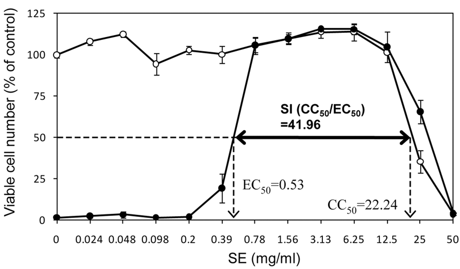
Anti-UV activity of SE. HSC-2 cells were treated without (○) or with UV (●) irradiation (6 J/m2/min) for 1 min in PBS(−) containing SE. Viable cell number determined by MTT method 48 h after irradiation, expressed as percent of control (without UV irradiation). EC50: 50% effective concentration, CC50: 50% cytotoxic concentration, SI: selectivity index (CC50/EC50). Mean±S.D. of triplicate determinations.
Statistical analysis. All the experiments were performed by triplicate assays, and each value represents mean±S.D.
Results
UV irradiation. UV irradiation (6 J/m2/min) for 1 min followed by 48 h culture resulted in extensive cell death. SE dose-dependently inhibited the UV-induced cytotoxicity in a bell-shaped fashion (Figure 1). The viability of the cells was recovered to 50% by the addition of 0.53 mg/ml SE (=EC50). From the dose-response curve without UV irradiation, CC50 of SE was calculated to be 22.24 mg/ml. From these values, SI (CC50/EC50) was calculated to be 41.96. Similar experiments were repeated three times to yield the mean value of SI=19.7±15.1 (mean of four independent experiments) (Table I). This value was slightly less than that of sodium ascorbate (SI=30.2±13.4) (mean of five independent experiments), but higher than that of gallic acid (SI=17.1), EGCG (SI=7.7), chlorophyllin (SI=0.53) and chlorophyll a (SI<0.24) (Table I).
Synergistic anti-UV activity of SE and sodium ascorbate. By the addition of 0.125 or 0.25 mM sodium ascorbate, the anti-UV activity of SE was increased from SI=13.8 to 20.5 and 70.6, respectively (Figure 2).
Anti-UV activity of SE fractions. When SE was fractionated by gel filtration chromatography, it was roughly separated into four fractions: polysaccharide (fraction 22-28 [66-84 ml]), lignin-carbohydrate complex (fraction 29-44 [87-132 ml] and 45-51 [135-153 ml]) and low molecular weight polyphenol (fraction 52-59 [155-177 ml] indicated by Fr. I) (Figure 3A). When each column fraction was added during UV irradiation, the viability of the cells was recovered up to nearly 50% of the control level, depending on fraction of the eluted material. Most of anti-UV activity was concentrated in the lower molecular weight fractions (Fr. I), which was eluted as a single peak on HPLC (Figure 3C). Noticeably less anti-UV activity was demonstrated by the two lignin-carbohydrate complex fractions, whereas no anti-UV activity was shown with the polysaccharide fraction (Figure 3B). Each of these fractions did not significantly affect the growth of the HSC-2 cells which were not exposed to UV irradiation (data not shown).

Synergistic anti-UV activity of SE and sodium ascorbate. HSC-2 cells were treated without (○) or with UV (●) irradiation with SE in the absence (A) or presence of 0.125 (B) or 0.25 mM (C) sodium ascorbate. Viable cell number determined by MTT method 48 h after irradiation, expressed as percent of control (without UV irradiation). Mean±S.D. of triplicate determination.
Discussion
The present study demonstrated for the first time that SE showed potent anti-UV activity (SI=19.7), which was slightly lower than that of sodium ascorbate (SI=30.2), but higher than that of (-)-epigallocatechin gallate (major green tea component), gallic acid (structural unit of tannin) (SI=17.1), chlorophyll a (SI<0.2) and chlorophyllin (SI=0.5) (that may be associated with lignin-carbohydrate complex) (Table I). The SI of SE was also higher than that of tea extracts (green tea, black tea, jasmine, oolong tea, barley tea, Kohki tea) (SI<3.4) (17). Since the anti-UV activity, evaluated by SI value, varied considerably from experiment to experiment (SI=9.5~42.1), it is essential to include a positive control, such as sodium ascorbate, that consistently produces high SI values (SI=13.9~44.7). The present study demonstrated for the first time that the combination of SE and sodium ascorbate produced synergistic anti-UV activity (Figure 2). These properties of SE further suggest its potential as an alternative medicine.

Fractionated SE. A: Fractionation of SE by gel filtration chromatography. The eluate was collected every 10 min and the elution curve made by plotting the eluate absorbance at 254 nm (monitored by mV, indicated by solid line). B: Anti-UV protective activity of each fraction. HSC-2 cells were exposed to 1 min-UV irradiation in PBS(−) with 70 % of each fraction. The viable cell number was determined by MTT method. C: The HPLC analysis of SE and Fr.I. The detection wavelength was set at 280 nm.
Gel filtration chromatography demonstrated that the SE was composed of only four major fractions, confirming our previous suggestion that SE is present as a biological complex associated with many components after extraction with alkaline solution (7). The major anti-UV activity was concentrated in the lower molecular weight fraction that contained only one major peak as judged by HLPC. Since the preparative method does not use organic solvent, this peak may not be flavonoids or tannins. Further structural analysis is required to identify this peak and elucidate the action mechanism of SE.
- Received March 22, 2011.
- Revision received May 23, 2011.
- Accepted May 25, 2011.
- Copyright © 2011 International Institute of Anticancer Research (Dr. John G. Delinassios), All rights reserved
References
In this issue


Jump to section
Related Articles
Cited By...
- Prominent Anti-UV Activity and Possible Cosmetic Potential of Lignin-carbohydrate Complex
- Synergism of Alkaline Extract of the Leaves of Sasa senanensis Rehder and Antiviral Agents
- Anti-Halitosis Effect of Toothpaste Supplemented with Alkaline Extract of the Leaves of Sasa senanensis Rehder
- Anti-UV Activity of Kampo Medicines and Constituent Plant Extracts: Re-evaluation with Skin Keratinocyte System
- Biological Interaction between Sasa senanensis Rehder Leaf Extract and Toothpaste Ingredients
- Structural Characterization of Anti-UV Components from Sasa senanensis Rehder Extract
- Anti-UV Activity of Lignin-Carbohydrate Complex and Related Compounds
- Pilot Clinical Study of Sasa senanensis Rehder Leaf Extract Treatment on Lichenoid Dysplasia
- Anti-UV/HIV Activity of Kampo Medicines and Constituent Plant Extracts
- Biological Activity of SE-10, Granulated Powder of Sasa senanensis Rehder Leaf Extract
- Comparative Study of Biological Activity of Three Commercial Products of Sasa senanensis Rehder Leaf Extract
- Quest for Anti-inflammatory Substances Using IL-1{beta}-stimulated Gingival Fibroblasts
- Biological Activity of Luteolin Glycosides and Tricin from Sasa senanensis Rehder