Article Text

Abstract
Background Mouse models have shown that interleukin (IL)6 stimulates survival, proliferation and progression to cancer of intestinal epithelial cells via activation of signal transducers and activators of transcription 3 (STAT3).
Objective To investigate the expression of IL6/phosphorylated STAT3 (p-STAT3)/suppressor of cytokine signalling 3 (SOCS3) in biopsy specimens from patients with ulcerative colitis (UC) and UC-related colorectal cancer (CRC) progression.
Methods Biopsy specimens from patients with inactive UC (n=18), active UC (n=28), UC with low-grade dysplasia (LGD) (n=9), UC with high-grade dysplasia (HGD) (n=7), UC-CRC (n=11) and sporadic CRC (n=14) were included. Biopsy specimens (n=9) from patients without colonic abnormalities served as control. The protein expression of IL6, p-STAT3 and SOCS3 was determined immunohistochemically.
Results Patients with active UC had significantly more IL6 and p-STAT3-positive epithelial cells than both patients with inactive UC and controls (strong positive IL6: 53.6%, 11.1% and 0%, respectively; p-STAT3: 64.3%, 22.2% and 11.1%, respectively; all p≤0.012). SOCS3-positive cells were significantly increased in colonic epithelium of both inactive and active UC compared with controls (strong positive: 94.4%, 96.4% and 11.1%, respectively; both p<0.001). In dysplasia and cancer, significantly more epithelial cells expressed IL6 and p-STAT3 compared with controls (strong positive IL6: 72.7% and 0% respectively; p-STAT3: 54.5% and 11.1%, respectively; both p<0.05), whereas the proportion of SOCS3-positive cells in this progression reduced (LGD 33.3%; HGD 14.3%; UC-CRC 9.1%). In addition, methylation of the SOCS3 gene was detected in epithelial cells from UC-CRC biopsy specimens.
Conclusion The importance of IL6/p-STAT3 in patients with inflammation-induced CRC was demonstrated. Moreover, SOCS3 may be involved in UC pathogenesis and the absence of SOCS3 seems critical for CRC progression.
- Ulcerative colitis (UC)
- colorectal cancer (CRC)
- interleukin 6 (IL6)
- signal transducers and activators of transcription 3 (STAT3)
- the suppressor of cytokine signalling 3 (SOCS3)
- carcinogenesis
- cell signalling
- inflammatory bowel disease
- intestinal epithelium
- UC
- ulcerative colitis
- CRC
- colorectal cancer
- IL6
- interleukin 6
- STAT3
- signal transducers and activators of transcription 3
- SOCS3
- suppressor of cytokine signalling 3
- IBD
- inflammatory bowel disease
- LGD
- low-grade dysplasia
- HGD
- high-grade dysplasia
- JAK
- Janus kinases
- sIL6R
- soluble IL6 receptor
- p-STAT3
- phosphorylated STAT3
- SH2
- Src homology 2
- HE
- haematoxylin & eosin
- IHC
- immunohistochemistry
- DSS
- dextran sulphate sodium
- MSP
- methylation-specific PCR
Statistics from Altmetric.com
- Ulcerative colitis (UC)
- colorectal cancer (CRC)
- interleukin 6 (IL6)
- signal transducers and activators of transcription 3 (STAT3)
- the suppressor of cytokine signalling 3 (SOCS3)
- carcinogenesis
- cell signalling
- inflammatory bowel disease
- intestinal epithelium
- UC
- ulcerative colitis
- CRC
- colorectal cancer
- IL6
- interleukin 6
- STAT3
- signal transducers and activators of transcription 3
- SOCS3
- suppressor of cytokine signalling 3
- IBD
- inflammatory bowel disease
- LGD
- low-grade dysplasia
- HGD
- high-grade dysplasia
- JAK
- Janus kinases
- sIL6R
- soluble IL6 receptor
- p-STAT3
- phosphorylated STAT3
- SH2
- Src homology 2
- HE
- haematoxylin & eosin
- IHC
- immunohistochemistry
- DSS
- dextran sulphate sodium
- MSP
- methylation-specific PCR
Introduction
Ulcerative colitis (UC) is a phenotype of inflammatory bowel disease (IBD) characterised by a chronic recurrent colonic inflammation, which is associated with an increased risk of developing colorectal cancer (CRC).1 2 Chronic inflammation in the intestine leads to damage of the epithelium. Locally produced cytokines cause inflammation and stimulate the proliferation of crypt cells to compensate for the loss of epithelial cells. This chronically stimulated state of the epithelium may eventually lead to the development of UC-CRC.3–5 Interleukin (IL)6 is a proinflammatory cytokine that has been detected in both colon biopsy specimens and serum samples of patients with UC, and the levels of IL6 correlated with the disease severity.6 7
Interestingly, unlike epithelial cells, most lamina propria T cells of patients with IBD do not express the membrane-bound IL6 receptor (IL6R). In addition to the membrane-bound IL6R, IL6 can act via soluble IL6 receptor in cells expressing gp130 in a process known as trans-signalling. Several observations support an important role of IL6 trans-signalling in the development and maintenance of IBD,8 9 and the specific inhibitors of soluble IL6R-signalling pathway are used to ameliorate IBD.10 The IL6 (trans)-signalling initiates the dimerisation and activation of receptor-associated Janus kinases (JAK). Subsequently, the activated JAK phosphorylates the tyrosine residues on the cytoplasmic tail of the receptor, providing a docking site for proteins with Src homology 2 domains like the signal transducers and activators of transcription (STAT)3 protein.8 Next, STAT3 binds to the receptor and is activated by phosphorylation through JAK, leading to homo- and heterodimerisation of phosphorylated STAT3 (p-STAT3) with translocation to the nucleus, where they interact with specific DNA sequences and induce transcription of target genes.
One of the targets genes of the IL6/p-STAT3 pathway is the suppressor of cytokine signalling 3 (SOCS3). SOCS are a family of proteins that regulate negative feedback to the JAK/STAT cytokine signalling cascade.11 12 There are eight members of the SOCS family: the cytokine inducible Src homology 2 domain-containing protein and SOCS1 to SOCS7.13 SOCS3 protein is expressed in intestinal epithelial cells and lamina propria in mouse colitis models and in patients with UC,14 15 suggesting a role in the pathogenesis of colitis.16
In addition to some human observations,17 18 there is strong experimental evidence from mouse studies that suggest a key role for IL6/STAT3/SOCS3 in regulating intestinal epithelial cell homoeostasis. These studies also imply that an imbalance between SOCS3 expression and IL6/p-STAT3 signalling will lead to inflammation10 and eventually to inflammation-induced carcinogenesis.19 The latter observations support the idea that IL6 signalling is a potential therapeutic target in IBD and IBD-related CRC.9 20 There is, however, a general lack of clinical data from patients with UC and UC-CRC to support this. We studied the expression of IL6/p-STAT3 and SOCS3 in colon biopsy specimens from patients with UC in different phases of the disease. Our study provides clinical data supporting animal model-derived mechanisms on IL6/STAT3, which suggest the involvement of SOCS3 in UC-related cancer, and support the IL6 signalling pathway as a therapeutic target in UC and UC-related CRC.
Patients, materials and methods
Patients and biopsies
To investigate the involvement of the IL6/STAT3/SOCS3 pathway in UC and UC-related carcinogenesis, we collected biopsy specimens from 9 healthy controls, 18 patients with inactive UC, 28 patients with active UC, 9 UC patients with low-grade dysplasia (LGD), 7 UC patients with high-grade dysplasia (HGD), 11 patients with UC-CRC and 14 patients with sporadic CRC. Colon biopsy specimens were obtained during colonoscopy performed for clinical care and surveillance studies after informed consent. The diagnosis of UC was based on conventional clinical, endoscopic and pathohistological criteria as described by Lennard-Jones.21 Colonic biopsy specimens from non-UC patients without abnormalities at colonoscopy and histories of gastrointestinal disease served as controls. An independent pathologist reconfirmed colitis, dysplasia (low grade or high grade) and CRC in the biopsy specimens of patients with UC based on histological scoring. In addition, the colitis severity was graded from 0 to 5 according to Geboes criteria.22 Patients' characteristics and histological data are shown in table 1.
Patients' characteristics
Immunohistochemistry
All the biopsy specimens were fixed in 10% formalin and embedded in paraffin. Haematoxylin eosin staining on paraffin-embedded biopsy specimens was performed to evaluate the severity of colitis. Sections of paraffin-embedded tissues (4 μm) were mounted onto slides of Superfrost Plus (Thermo Scientific, UK), deparaffinised in xylene and rehydrated. The endogenous peroxidase activity was blocked by 3% H2O2 in methanol for 15 min at room temperature. Antigen retrieval was performed by boiling in preheated buffer (Tris/EDTA pH 9.0 for SOCS3 or IL6 and EDTA pH 8.0 for p-STAT3) for 15 min at 200 W in a microwave. Next, slides were transferred to Shandon chambers and blocked by 10% normal human serum, 10% goat serum in phosphate-buffered saline pH 7.4 for SOCS3 and IL6, 5% goat serum in Tris-buffered saline and 0.1% Tween 20 for p-STAT3 for 1 h at room temperature. Monoclonal rabbit anti-p-STAT3 tyrosine 705 (cell signalling, #9145), polyclonal rabbit anti-SOCS3 and polyclonal rabbit anti-IL6 (both abcam ab53984, ab6672) were diluted in 10% blocking buffer at 1:50; 1:200; 1: 200, respectively, and incubated at 4°C overnight. Envision goat anti-rabbit-HRP (DAKO, Denmark) was used as secondary antibody. Immunoreactions were detected using 3-3-diaminobenzidine (10 mg/ml) in Tris-HCl 0.05 M pH 7.6 containing imidazole stock (0.068 g/10 ml) and 0.03% H2O2, followed by counterstaining with haematoxylin. Negative controls were performed using only the secondary antibody.
Immunohistochemical scoring
Two experienced people scored the staining of epithelial cells in a blinded manner. The percentage of cells that stained positive (immunoreactivity above background) in the area consistent with that used for diagnoses was quantified. The immunoreactivity was defined for SOCS3 as: strong positive (>60% of the cell population in the biopsy specimen stains positive), positive (30–60%) or mild (<30%) cytoplasm staining; for p-STAT3 as: strong positive (>20%), positive (10–20%) or negative (<10%) nuclear staining in epithelial cells. The expression of IL6 cytoplasm staining was evaluated in both epithelial and non-epithelial cells, and defined as strong positive (>60%), positive (30–60%) or mild (<30%) in epithelial cells and strong positive (>70%), positive (40–70%) or mild (<40%) in non-epithelial cells.
Macro-dissection and DNA processing
Intestinal epithelial cells and/or adenomatous tissue were isolated from paraffin-embedded intestinal biopsy specimens (three controls, three inactive UC, three active UC and four UC-CRC) using macro-dissection.23 DNA was isolated using a Fixed-Tissue Genomic DNA Purification kit (Cat. #MD1180, Promega, USA) and bisulphate-modified using the EZ DNA methylation kit (ZYMO Research, USA). Universal methylated and unmethylated DNA (Millipore, USA) were used as controls.
Methylation-specific PCR (MSP)
MSP was performed on bisulphite-treated genomic DNA using specific primers as previously described.24 Sequences of methylation-specific SOCS3 primers were 5′-GGAGATTTTAGGTTTTCGGAATATTTC-3′ (forward) and 5′-CCCCCGAAACTACCTAAACGCCG-3′ (reverse), corresponding to -525 through -499 and -384 through -406, respectively. Sequences of the unmethylation-specific primers were 5′-GTTGGAGATTTTAGGTTTTTGGAATATTTT-3′ (forward) and 5′-AAACCCCCAAAACTACCTAAACACCA-3′ (reverse). A PCR was performed in a 25 μl volume containing 40 ng bisulphate-modified DNA, 5× Gold Taq buffer, 0.2 mM dNTPs, 10 pmol specific primer mix (forward and reverse primers) and one unit Gold Taq enzyme (Promega). The PCR condition was set up as 95°C for 5 min, followed by 40 cycles of 95°C for 30 s, 57°C for 30 s and 72°C for 30 s, and ending with a final extension of 72°C for 5 min. The PCR products were visualised on a 2% agarose gel using ethidium bromide and UV illumination.
Statistical analysis
Statistical analysis was performed using SPSS software version 15.0. Data were presented as mean±SD. The relationship between each group for SOCS3, p-STAT3 and IL6 was determined by χ2. The association between IL6/p-STAT3-positive cells and severity of disease was demonstrated by Spearman correlation. P value <0.05 was considered statistically significant.
Results
Patients' characteristics
To investigate the possible involvement of the IL6/STAT3/SOCS3 signalling pathway in UC and UC-related carcinogenesis we studied the expression of these proteins in biopsy specimens from patients with UC in different stages. As shown in table 1, both the mean age and duration of disease were significantly higher in the UC-CRC group than in the other disease status groups (p<0.01), which is in accordance with the increasing risk of developing UC-CRC over time. In addition, the mean age of patients with sporadic CRC was also significantly higher than those of patients in other groups (p<0.01). There were no other significant differences among the other groups, including treatment. Inactive UC defines patients in long-term remission with no endoscopic signs of colitis and a Geboes histology score of 0 or 1. Active UC defines patients in the acute phase of the disease with endoscopically visible signs of colitis and a Geboes histology score of ≥2.
IL6 expression in patients with active UC and inactive UC
Immunohistochemistry was performed to determine the protein expression of IL6 in different cells and in different phases of disease (figure 1a). The colonic epithelial cells revealed significantly more IL6-positive cells in the biopsy specimens from patients with active UC than in patients with inactive UC (p=0.011) or controls (p<0.001) (figure 2a). In addition, there were significantly more IL6-positive cells in the non-epithelial cells in biopsy specimens from patients with active UC than in those with inactive UC (p<0.001) or controls (p<0.001) (figure 2b). Figure 2c, d showed a positive correlation between IL6 expression and the severity of colitis in both epithelial (r=0.56, p=0.0001) and non-epithelial cells (r=0.62, p<0.0001) when biopsy specimens with inactive and active UC were combined.

Immunohistochemical staining of (a) interleukin (IL)6 (×400, cytoplasmic staining); (b) phosphorylated signal transducers and activators of transcription 3 (p-STAT3) (×400, nuclear staining); (c) suppressor of cytokine signalling 3 (SOCS3) (×400, cytoplasmic staining) in colonic biopsy specimens from controls, patients with inactive ulcerative colitis (UC) and patients with active UC.

Analysis of the immunohistochemical interleukin (IL)6 staining in (a) colonic epithelial cells and (b) non-epithelial cells from biopsy specimens of controls, patients with inactive ulcerative colitis (UC) and patients with active UC. *Significantly more IL6-positive cells in were found in active UC than in both inactive UC and control groups. In epithelial cells: active UC versus inactive UC: p=0.011; active UC versus normal controls: p<0.001. In non-epithelial cells: active UC versus inactive UC: p<0.001; active UC versus normal controls: p<0.001. Correlations between IL6-positive (c) epithelial/(d) non-epithelial cells and the severity of colitis in patients with UC (r=0.56, p=0.0001/r=0.62, p<0.0001).
P-STAT3 and SOCS3 expression in patients with active UC and inactive UC
To see whether IL6 expression correlates with the activation of the STAT3/SOCS3 signalling pathway in the different biopsy specimens, we investigated p-STAT3 and SOCS3 expression (figure 1b, c). As shown in figure 3a, significantly more p-STAT3-positive epithelial cells could be seen in biopsy specimens of patients with active UC than in both inactive UC (p=0.007) and controls (p=0.012). The number of p-STAT3-positive epithelial cells and non-epithelial cells both correlated positively with the severity of colitis (figure 3b, r=0.44, p=0.001; figure 3c, r=0.51, p<0.001).
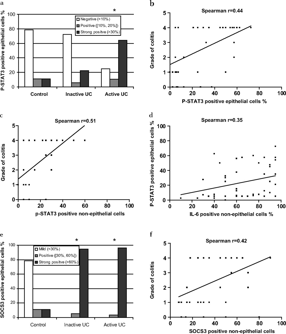
(a) The differences in phosphorylated signal transducers and activators of transcription 3 (p-STAT3) protein expression in epithelial cells from biopsy specimens of controls, patients with inactive ulcerative colitis (UC) and patients with active UC . *p-STAT3 expressed on significantly more cells in active UC than in both inactive UC and control groups (active UC vs inactive UC: p=0.007; active UC vs normal control: p=0.012). (b) The correlation between p-STAT3-positive epithelial cells and severity of colitis of patients with UC (r=0.44, p=0.0009). (c) The correlation between p-STAT3-positive non-epithelial cells and severity of colitis of patients with UC (r=0.51, p<0.001). (d) The correlation between interleukin (IL)6-positive non-epithelial cells and p-STAT3-positive epithelial cells (r=0.35, p=0.016). (e) The differences in suppressor of cytokine signalling 3 (SOCS3) protein expression in epithelial cells from control, inactive UC and active UC groups (in epithelial cells). *SOCS3 expressed on significantly more cells in both inactive and active UC in comparison with those of controls (inactive UC vs normal control: p<0.001; active UC vs normal control: p<0.001). (f) The correlation between SOCS3-positive non-epithelial cells and severity of colitis of patients with UC (r=0.42, p=0.005).
Since non-epithelial cells (eg, leucocytes) have been suggested to be the major source of IL6 in the inflamed intestine, we correlated the expression of IL6 in non-epithelial cells to the p-STAT3 expression in epithelial cells. As shown in figure 3d, IL6-positive non-epithelial cells correlated with p-STAT3-positive epithelial cells (r=0.35, p=0.016).
SOCS3-positive epithelial cells were uniformly increased in both inactive and active UC biopsy specimens compared with those of the control (inactive UC vs normal control: p<0.001; active UC vs normal control: p<0.001) (figure 3e). So, whereas no positive correlation between SOCS3 expression and severity of colitis was found in the epithelial cells, a positive correlation was found between SOCS3-positive non-epithelial cells and the severity of the colitis (figure 3f, r=0.42, p=0.005).
IL6 expression in colonic epithelial cells of patients during UC-CRC progression
Since IL6 has both proinflammatory as well as carcinogenic potential, we investigated IL6 expression in biopsy specimens from patients in different stages of progression to UC-related CRC (LGD, HGD and UC-CRC) (figure 4a), and analysed IL6 levels in epithelial and non-epithelial cells. Significantly more IL6-positive epithelial cells were observed in all stages of progression to UC-related CRC than in the controls (p<0.001) (figure 5a). Although a trend was observed for the non-epithelial cells, there was no significant difference in IL6 expression between biopsy specimens from patients with UC-CRC progression and controls (data not shown).
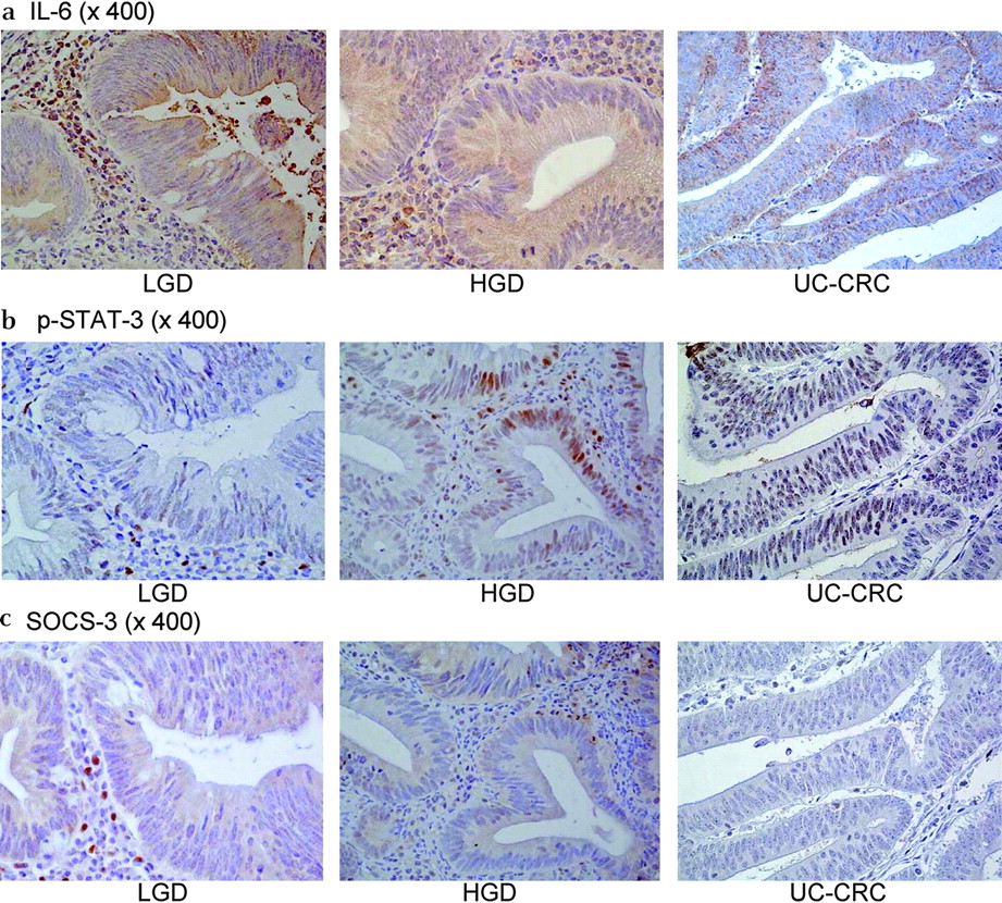
Immunohistochemical staining of (a) interleukin (IL)6 (×400, cytoplasmic staining); (b) phosphorylated signal transducers and activators of transcription 3 (p-STAT3) (×400, nuclear staining); (c) suppressor of cytokine signalling 3 (SOCS3) (×400, cytoplasmic staining) in colonic biopsy specimens from patients with UC with low-grade dysplasia (LGD), high-grade dysplasia (HGD) and colorectal cancer (CRC).
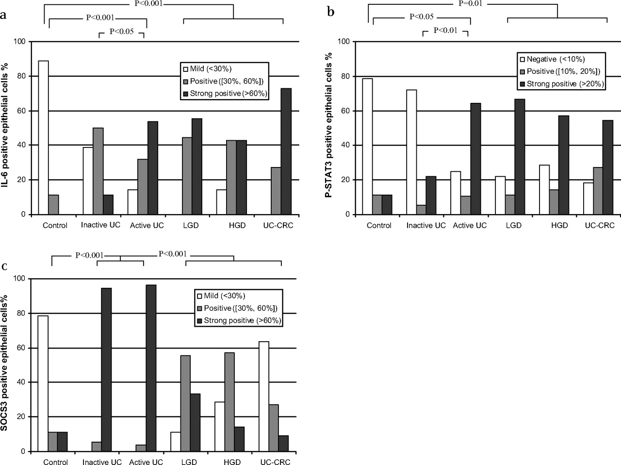
(a) Analysis of the interleukin (IL)6 protein expression in epithelial cells in the progression to ulcerative colitis colorectal cancer (UC-CRC). Significantly more IL6-positive cells were seen in UC-CRC progression than in controls (p<0.001). (b) Phosphorylated signal transducers and activators of transcription 3 (p-STAT3) protein expression in epithelial cells in the progression to UC-CRC. Significantly more p-STAT3-positive cells were seen in UC-CRC progression than in controls (p=0.010). (c) Suppressor of cytokine signalling 3 (SOCS3) protein expression in epithelial cells in the progression to UC-CRC. SOCS3 expression was significantly reduced in the progression to UC-CRC (low-grade dysplasia (LGD), high-grade dysplasia (HGD) and UC-CRC) compared with patients with (inactive and active UC) (p<0.001).
P-STAT3 and SOCS3 in colonic epithelial cells of patients during the UC-CRC progression
Since chronic inflammation in UC, partially mediated by IL6, can progress to CRC, we studied the downstream p-STAT3 and SOCS3 expression in the progression to UC-CRC (LGD, HGD and UC-CRC) and compared those data with the UC and normal control biopsy specimens (figure 4b,c). The number of p-STAT3-positive cells was, as in active UC, significantly increased in colonic epithelial cells during progression (LGD, HGD and UC-CRC) in comparison with the control group (p=0.010) (figure 5b). In contrast, the number of SOCS3-positive epithelial cells was significantly reduced during UC-CRC progression compared with patients with UC (inactive and active UC) (p<0.001) (figure 5c). SOCS3 expression was reduced during UC-CRC progression from LGD to CRC, and the reduced expression was strictly found at the dysplastic and cancer areas of the patients.
P-STAT3 and SOCS3 expression in UC-CRC and sporadic CRC
Although p-STAT3 expression was lower and SOCS3 expression was higher in the group with sporadic CRC than in UC-CRC group, no significant difference was detected for SOCS3 and p-STAT3 expression in these two groups (data not shown).
SOCS3 gene methylation in UC-CRC
To assess whether SOCS3 methylation is involved in the different phases of disease, we performed MSP on epithelial cell-derived and bisulphate-treated genomic DNA in a subset of our samples. As shown in figure 6, SOCS3 methylation was detected in all the investigated samples of patients with UC-CRC. SOCS3 methylation was not detected in the normal control biopsy specimens and in biopsy specimens from patients with inactive UC. In active UC, SOCS3 methylation was detected in only one of the biopsy specimens.

Methylation-specific PCR analysis of the suppressor of cytokine signalling 3 (SOCS3) gene in epithelial cells from healthy controls and from patients with inactive ulcerative colitis (UC), active UC and UC-colorectal cancer (CRC). PCR products in lanes marked “M” and “U” indicate the use of methylation and unmethylation-specific primers, respectively. Universal methylated and unmethylated DNA were used as a positive and negative controls. All the samples of normal control, inactive UC and active UC showed unmethylated alleles. One sample of active UC and all the samples of patients with UC-CRC showed alleles on both methylated and unmethylated lanes.
Discussion
The IL6/p-STAT3 pathway has a crucial role in maintaining gastrointestinal homoeostasis by regulating epithelial turnover and mucosal healing.25 Thus, inappropriate stimulation and (or) inhibition of this pathway may lead to dysregulation of gastrointestinal homoeostasis causing colitis or even CRC.
We demonstrated significantly more IL6/p-STAT3 expression in epithelial cells of biopsy specimens from patients with active UC than in patients with inactive UC and controls, which is in accordance with earlier studies.7 14 26 We further show that the IL6/p-STAT3-positive staining on both epithelial and non-epithelial cells correlates with the severity of the colitis. As demonstrated earlier, non-epithelial cells have an important role in the pathogenesis of chronic inflammation, as IL6 signalling via p-STAT3 can potentially affect pathogenic lamina propria in IBD by inducing T-cell apoptosis resistance.27 This increased IL6 signalling in active UC supports the potential of IL6 signalling blockade as a therapeutic strategy for IBD.7 9 28 SOCS3 levels in biopsy specimens from patients with active UC were higher than levels in healthy controls. Hypothetically, the increased SOCS3 expression in these biopsy specimens may be the downstream effect of increased IL6/p-STAT3 signalling. In this phase, SOCS3 activity may not be sufficient to completely inhibit IL6/STAT3 activation. This phase with overactive IL6 signalling may be an ideal time point for therapeutic blockade of the IL6 signalling pathway.
In mouse models, it was shown that the absence of gp130/IL6/p-STAT3 signalling in intestinal epithelial cells does increase the sensitivity to dextran sodium sulphate (DSS)-induced colitis, possibly owing to the reduced epithelial cell survival and proliferation.29 30 Thus, the high constitutive expression of SOCS3 we found in inactive UC epithelial cells could prevent normal IL6/p-STAT3-mediated epithelial cell homoeostasis and lead to enhanced sensitivity of these epithelial cells to inflammatory damage. Since IL6/p-STAT3 are hardly detected in inactive UC, they cannot explain the high SOCS3 expression in the epithelial cells of patients with inactive UC; however, SOCS3 has been shown to be inducible in various tissues by a range of stimuli in addition to IL6, including lipopolysaccharide, tumour necrosis factor α, interferon and IL10.31 32–35 We are currently conducting experiments to determine whether stimulators like lipopolysaccharide and IL1β are involved in the increased SOCS3 expression in inactive UC. The lack of IL6 signalling in inactive UC suggests that targeting IL6 signalling pathway may not be effective as a remission or maintenance treatment in patients with UC. In addition, the possible increase of the vulnerability of intestinal epithelial cells in the absence of IL6 signalling further support this idea.
Studies of UC-related carcinogenesis have shown the involvement of IL6 in the development of this cancer.30 36 37 In the epithelial cells of UC-CRC biopsy specimens, we found IL6/p-STAT3 expression, this suggests that the epithelial cells are an important source of IL6. In vitro data showed that IL6 stimulates colonic epithelial cell proliferation and upregulates anti-apoptotic molecules.37 The IL6 expression in epithelial cells that we observed may stimulate survival and tumour progression in an autocrine way. This idea is further supported by recent evidence that IL6/p-STAT3 has a key role in the DSS-induced tumour formation in mice models,36 and that IL6/p-STAT3 are involved in epithelial cell survival and tumour progression using conditional deletion of p-STAT3 in epithelial cells in DSS-treated mice.30 Additionally, it was demonstrated that the absence of the negative regulator of IL6/p-STAT3 signalling, SOCS3, enhanced the sensitivity of mice to DSS-induced colon carcinoma.38 All these data make intervention in IL6 signalling a feasible therapeutic target for preventing cancer in patients with UC.
The major difference between patients with UC and those with UC-CRC in our study was the expression of SOCS3 in intestinal epithelial cells. Whereas there was a high expression of SOCS3 in UC, this expression gradually declined during progression to UC-CRC.
Several studies have shown that SOCS3 is silenced in other non-CRCs by promoter hypermethylation.39–41 Preliminary data demonstrated SOCS3 methylation in all epithelial genomic DNA of our UC-CRC samples, and in one of the samples from patients with active UC. Therefore, the methylation silencing of SOCS3 might explain the observed lack of SOCS3 expression in our UC-CRC biopsy specimens. The absence of this negative regulator of IL6/pSTAT3 signalling, SOCS3, can enhance the sensitivity of mice to DSS-induced colon carcinoma,38 and the restoration of SOCS3 reduces STAT3 activation, induces apoptosis and decreases tumour growth.
Although p-STAT3 and SOCS3 were expressed uniformly between UC-CRC and sporadic CRC, other factors, which we did not investigate, can affect the pathogenesis of both disorders in different ways.
In conclusion, our study provides clinical support for the importance of IL6/p-STAT3 and SOCS3 expression in UC and UC-induced carcinogenesis. Thus, our data support the IL6 signalling pathway as an interesting therapeutic target in active UC and UC-CRC. Moreover, the high expression of SOCS3 in inactive UC compared with non-disease controls suggests the involvement of this protein in UC development, while the absence of SOCS3 expression in UC-CRC may be a critical factor in the progression towards CRC development.
Already known
The IL6/p-STAT3/SOCS3 pathway is involved in inflammatory bowel disease
The IL6 and gp130-mediated STAT3 pathway is required for survival of intestinal epithelial cells and development of colitis-associated cancer (mouse models)
Methylation of SOCS3 is shown in various cancers (eg, Barrett's adenocarcinoma)
New insights
SOCS3 expression is increased in inactive inflammatory bowel disease
IL6/p-STAT3 signalling seems highly active in dysplasia and cancer
SOCS3 expression is reduced during progression from active ulcerative colitis (UC) to UC-colorectal cancer (CRC)
The methylation of SOCS3 may also be involved in UC-CRC
Impact on clinical practice
Further studies on the observed high expression of SOCS3 in epithelial cells from inactive ulcerative colitis (UC) could provide new insights into the role of epithelial cells in UC that may redefine remission in UC and may suggest new treatment strategies for maintaining remission
Furthermore, our data provide the first evidence for the role of SOCS3 methylation in the development of UC-colorectal cancer (CRC). These data suggest that inhibiting the STAT3 signalling pathway could be a potential therapeutic target for UC-CRC
Acknowledgments
Part of this study was supported by the State Scholarship Fund from the Chinese Scholarship Council (No 2007101714).
The authors thank Professor Gregory J Gores (Mayo Clinic, Rochester, Department of Gastroenterology and Hepatology) for advice and discussion.
References
Footnotes
YL and CdH contributed equally to this paper.
Funding State Scholarship Fund, China.
Competing interests None.
Ethics approval This study was conducted with the approval of the Netherlands ethics committee.
Provenance and peer review Not commissioned; externally peer reviewed.
Linked Articles
- Digest