

Abstract
Background/Aim: Gastric acid reflux into the esophagus can cause irritation and inflammation of the esophagus and progress to reflux esophagitis (RE). Vitamin D3 (VitD3) has anti-inflammatory effects and plays an important regulatory role in adaptive and innate immunity. We hypothesized that VitD3 may play a protective role in RE. Materials and Methods: Seventy male Sprague–Dawley rats were used, and acute RE (n=35) or chronic RE (n=35) were surgically induced. The effects of different doses of VitD3 on morphological changes and alteration of pro-inflammatory cytokine levels were examined in the rat models. Western blot analysis was performed to determine protein expression levels of IL-1β, IL-6, and IL-8 in esophageal tissues. Serum levels of VitD3 and calcium were determined using enzyme-linked immunosorbent assays. Results: The protein expression of pro-inflammatory cytokines IL-1β, IL-6, and IL-8 was found significantly increased in RE. VitD3 treatment significantly reduced the levels of these pro-inflammatory cytokines in both the low-dose and high-dose VitD3 groups compared to control groups in acute RE, but not chronic RE. Macrographic and histopathological examination revealed various degrees of esophageal impairment in rats following surgical induction of acute or chronic RE in rats. These impairments were not improved by VitD3. Morphological grading of esophageal mucosa showed no significant differences between acute and chronic RE. Elevated serum levels of calcium were observed after VitD3 treatment. Conclusion: IL-1β, IL-6, and IL-8 levels were significantly elevated in RE. The abnormal increase in these important pro-inflammatory cytokines was suppressed by VitD3 in the rat models of acute RE. These novel findings suggest a potential protective role of VitD3 in early-stage RE.
- Reflux esophagitis
- rat models
- vitamin D
- inflammatory cytokines
- histology
- protective effects
Gastroesophageal reflux disease (GERD) is the most common upper gastrointestinal (GI) disorder, with a high prevalence across the world. Reflux of gastric acid into the esophagus can cause irritation and inflammation of the esophagus and progress into reflux esophagitis (RE). If left untreated at an early stage, it can cause heartburn, difficulty and pain in swallowing, and other uncomfortable symptoms, and progress into more severe clinical conditions, including erosive esophagitis, hemorrhagic necrosis, and even esophageal cancer. It has been noted that there is a seasonal variation in the incidence of GERD, with a peak in winter (1). In addition, higher-latitude climate is associated with a greater incidence of GERD. Vitamin D (VitD) in the skin is synthesized from exposure to sunlight, mainly ultraviolet light B (UVB) radiation. Insufficient UVB exposure in winter and in a high-latitude climate may cause VitD deficiency. VitD deficiency has been proposed to play a role in GERD (2-4). However, at present, it remains to be elucidated whether VitD is involved in RE.
RE involves inflammation of the esophagus in the early stages. As such, prevention of RE could be achieved by reducing the levels of key pro-inflammatory factors. Previous studies have shown that VitD3 possesses anti-inflammatory activity (5). For example, combined supplementation with Vit D and l-cysteine were effective in lowering the risk of oxidative stress and inflammation associated with type 2 diabetes or COVID-19 infection (6). Down-regulation of IL-6 and IL-β levels in adults after ultra-marathon running with VitD supplementation was demonstrated (7-10). In vivo studies indicated that VitD supplementation suppressed the formation of reactive oxygen species (ROS), thereby enhancing the function of vascular endothelial cells (11) and reduced the mortality of cardiovascular diseases (CVD) (12). Progress has also been made in understanding the molecular mechanisms underlying the anti-inflammatory action of VitD3, with studies suggesting that the nuclear factor-kappa B (NF- B) signaling pathway, through the Toll-like receptors 2 and 4 (TLR2/4) or Janus kinase (JAK), are likely to play an important role, at least in part, in VitD3-modulated inflammation (13, 14). Additionally, the anti-inflammatory activity of VitD3 can be attributed to its regulation in the NF-
 B and other molecular pathways (15). Although the anti-inflammatory activity of VitD3 has been demonstrated, the role of VitD3 in RE remains largely unclear.
Based on these important findings, including those of our previous studies (16, 17), we hypothesized that VitD3 may play a protective role in RE. In the present study, we examined the protective effects of VitD3 treatment for the attenuation of esophageal inflammation in rat models of acute and chronic RE.
Materials and Methods
Experimental animals and study design. A total of 70 male Sprague–Dawley rats, weighting 180-220 g (6-7weeks old, were purchased from the experimental animal center of Fujian Medical University Fuzhou, Fujian, PR China. [Animal License No. SCXK (Min) 2012-0001]; All rats were kept in cages and maintained in a vivarium under standard conditions with constant temperature of 22±2°C, humidity of 50±5%, 12-h light-dark cycle, and AIN-93M standard pellet diet (Table I) with drinking water. The establishment of rat models of acute and chronic RE and treatment with VitD3 are described below. The study protocols involving rats were reviewed and approved by the Laboratory Animal Welfare & Ethics Committee of Fujian Medical University with approval numbers FJMU IACUC 2019-0050 and FJMU IACUC 2021-0032.
Ingredient composition of the diets fed, and used for esophageal perfusion of rats.
Induction of RE in rats. RE was surgically induced using the pyloric nylon loop-induced chronic acid reflux esophagitis (PNL-CARE) technique, as previously reported (18). A schematic of the procedure is presented in Figure 1. In brief, rats were anesthetized by intraperitoneal injection of 2% pelltobarbitalum natricum (0.2 ml/100 g). The limiting ridge or transitional region between the forestomach and glandular stomach was ligated with a 3-0 non-absorbable suture (Johnson Medical Ltd. Shanghai, PR China). Subsequently, the pyloric ring was occluded using home-made pyloric clips to accomplish partial obstruction of the pylorus. Pyloric rings of different sizes were made following occlusion: 4.2 mm in diameter for establishment of the acute RE model and 4.5 mm in diameter for establishment of the chronic RE model. In the sham operation group, the duodenum and the stomach were dissociated for 2 min without ligating the pylorus and the duodenum. After successful establishment of acute and chronic RE rat models, the rats were deprived of food for 48 h, but allowed free access to drinking water. The rats were then given an intraperitoneal injection of gentamicin (0.5 ml/D) to prevent infection. The occurrence and severity of acute or chronic RE were examined morphologically and pathologically.

Schematic procedures for induction of RE in rats. RE was induced by surgery. Upper left: The limiting ridge or transitional region between the forestomach and glandular stomach of the rat. Upper right: Occlusion of forestomach using home-made pyloric clips to accomplish partial obstruction of the pylorus. Lower: Visual identification of forestomach and pylorus. RE: Reflux esophagitis.
Treatment with VitD3. VitD3 powder was purchased from Aladdin (Shanghai, China). To prepare injectable VitD3 solution for treatment, VitD3 powder was dissolved in dimethyl sulfoxide (DMSO). The dose of VitD3 was calculated on the basis of that for human adults (12).
A total of 70 male Sprague–Dawley rats were randomly divided into the acute RE and chronic RE groups (n=35 in each group). Rats in both groups received treatment with high concentration VitD3 supplementation (0.125 mg/d) (HVD group, n=10); low concentration VitD3 supplementation (0.0312 mg/d) (VD group, n=10); or 0.1 ml 10% dimethyl sulfoxide (DMSO) as a control (n=10). Five rats underwent the sham procedure. Prior to model establishment in the acute RE group, the esophagus of the rats was perfused with different doses of VitD3 or 10% DMSO for 14 consecutive days. At 72 h after surgery, esophageal specimens were collected from each group. In the chronic RE group, the esophagus of rats was perfused with different doses of VitD3 or 10% DMSO solution as a control for 14 days, beginning 24 hours after model establishment. Esophageal specimens were collected for subsequent macrographic and histopathological examinations as well as western blot analysis.
Macrographic and histopathological examinations. Macrographical examination and assessment of esophageal tissues were based on the Kuwahta grading system (19). For histopathological examination, esophageal specimens were fixed in 10% formaldehyde solution and sectioned at 5 mm thickness. The resulting tissue sections were stained with hematoxylin-eosin (H&E) and evaluated by two independent pathologists who were blinded to the information of the experimental and control groups. The degree of esophageal inflammation and mucosal barrier impairment (severe, moderate, and mild) were assigned in accordance with the Chinese Society of Digestive Endoscopy GERD Guidelines. Briefly, the occurrence of mucosal ulcers was defined as a severe lesion, mucosal erosion as a moderate lesion, and squamous hyperplasia and inflammatory infiltration as mild lesions (20).
Western blot analysis. Western blot analysis was performed to examine protein expression levels of IL-1β, IL-6, and IL-8. The primary antibodies included rabbit anti-rat IL-1β antibody (Wuhan Sanying Biotechnology Co., Ltd., Wuhan, Hubei, PR China), rabbit anti-rat IL-6 antibody (Baaode Biotechnology Co., Ltd., Shanghai, PR China), and rabbit anti-rat IL-8 antibody (Abcam, Cambridge, MA, USA). Horseradish peroxidase (HRP)-labeled goat anti-rabbit IgG (Xiamen Lulong biotechnology, Xiamen, Fujian, PR China) was used as the secondary antibody. The protein expression levels were normalized to that of β-actin as a loading control.
Measurement of serum calcium and VitD3 levels. A calcium ion test kit was purchased from Nanjing Jiancheng Bioengineering Research Institute (Nanjing, Jiangsu, PR China) and used for measurement of serum calcium levels. A VitD3 enzyme-linked immunosorbent assay (ELISA) kit was obtained from Nanjing Senbeijia Biotechnology (Nanjing, Jiangsu, PR China). Serum calcium and VitD3 levels were determined with the respective kits following the manufacturers’ instructions.
Statistical analysis. Statistical analysis was conducted using the SPSS 22.0 statistical software (IBM, Chicago, IL, USA). Data with a normal distribution were expressed as mean±standard deviation (SD). The categorical data were analyzed with the chi-square test. The quantitative data were analyzed with the Student’s t-test. One-way analysis of variance (ANOVA) was used for comparisons among multiple groups, and the rank sum test was used for analysis of rank data. p≤0.05 was indicative of a statistically-significant difference.
Results
Effects of VitD3 on IL-1β, IL6, and IL-8 protein expression in rats with acute or chronic RE. The results of inflammatory cytokine expression measurements are presented in Figure 2 and Figure 3. Relative to the sham surgery group, the surgery-induced acute REHVD, and VD groups had elevated IL-1β, IL6, and IL-8 protein expression (p<0.05), but were decreased in comparison with the control group (DMSO) (p<0.05). No significant difference was observed between the HVD and VD groups (p>0.05) (Figure 2 and Figure 3). For chronic RE, VitD3 treatment appeared to slightly inhibit the IL-1β, IL6, and IL-8 protein expression compared with the control group (DMSO), but the differences were not significant (p>0.05) (Figure 2 and Figure 3).

Western blot analysis of the effects of VitD3 on IL-1β, IL6, and IL-8 protein expression in rats with chronic or acute RE. VitD3: 1,25 dihydroxy vitamin D3; IL: interleukin.
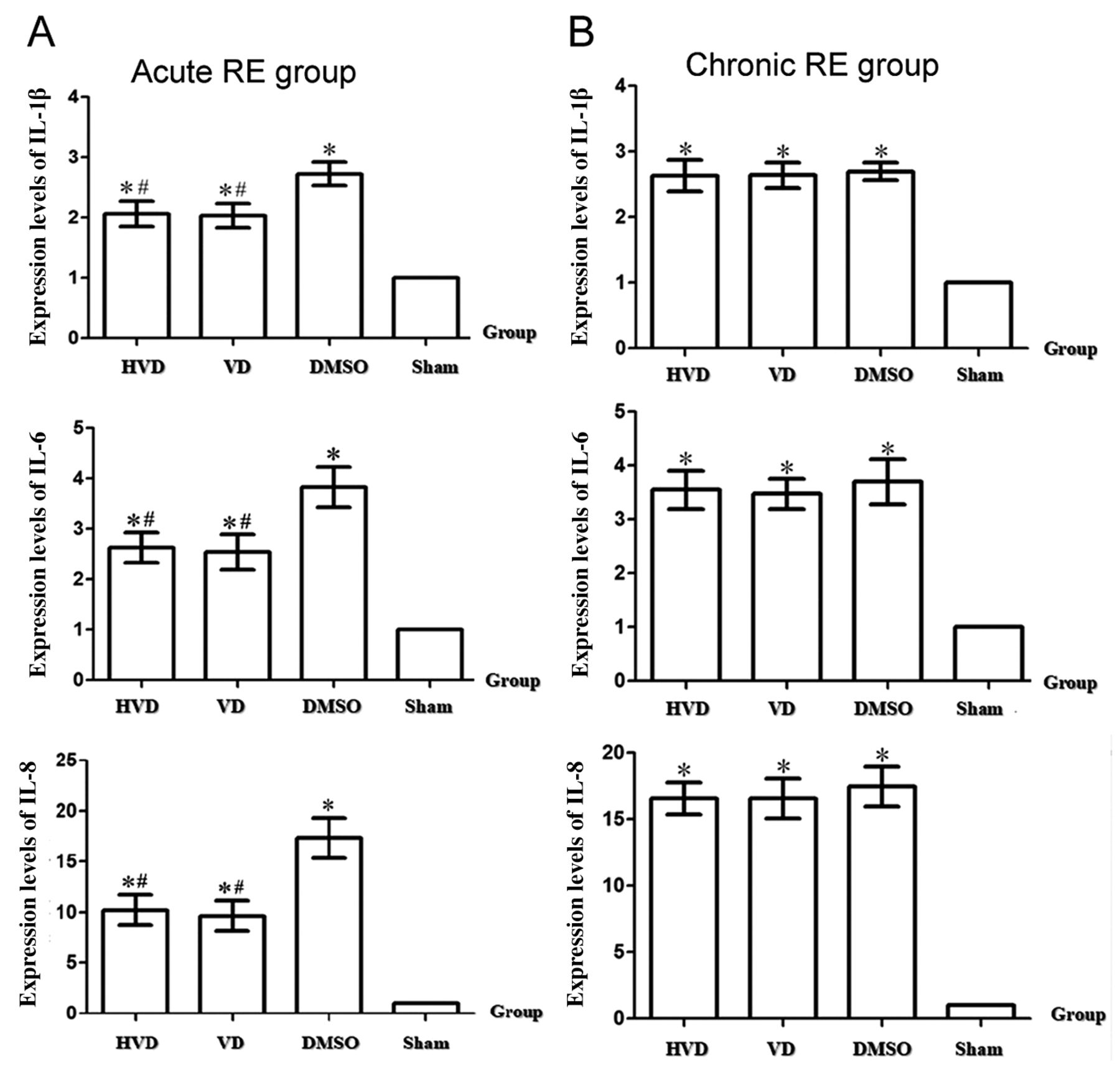
Relative protein expression levels of IL-1β, IL6, and IL-8 in rats with chronic or acute RE treated with VitD3. In acute RE (A), VitD3 treatment significantly suppressed the IL-1β, IL6, and IL-8 protein expression levels, whereas no significant difference was observed between the HVD and VD groups. For chronic RE (B), VitD3 treatment appeared to slightly inhibit IL-1β, IL6, and IL-8 protein expression compared to that in the control group (DMSO), but the difference was not significant. *p<0.05 vs. sham group; #p<0.05 vs. DMSO group. VitD3: 1,25 dihydroxy vitamin D3; IL: interleukin.
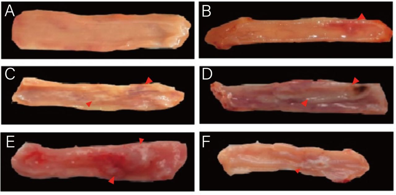
Macroscopic assessment of esophagus tissues in rats. The various degrees of esophageal morphologic alterations are displayed in Figure 4. Esophageal erosion, ulcer, and hemorrhagic necrosis were observed as common lesions in the mucosa of the esophagus in the RE models. VitD3 treatment did not attenuate esophageal lesions in rats with chronic RE or acute RE. In the HVD group of the chronic RE model, one rat showed distention or enlargement of the upper esophagus and suspected stenosis or narrowing of the esophageal ulcer of the middle and lower segments of the esophagus. In the VD group of the chronic RE model, thickening of the esophageal mucosa layer was observed in one rat. The Kruskal–Wallis H test (rank sum test) showed that there was no significant difference in the morphologic grading of esophageal mucosa between the acute and chronic RE rat models (p>0.05) (Table II). These macroscopic assessments of esophageal tissues in rats with and without VitD3 treatment indicated no significant morphological effect of VitD3 in chronic or acute RE in rats (Figure 4, Table II).

Macroscopic findings of esophageal tissues in rats with RE. Macroscopic examinations were performed in rats undergoing sham-surgery (A) and rats with acute or chronic reflux esophagitis (RE) (B-F). Macroscopic findings (red arrow) revealed erythematous mucosa (B), erosion (C), ulcer (D), thickening (E), and stenosis or narrowing of the esophagus (F) in rats with chronic or acute RE. RE: Reflux esophagitis.
Macroscopic assessment of esophageal inflammation in rats with acute or chronic RE.
Histopathological examination of esophageal tissues in the rat model. Histopathological examination revealed various degrees of inflammation of the esophageal tissues in rats with chronic or acute RE. H&E staining of the esophagus specimens from rats with acute RE demonstrated a considerable enhancement in neutrophil infiltration in the three layers of the submucosa, muscularis mucosae, and lamina propria. More severe lesions, such as loss of esophageal mucosa, necrotic tissues, and red blood cells, were also observed in acute RE. Compared to acute RE, there was not only a large amount of neutrophil infiltration in the three layers of submucosa, muscularis mucosae, and lamina propria, but also a few infiltrating lymphocytes and monocytes in chronic RE. The Kruskal–Wallis H test showed that there was no significant difference in HE staining-based pathological inflammatory grading of the esophageal mucosa between the acute and chronic RE rat groups (p>0.05) (Table III). Notably, the histopathological examination of esophageal tissues from rats treated with VitD3 did not show any significant differences in the histopathological changes in the chronic or acute RE rat groups (Figure 5).
Histopathological assessment of esophageal inflammation in rats with acute or chronic reflux esophagitis.
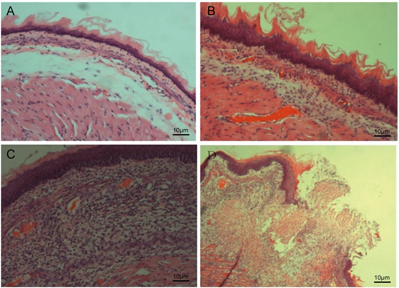
Histopathological examination of esophageal tissues in rats with RE. H&E staining of the esophagus specimens from rats in the sham-surgery control group (A) (×100 magnification) and those with acute or chronic RE (B-D) (×100 magnification). Histopathological examination revealed (B) a slight enhancement of neutrophil infiltration in the mucosa of the esophagus, (C) high levels of enhancement in neutrophil infiltration in the mucosa of the esophagus, and (D) esophageal ulcer. RE: Reflux esophagitis.
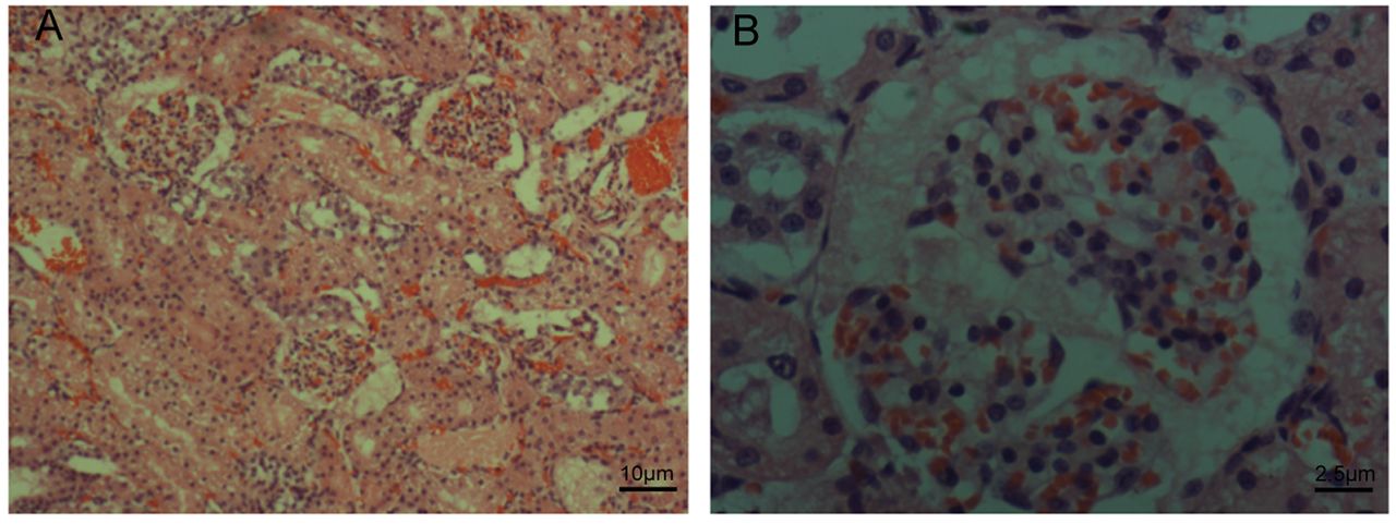
Effects of VitD3 on serum calcium levels in the RE model rats and the association with morphological changes of the kidney. The major consequence of high-dose VitD3-induced toxicity is hypercalcemia, leading to various clinical symptoms (e.g., nausea, vomiting, frequent urination) and can even progress to more serious kidney problems. We therefore examined serum levels of calcium as well as performed histological examination of the kidney after VitD3 treatment of acute or chronic RE model rats. As shown in Table IV, the serum levels of VitD3 and calcium were significantly higher in the HVD group than in the VD group, sham-surgery group, and the control group (p<0.05), in both the acute and chronic RE groups. Histological examination revealed no abnormal morphologic changes of the kidney in rats treated with either low- or high-dose VitD3 (Figure 6).
Serum levels of VitD3 and calcium, and their association with acute or chronic reflux esophagitis in rats.

Histopathological examination of kidney tissues in rats with RE. H&E staining of kidney tissues in RE rats treated with VitD3 (×100 magnification) (A) and (×400 magnification) (B). Histological examination identified no abnormal morphological changes of the kidney in rats treated with VitD3. RE: Reflux esophagitis; VitD3: 1,25 dihydroxy vitamin D3.
Discussion
The novel finding of the present study is that VitD3 plays a potential protective role in RE at the early stages. Studying the effects of VitD3 treatment in rat models of acute and chronic RE presented the following findings: 1) esophageal impairment occurred in rats with surgery-induced acute and chronic RE, as evidenced by macrographic and histopatho-logical findings; 2) no significant differences in morphological grading of esophageal mucosa were observed after VitD3 treatment in RE rats; 3) the protein expression levels of pro-inflammatory cytokines, including IL-1β, IL-6, and IL-8, were significantly increased in both the acute and chronic RE groups, compared to the sham-surgery group; 4) VitD3 treatment significantly reduced the levels of these pro-inflammatory cytokines in both low-dose and high-dose VitD3 acute RE groups compared to the DMSO control group; and 5) serum calcium levels increased significantly in response to VitD3 treatment, while no abnormal alterations were observed on histological examination of kidney tissues of these rats.
RE is considered a consequence of chronic inflammation of the esophagus occurring mainly when the tissue is exposed to reflux of gastric acid. In the present study, rat models of surgically induced acute and chronic RE were generated following modified procedures, namely PNL-CARE, as we previously reported (16-18). Macrographic and histopathological findings showed characteristic esophageal impairment in rats after surgical induction of acute or chronic RE. In addition, a number of key pro-inflammatory cytokines, specifically IL-1β, IL-6, and IL-8, were significantly elevated in the rat models of acute and chronic RE, which may be attributed to the inflammatory response in the esophagus after it is irritated by gastric acid reflux (20, 21). Furthermore, using the rat models we previously established (16-18), in the present study we tested our hypothesis that VitD3 treatment could exert a beneficial effect in the inflammatory process occurring in acute or chronic RE.
However, VitD3 affected the levels of IL-1β, IL6, and IL-8 protein expression differently between acute and chronic RE. In the chronic RE rats, VitD3 treatment slightly diminished the expression levels of IL-1β, IL6, and IL-8 protein in contrast to the control groups, and no difference between groups was observed. This may be due to the fact that the timing of VitD3 supplementation differs. Acute RE models are first supplemented with VitD3 and then induced to establish the model. In contrast, the chronic RE model is symptomatic first, followed by VitD3 supplementation. There is a certain delay in the rise of VitD3 serum levels, and the inflammatory response may appear earlier than the rise of VitD3 levels, thus affecting the role of VitD3 in regulating the early inflammatory response in chronic RE. The esophageal mucosal barrier was disrupted in the chronic RE model, where the perfusion of DMSO solution for 14 consecutive days may aggravate esophageal inflammation, thus affecting some of the inflammatory effects of VitD3. While in the acute RE model, normal esophageal mucosal epithelium with mucus and intact complex squamous epithelium existed, which can reduce the effect of DMSO solution on it. Although VitD3 treatment significantly suppressed the expression of pro-inflammatory factors (IL-1β, IL6, and IL-8), in acute RE there was no significant difference in the morphologic grading of the esophageal mucosa between the VitD3 treatment groups and control groups of rats with acute RE. The present results indicate VitD3 alone may not improve the esophageal impairment arising from the reflux of gastric acid into the esophagus in RE.
We also observed that the serum levels of calcium were markedly elevated after VitD3 treatment in rats, while histological findings of kidney tissues detected no abnormality, indicating no renal toxicity in relation to excessive serum calcium.
Collectively, excessive secretion of the pro-inflammatory factors is proposed as an early event in the development of RE (22), and our findings suggest that VitD3 has the potential to slow RE development.
In conclusion, IL-1β, IL-6, and IL-8 were markedly elevated in RE, and the abnormal increase of these pro-inflammatory cytokines was suppressed by VitD3 in rats with acute RE. Although no morphological and histopathological changes were observed with VitD3 treatment, these novel findings suggest a potential protective role for VitD3 in the early stages of RE. VitD3 holds promise to slow down the progression of RE or prevent RE, but further research is required on this topic.
Acknowledgements
This research was supported by the Nature Science Foundation of Fujian province (Grant number: 2020J01974), Fujian Provincial Finance Project (Grant number: 22SCZZX012), the Startup Fund for Scientific Research of the Fujian Medical University (Grant number: 2021QH1058). All Institutional and national guidelines for the care and use of laboratory animals were followed.
Footnotes
Authors’ Contributions
JJW, and ZHZ designed this experiment. JJW and JZ conducted experiments. YJ and TTL analyzed data. PHZ and TTL explained the experimental results. JJW, JZ, and YJ prepared these figures. JJW and GWZ wrote the manuscript. JJW and GWZ contributed to manuscript editing. RMH revised the manuscript. All Authors participated in reading and discussing the manuscript.
Conflicts of Interests
The Authors declare no conflicts of interest.
- Received November 30, 2022.
- Revision received December 10, 2022.
- Accepted December 16, 2022.
- Copyright © 2023, International Institute of Anticancer Research (Dr. George J. Delinasios), All rights reserved
This article is an open access article distributed under the terms and conditions of the Creative Commons Attribution (CC BY-NC-ND) 4.0 international license (https://creativecommons.org/licenses/by-nc-nd/4.0).