

Abstract
Background/Aim: Canine Cutaneous Histiocytoma (CCH) is a Langerhans' cells benign tumour that undergoes spontaneous regression. The aim of the present study was to investigate the role of angiogenesis, a key step for tumour development, in CCH regression. Materials and Methods: 50 CCH samples were classified into 4 histological groups according to a regression scale, and evaluated for expression of vascular endothelial factor-A (VEGF-A) and its receptor VEGFR-2 as well as microvessel density (MVD). Results: Tumours during early stages of the regressive process had a lower MVD compared to later stages, while CCH tumoural cells showed a limited production of VEGF, but higher levels of VEGFR-2. On the contrary, tumours in advanced phases of regression showed a higher number of neovessels, probably associated with the inflammatory state and the healing process. Conclusion: Our results suggest that angiogenesis may be compromised at early stages of histiocytoma development and this may be a determinant of regression in this tumour.
- Canine cutaneous histiocytoma
- angiogenesis
- VEGF
- CD31
Canine cutaneous histiocytoma (CCH) is a benign tumour on the dog's skin that, in almost all cases, undergoes spontaneous regression. It can occur anywhere on the dog's skin surface as a result of neoplastic proliferation of epidermal dendritic cells, called the Langerhans cells. It is classified as a tumour of round cells, and is reported as the most frequent cutaneous neoplasm in dogs (1-3). CCH has similarities to some forms of human Langerhans cell histiocytosis (LCH), nevertheless, LCH has distinct clinical behaviour between dogs and humans (4). The regression phenomenon, typical of this disease, makes CCH an attractive system for the study of human Langerhans cell histiocytosis and has been considered as an unique model to understand the pathogeny of this enigmatic human disease (5-7).
Spontaneous tumour regression has been described for different types in both humans and animals, and is possibly associated with an efficient immune response (8-10). Several pathways are proposed for CCH regression namely the loss of the adhesion molecule E-cadherin, rise of Th1 pro-inflammatory cytokines such as IL-2, tumour necrosis factor-α (TNF-α), interferon-γ (IFN-γ), up-regulation of the nitric oxide synthase (iNOS) expression, the enzyme that produces the cytotoxic metabolite nitric oxide, and changes in the expression of extracellular matrix metalloproteinases, such as MMP-9 (6, 11-13). Major Histocompatibility Complex MHC class II is also related to tumour regression, the migration of MHC molecules in the cell periphery is a significant factor for the regression in CCH (4). Also, an imbalance between cell proliferation and apoptotic cell death, rather than a decrease in cell proliferation or an increase of apoptosis alone, is a driving force leading to CHH regression (10). Despite all this data, regression mechanisms are not yet fully understood.
Angiogenesis, as defined by the occurrence of neovessels and estimated by the microvessel density, is a crucial process to the growth, development and metastasis of several tumours in mammals (14). Either normal angiogenesis or tumoural angiogenesis are controlled by growth factors and their respective receptors. The vascular endothelial growth factor (VEGF-A) and its receptor VEGFR-2 (FIK-1) are amongst the most studied molecules that have been related with increased angiogenesis in several cancer types in humans and animals (15, 16).
Considering the high cellularity and proliferation rate of the histiocytoma, as well as the levels of apoptosis and necrosis usually observed (10), the aim of our work was to determine if a lack of an appropriated vascular supply, due to an insufficient production of VEGF-A, VEGFR-2 or poor microvessel formation could be associated with its spontaneous regression.
Materials and Methods
Tissue samples. Fifty CCHs were obtained from the archive of the Histopathology Laboratory of the University of Trás-os-Montes and Alto Douro (UTAD), Vila Real, Portugal. Paraffin sections of skin tumours samples were sectioned at 3 μm, and stained with Hematoxylin and Eosin (H&E). The diagnosis and classification of histiocytomas were performed by two independent observers according to the world health organization (WHO) criteria. All histological preparations were observed systematically, according to the degree and location of lymphocytic infiltration being allocated to one of four groups representing different stages of tumour regression, according to previous studies (6, 17). Briefly, i) in group I, lymphoid inflammatory infiltration was absent or minimal in periphery, ii) in group II it was moderate, peripheral, and with formation of nodular structures, iii) in group III it was abundant both in the periphery and the centre of the tumour, and iv) in group IV it was diffuse, extending to the epidermal surface. Ulceration (absent, present), necrosis (absent, present), stroma (scarce, moderate, abundant), mitotic count (n<2, 2-5 and >5 in high power field; 400×; Nikon® Eclipse E600 microscope, Nikon Instruments Inc., Melville, New York, USA), counting at least 10 fields) were also evaluated.
Immunohistochemical study. Immunohistochemistry was performed using the indirect staining method of Streptavidin-Biotin-Peroxidase, using the UltraVision Detection System, HRP, (Thermo Fisher Scientific®, Runcorn, Cheshire, UK). As a method of antigen recovery, the histological preparations were subjected to a microwave thermal pre-treatment, which involved three 5-min cycles in the microwave at 750 Watts in citrate buffer (pH=6.0±0.2). The antibodies used were VEGF (Ab-3 JH121, NeoMarkers®, Fremont, California, USA; 1:100) and its receptor Flk-1 (A-3, Santa Cruz Biotechnology®; California, USA, 1:100), and CD31 (Clone JC70A, Dako®, Glostrup, Denmark; 1:20). All incubations were made in a humid chamber overnight at 4°C. The reaction sites were developed with the addition of a 3,3’-diaminobenzidine tetrahydrochloride (DAB) solution (Thermo Fisher Scientific®, Runcorn, Cheshire, UK). The sections were immersed in a solution of Gill's Hematoxylin, dehydrated, cleared and mounted with Entellan synthetic resin (Merck® Merck KGaA, Darmstadt, Germany). In each set of samples for each procedure, a positive control (canine angiosarcoma or breast carcinoma) and a negative control (an HCC sample in which the incubation of the primary antibody was performed using PBS only).
Evaluation of immunoreactivity. In order to evaluate the VEGF-A and VEGFR-2 (Flk-1) expression, a semi-quantitative analysis was performed according to the percentage (%) of positively-stained tumour cells: i) 0-5% as (negative), ii) 5-25% as + (focal), iii) 25-50% as ++ (multifocal), iv) >51% as +++ (diffuse). The intensity of marking was also evaluated: i) weak (+), ii) moderate (++), iv) strong (+++). In the other hand, to determine the microvessel density, the counts were performed in the central region of the tumour, in order to avoid the ulcerated and necrotic regions, without prior knowledge of the sample's tumour group. From each histological preparation, two hotspots were identified under low-power magnification (40×). Then, under high power (200×), 3 fields were randomly selected in each selected area. Images were captured using a Nikon Eclipse E600 microscope with a Nikon DXM1200 digital camera (Nikon Instruments Inc., Melville, New York, USA) from each of these fields in JPEG format, at a 200× magnification (surface area=28.96 cm2). The number of new vessels was subsequently digitally counted. Positive cells or cell aggregates that were clearly separated from adjacent neovessels, tumour cells and connective tissue elements were considered as individual neovessels. The average number of new vessels was calculated and expressed per 200´ field.
Statistical analysis. The statistical analysis of the data was performed using the IBM® SPSS® Statistics program (Version 24.0, Version 24.0. Armonk, NY, USA). A descriptive analysis of categorical variables was performed, obtaining the absolute and relative frequencies of the categories under study. The microvessel density values were described according to the mean, median, standard deviation, minimum and maximum values. In order to evaluate the association between VEGF and its receptor with the histological groups, the chi-square test was performed. The associations between the microvessel density measured by CD31 and the histological group were determined by the Kruskal-Wallis non-parametric test, after checking the non-normal distribution of the DMV values using the Kolmogorov-Smirnov test. When the microvessel density values were different between the categories, the Bonferroni test was performed to find out between which categories the differences were statistically significant. Associations were considered significant for values of p<0.05.
Results
Histopathological evaluation. Tumours formed nodular formations, exophytic proliferations, or “button”-shaped lesions. All lesions were unique, alopecic and often ulcerated. Neoplastic cells have an oval, round or reniform nucleus, usually eccentric, with an evident nucleolus and acidophilic cytoplasm that varies from moderate to abundant (17). Ulceration of the epidermis was present in 38 cases (76%). Mitosis figures ranged from 0 to 5 in the high-power field: 11 tumours (22%) had a mitosis count (MC) of less than 2, 20 cases had an MC between 2 and 5, and 19 cases (38%) had an MC greater than 5. The tumour stroma was generally scarce (n=38; 76%), while its abundance was only verified in 2 cases, being moderate in the others (n=10). In more than half of the cases (n=34; 63.3%) necrosis was not observed. Of all, 18 CCHs were classified as group I (36%), 13 as group II (26%), 12 as group III (24%), and 7 as group IV (14%).

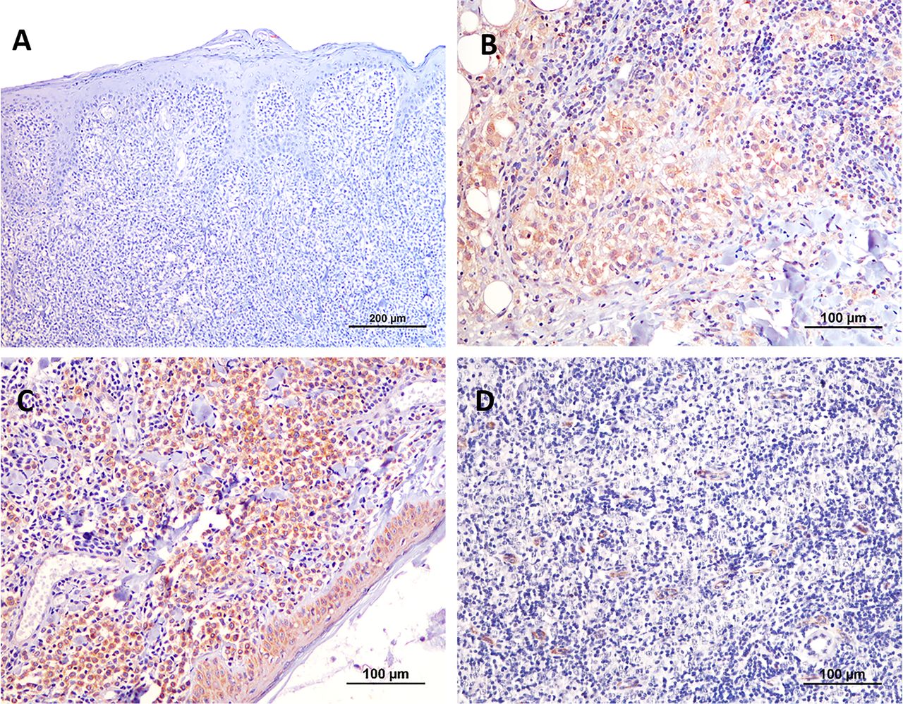
VEGF-A expression. A) Absence of VEGF-A immunoreactivity in a group I canine cutaneous histiocytoma; B) VEGF-A positive cells with a diffuse and strong labelling in a group IV canine cutaneous histiocytoma. C) VEGFR-2 positive cells with a diffuse and strong labelling in a group I canine cutaneous histiocytoma with a diffuse and strong labelling. D) CD31 staining of endothelial cells in a group IV canine cutaneous histiocytoma.
Expression of VEGF-A. Expression of VEGF-A was detected in the cytoplasm of tumour cells with a granular or diffuse pattern, as well as in macrophages (positive internal control). Immunoreactivity was not uniform throughout the preparation, with a greater expression in peripheral tumour cells of the deeper skin layer. Regarding the percentage of VEGF-A positive cells, most tumours were negative (n=31, 62%) or had focal positivity (n=6, 12%) and 7 cases (14%) presented diffuse expression. About the intensity of the immunostaining, 14% of the cases (n=7) showed a strong reaction, 2 cases (4%) had a low intensity of marking, and 10 cases (20%) had a moderate intensity (Figure 1A and 1B).
A statistically significant association was observed between the percentage of VEGF-A positive cells and abundant tumour stroma (p=0.039), and the presence of necrosis (p=0.014). Thus, sparse stromal tumours were mostly negative for VEGF-A, while moderate stromal tumours were positive in more than half of the cases. Most tumours with evident necrotic areas did not express VEGF-A. Labelling intensity showed a statistically significant association with necrosis (p=0.016), similar to that was described for the extent of VEGF-A labelling.
VEGF-A expression was significantly associated with the tumour group [both for the percentage of VEGF-A positive cells (p=0.002) and VEGF-A intensity of immunolabelling p=0.009)]. VEGF-negative tumours generally belonged to the histological groups I and II. Cases with a higher VEGF-A expression belonged mainly to the histological group III (a more advanced stage of the regressive process).

Microvascularization density in the four histological groups considered (Group I: scarce and peripheral lymphocytic infiltrate; group II: moderate and peripheral lymphocytic infiltrate; group III: abundant infiltrate at the periphery and in the center; group IV: diffuse lymphocytic infiltrate) (*p<0.05; ***p<0.001).
Immunohistochemical expression of VEGFR-2. VEGFR-2 was also expressed in the cytoplasm of tumour cells with a granular or diffuse pattern as well as in macrophages (positive internal control). VEGFR-2 expression was observed in most of the analysed cases (39/50 cases; 78%), of which: i) 7 cases had a focal marker (5-25% of the tumour cells), ii) 17 cases a multifocal marker and iii) 15 cases a diffuse marker. Regarding the intensity of the immunoreaction, i) 12 tumours (24%) had a weak intensity, ii) 19 tumours (38%) a moderate intensity and iii) 8 cases (16%) a strong intensity. A diffuse, moderate marking on the epidermis was also observed (Figure 1C).
VEGFR-2 intensity was significantly associated with the mitotic index (p=0.012). Regarding the stroma, tumours with abundant stroma were negative, whereas tumours of sparse and moderate stroma were mostly positive. This association was also statistically significant (p=0.045).
VEGFR-2 positive cells were associated with the tumour group both for the extent of labelling (p<0.001) as well as the intensity (p=0.003). All tumours in group I expressed VEGFR-2 with a multifocal (n=12) or diffuse (n=6) pattern, while group IV tumours were mostly negative for the receptor.
Immunohistochemical assessment of microvessel density (MVD). The microvessel density, estimated by CD31 staining of endothelial cells (Figure 1D), showed an average of 9.08±6.58 microvessels, a median of 6, ranging from 2 to 28 vessels per 200× field. MVD varied in the different categories of the mitotic index (p<0.001). Tumours with fewer mitoses had more microvessels and tumours with more mitoses had fewer microvessels. MVD also varied between the different stroma categories (p=0.007), so that tumours with abundant stroma had more microvessels compared to tumours with scarce stroma.
MVD was lower in CCH from groups I (median=5.5, range=2-10) and II (median=5.0, range=3-6) and higher in groups III (median=11.0, range=4-28) and IV (median=22.0, range=18-24), with p<0.001. Considering the comparison between the different groups, we can see that the differences in MVD were significant between groups: i) I and III, ii) I and IV, iii) II and III, and iv) II and IV. There are no differences between groups I and II and groups III and IV (Figure 2).
Discussion
Tumour growth and metastasis are dependent, in part, on their capacity to induce the development of new blood vessels. In reverse, angiogenesis is one of the mechanisms that could has been involved in the spontaneous regression described in several tumours (18, 19).
Canine cutaneous histiocytoma, one of the most frequent tumours of the skin in young dogs, presents a rapid growth, however, after a few weeks its size decreases and the tumour mass regresses (20). In order to estimate angiogenesis in CCH, we evaluated the expression of VEGF-A, VEGFR-2 and MVD in 50 tumours categorized in four histological groups, each corresponding to different stages of CCH regression. Our results indicate that angiogenesis, estimated by the angiogenic factor VEGF-A and its receptor, and by the CD31-positive microvessel density, seems to be a dynamic process along CCH regression. During early stages of canine histiocytoma development (groups I and II) there are only few blood vessels, most probably due to low levels of VEGF-A. In spite of tumour cells producing VEGFR-2 during these stages, since the signalization complex VEGF-A/VEGFR-2 is a major regulator of blood vessel formation (21), the absence of VEGF-A can compromise CCH angiogenesis and tumoural growth. The scarcity of microvessels causes ischemia and cell hypoxia and could lead to the death of tumour cells by necrosis or apoptosis (22, 23). Considering the tumours in groups III and IV, which are more advanced with regards to the regression process, the abundant inflammatory response may be a stimulus that triggers the formation of microvessels through the production of angiogenic factors by the immune cells (24) and justify the greater number of vessels and higher VEGF expression in those tumours. There are also other circumstances that, theoretically, can contribute to angiogenesis in tumours in an advanced stage of the regression process, e.g., an increased infiltration of macrophages (10), or even hypoxia inducing a fast and strong increment in VEGF mRNA (25).
Our results are not similar to studies about angiogenesis in Langerhans cells human histiocytosis. These tumour cells express VEGF especially in disseminated lesions (26-28). This could be justified partially by the distinct clinic behavior and aggressiveness of these tumors when compared to canine cutaneous histiocytoma. Despite the similarities between human and canine histiocytosis, the human pathology in its disseminated form has a rapidly progressive multisystem involvement and an unfavorable prognosis (29). Canine cutaneous histiocytoma is usually unique, without a tendency for metastasis and with a favorable prognosis (1). Moreover, our results could emphasize the importance of VEGF as a therapeutic target during the early phases of malignancy in humans by inhibiting tumoral angiogenesis and early invasion of malignant cells (26, 28).
In conclusion, low expression of VEGF-A during early stages of HCC development, and consequently reduced microvessel density, may be crucial for initiating regression. The scarce production of VEGF in CCH by tumour cells does not justify the CCH regression, but the interaction of the tumour cells with the stroma and other systems, such as circulating immune cells, may contribute or even be the switch igniting the regression process.
Acknowledgements
The Authors would like to thank the financial support of project UIDB/00211/2020 funded by Fundação para a Ciência e Tecnologia/Ministério da Ciência Tecnologia e Ensino Superior (FCT/MCTES) through national funds. Authors would also like to thank the support received by projects UIDB/04033/2020 and UIDB/CVT/00772/2020, from FCT/MCTES.
Footnotes
Authors' Contributions
IP conceived and designed the study; DC, RF, IP and PR conducted the experiments, IP, JP, PR and FS analyzed the data; DC, RF, IP and FQ wrote the paper, IP, JP, FQ, FS discussed the manuscript.
This article is freely accessible online.
Conflicts of Interest
The Authors declare no conflicts of interest with regard to this study.
- Received August 7, 2020.
- Revision received September 6, 2020.
- Accepted September 16, 2020.
- Copyright© 2020, International Institute of Anticancer Research (Dr. George J. Delinasios), All rights reserved