

Abstract
Experimental urinary bladder tumours have been proposed as a useful model for the study of urinary bladder carcinogenesis, as well as for evaluating new therapeutic strategies. Consequently, the administration of chemical carcinogens is one of the most commonly used methods for inducing urinary bladder tumours. N-butyl-N-(4-hydroxybutyl)nitrosamine (BBN) is, undoubtedly, the most-used urothelial chemical carcinogen. BBN belongs to the nitrosamine family, a wide group of alkylating agents that are able to induce bladder tumours in laboratory animals. Depending on the animal species, the spectrum of urothelial lesions induced by BBN varies, but is similar to those observed in humans. BBN has a high propensity to induce mutations affecting the expression of genes such as p53, RAS and H19 among others. The aim of this study was to review BBN as a urothelial tumour inducer, taking into consideration its chemical characteristics, properties and spectrum of lesions induced, as well as its possible applications.
- Urothelial
- carcinogenesis
- N-butyl-N-(4-hydroxybutyl) nitrosamine
- tumours
- review
Bladder cancer is among the most common malignancies worldwide, and it assumes greater prominence in developed countries. The most recent data, published in 2011, showed an estimated 386,300 new cases and 150,200 deaths in 2008. Bladder cancer is the second-most common tumour of the genitourinary tract and the second-most common cause of death in patients with genitourinary tract malignancies (1). In humans, the most common cell type of bladder cancer is transitional cell carcinoma, although adenocarcinomas, squamous cell carcinomas and sarcomas can also occur (2). The majority of patients with bladder cancer, 75-85%, are diagnosed with superficial tumours, while the remaining 15% to 25% have invasive ones (2, 3). The superficial ones are low-grade, well-differentiated papillary tumours that do not invade or metastasize. Histological evolution in this case starts as hyperplasia, evolving to papilloma, papillary urothelial neoplasms of low malignant potential and papillary carcinoma (low- and high-grade) (4). The invasive tumours originate as flat lesions such as hyperplasia, dysplasia and carcinoma in situ that then evolve into invasive carcinomas (5). These are high-grade tumours that frequently have areas of squamous cells or glandular differentiation or areas of undifferentiated tumour, including small-cell or spindle-cell variations. Invasive tumours are devastating, since over 50% of the patients will die from metastatic disease (2).
Experimental Models of Bladder Cancer
The need for experimental models of tumours that are as similar as possible to those in humans has prompted researchers to attempt various approaches. Animals such as dogs, rabbits, guinea pigs and hamsters have all been used in the past to study urinary bladder carcinogenesis (6). However, for ethical reasons and due to the conditions in animal facilities, dogs cannot be used in experimental models of carcinogenesis. Animal facilities in order to maintain guinea pigs and rabbits also require special equipment, making models difficult to apply. For these reasons, small rodents, such as mice and rats, are the most common species used. These animals have a lower urinary tract that is comparable to that of humans, and neoplasms in their bladders are morphologically very alike (7). On the other hand, in most strains of rodents, bladder cancer is not commonly found unless a chemical or other treatment is applied (8). Another advantage is associated with the fact that these are small animals and there is an abundance of information concerning the biological characteristics of neoplastic development in these species (6). Mouse strains such as BALBC, C57BL and ICR, and rat strains such as Wistar, Sprague-Dawley and Fisher, are among the animals most used in bladder cancer research. The Brown Norway and the DA/Han rat are two rat strains with a high incidence of spontaneously occurring urothelial tumours of the bladder, and for this reason these models can be used in experimental protocols without the application of chemical carcinogens (9). Besides these, there are also genetically altered animals (that lack genes, express genes or have mutated genes) (10, 11) that exhibit increased susceptibility to developing urothelial lesions and even cancer (6). Examples of these animals are presented in Table I. These genetic alterations provide an ideal system for understanding the roles of molecular events wich take place in bladder carcinogenesis (11, 12, 29, 30), allowing it to be determined if genetic changes, including oncogene overexpression, mutation, or tumour-suppressor gene loss, can increase the risk of neoplastic progression (29, 31).
Genetically altered animals used in carcinogenesis studies.
These models are useful for extrapolating the results from animal carcinogenicity studies into human risk assessment (31) and also provide in vivo platforms for testing strategies for preventing bladder cancer (30, 32). There are several disadvantages associated with the use of transgenic models in cancer research, one being that mutation induction in a given tissue does not always lead to tumour development in that tissue. Moreover, these models also require a significant investment of time and resources towards tumour establishment (31, 33, 34).
Over the years, different methods have been developed to induce tumours in the mouse and rat urinary bladder:
Foreign bodies: In 1951, Jull successfully induced the development of urinary bladder tumours via surgical implantation of pellets containing cholesterol, paraffin and several chemicals (35, 36). Later, in 1966, Bryan and Springberg intravesically implanted other compounds such as arachic acid, hexaethylbenzene, hexamethylbenzene, palmitic acid and stearamide, individually compressed as pellets, into the mouse bladder and verified that all these compounds were associated with the development of bladder tumours. Foreign bodies within the lumen of the urinary bladder play a role in promoting carcinogenesis, since they can irritate or traumatize the urothelium, stimulating mitotic activity and thereby causing the development of nodular and papillary hyperplasia (35-37).
Urinary calculi: Urinary tract calculi represent foreign bodies, similar to pellets, but do not require surgical implantation into the bladder lumen (3, 8, 38). They may act as a mitotic stimulus that makes calculus-bearing rodent bladders more susceptible to tumorigenesis (39). Numerous chemicals such as uric acid, calcium oxalate, uracil, melamine, among others, have been associated with the development of urinary calculi (8, 40-43). The calculi can arise from the administered chemical itself or from an endogenous metabolic product that is caused by the administration of the chemical. Lalich first reported the induction of stone formation in the urinary bladder of rats via oral administration of uracil (41). The administration of 3% uracil in the rat diet for a period of two weeks induced urolithiasis and mechanical irritation of the rat bladder urothelium, which at first responded by the development of papillomatosis and papillary/nodular hyperplasia. These lesions gradually disappeared after cessation of dietary uracil, through an enhanced apoptotic process (40). Nevertheless, uracil administered to mice and rats at low dosages does not produce calculi or tumours, which shows that a threshold effect for secondary carcinogenesis is necessary. This high-dose (threshold) phenomenon appears to occur more readily in rodents than in humans. In fact, urolithiasis is not a cause of bladder cancer in humans (44). Urothelial carcinogenesis related to calculus formation is also influenced by factors such as urine pH, volume, osmolality, cationic and anionic concentration of minerals and quantitative and qualitative differences in the presence of urinary protein (8, 45). An increased urinary pH and an elevated sodium concentration increase cell proliferation in the urothelium and promote tumorigenesis (39, 45-47).
Irradiation: Radiotherapy not only results in beneficial effects regarding tumour control, but is also associated with adverse effects in normal tissues. For this reason, irradiation is one of the most widely researched carcinogens (48). The urinary bladder, due to its location, is an organ that is frequently affected by pelvic radiotherapy (49-52). In man, bladder cancer tends to occur 5 to 10 years after radiation and is characteristically of high grade and locally advanced at diagnosis. The relative risk of secondary bladder malignancy ranges from 1.5- to 4-fold and is likely to be proportional to the dose of radiation given (49). In experimental studies with female mice of the inbred C3H/Neu strain, a single dose of irradiation with x-rays (20 Gy) induced bladder dysfunction, with early effects, observed between 0 and 31 days afterwards, and later effects, seen between 120 and 360 days (50). Another study, using female F344 Fischer rats, showed the development of preneoplastic urothelial hyperplasia and urothelial carcinoma, when rats were observed 20 months post-irradiation (53). Several experimental studies showed that the rate of bladder tumours achieved with irradiation depends on the dosage, the period of exposure and the follow-up time (49-55). Due to this low incidence and the long delay before a tumour appears, which are related to the slow cell-turnover rate of the urothelium, this is not considered a productive model. Moreover, an x-ray source and a authorized facility where it can be used are required (56).
Administration of chemical substances: This remains the most important method used to induce tumours in rodents (40, 56, 57). The discovery that human bladder cancer might be related to chemical exposure dates back to the 19th century (58). In 1895, Dr. Ludwig Rehn reported bladder cancer in german dye workers who manufactured aniline dyes. Later, this was shown to be related to the presence of 2-naphthylamine in the dyes. Since that time, many other chemicals and environmental agents have been associated with bladder cancer development (59-61). The administration of chemicals to induce tumours in mouse and rat is characterized by a long delay in tumour development and a high incidence of tumours. However, chemical substances should be carefully chosen, in order to avoid non-organ specificity of the carcinogenic/mutagenic effect. In recent decades, several chemical compounds have been proven to be particularly effective in induction of bladder cancer. When administered via an appropriate route, at the appropriate doses and in the appropriate strain of animal, all produce 100% incidence of bladder tumours.
Bladder Tumour Cell Lines
Bladder tumour cell lines are also valuable as in vitro models that enable the study of the underlying biology and the identification of potential therapeutic targets, from which promising agents would ideally be advanced into translational studies. Various in vitro models have been suggested, such as the three-dimensional spheroid models, which attempt to simulate the behaviour of cancer towards therapeutic agents (62). Bladder tumour cell lines, although not suitable for studying the evolution of a tumour, unlike animal models, are frequently used to establish orthotopic tumour implantation. Similar to animal models, bladder cancer cell lines can also be used to test the therapeutic and preventive actions of experimental drugs (63, 64). There are approximately 50 human bladder cancer cell lines established to date (64). Mouse cell lines availability for the study of bladder cancer is considerably lower. An example is BTT-T739, a transitional cell carcinoma cell line derived from bladder tumours that are chemically induced by BBN in T-739 mice (65, 66). However, some authors question the relevance of rodent cell lines to normal and malignant human urothelial cells (67).
Nitrosamines
Nitrosamines were first reported by Geuther in 1863; however, they remained a group of compounds considered to be of no great importance for almost a century. This was until 1954, when Barnes and Magee reported nitrosodimethylamine liver toxicity in rats and humans, and later in 1956, when the same investigators reported hepatic cancer in rats fed with the same compound (68). The description of the carcinogenic activity of nitrosamine compounds, and their use in inducing tumours in the urinary bladder, in 1964 by Duckrey and collaborators provided experimental urinary tract pathology with a useful research instrument. Since then, N-butyl-N-(4-hydroxybutyl) nitrosamine (BBN) has become established as one of the most, if not the most, suitable carcinogen for induction of urinary bladder carcinoma (68, 69).
BBN
BBN is a clear yellow to reddish-yellow colour liquid. BBN is also a carcinogen that is metabolically derived from an N-nitroso compound found in cigarette smoke (19). BBN is a metabolite of dibutylnitrosamine (DBN). Both, DBN and BBN induce tumours of the urinary bladder, but DBN can also induce liver and oesophageal tumours (69-71).
BBN has been used as a representative carcinogen that efficiently induces bladder cancer in laboratory animals. BBN is commonly administered via the oral route, in drinking water or by gavage, at a dose that ranges from 0.01-0.05% (72-74). However, it can also be administered subcutaneously and introduced directly into the urinary bladder by intravesical instillation (6, 70, 75-78). When BBN is administered in drinking water, the use of opaque bottles is necessary, since this is a photosensitive compound (79). Gavage administration, although highly effective, is not frequently used because aspiration pneumonia can occasionally occur as a consequence of tracheal intubation. It is also possible that oesophageal injury and gastric rupture may occur, as well as restraint-associated distress, particularly with repeated use. All of these undesirable effects can increase rodents' morbidity and mortality (80, 81). Subcutaneous administration performed in infant mice resulted not in bladder tumours but mainly in lung and liver tumours (76). Intravesical instillation of BBN with successful induction of papillomas and carcinomas in female ACI/N rats was reported by some authors (75). Others using the same route of administration of BBN studied the damage and repair of DNA in the urinary bladder epithelium of Wistar rats (77). For intravesical instillation, anaesthesia is necessary to prevent distress, to carry out the technique safely and to avoid animal pain. Females are used rather than males, given the anatomical differences that render this technique impossible in males. This route of administration also has some advantages, namely the fact that the compound is only contained within the environment of the urothelium, therefore protecting the animals from systemic exposure (82). Regardless of its disadvantages, oral administration remains the most common route for BBN administration.
BBN is an indirect carcinogen: after ingestion it is metabolized mainly in the liver, but also in the bladder, into several metabolites. The alcoholic group of BBN is oxidized into a carboxylic group by the enzymatic system alcohol/aldehyde dehydrogenase, resulting in N-butyl-N-(3-carboxypropyl)nitrosamine (BCPN) (Figure 1) (83-85). BBN is also converted to BBN-glucoronide by uridine diphosphate-glucuronosyltransferase-catalyzed conjugation, but this metabolite does not possess carcinogenic properties. BCPN, unlike BBN-glucoronide, is a bladder carcinogen. These metabolites reach the urinary bladder through urine and come into contact with the urothelium, binding covalently to cellular macromolecules and initiating the carcinogenic process (78, 85-88). Several other minor metabolites, resulting from beta-oxidation according to the Knoop mechanism, can also be detected, such as N-butyl-N-(2-hydroxy-3-carboxypropyl)nitrosamine, N-butyl-N-(carboxymethyl)nitrosamine and N-butyl-N-(2-oxopropyl)nitrosamine (86, 87, 89). Due to its mechanism of action, BBN is considered to be a genotoxic or DNA-reactive carcinogen (88); it causes DNA damage in the bladder epithelium and selectively induces urinary bladder tumours in mice and rats (78, 90). In BBN-induced bladder tumours, the clonal mutations detected were predominantly G-A or C-T transitions (15/27, 56%) and substitutions of T (10/27, 37%) (90).

Metabolism of N-butyl-N-(4-hydroxybutyl)nitrosamine (BBN). (BBN-G: BBN-glucoronide; BCPN: N-butyl-N-(3-carboxypropyl) nitrosamine; BCPNG: BCPN-glucoronide; BHCPN: N-butyl-N-(2-hydroxy-3-carboxy-propyl)nitrosamine; BCMN: N-butyl-N-(carboxymethyl)nitrosamine; BOPN: N-butyl-N-(2-oxopropyl)nitrosamine (64, 81).
Urothelial Lesions Induced by BBN
BBN-induced urothelial lesions in rodents resemble human urothelial lesions in their morphological characteristics. However, susceptibility to BBN-induced bladder tumours differs significantly among mouse and rat strains. From our experience, we can say that the mouse and rat spectrum of urothelial lesions depends on the sex, strain, age and period of exposure to BBN. Macroscopic lesions observed in mice and rats are dependent on the length of exposure to the compound. Bladder cancer in mice develops relatively early after BBN exposure and macroscopic lesions are observed after 12 weeks of exposure to BBN (0.05%). In rats, the exposure period to BBN (0.05%) must be at least 20 weeks, to enable the development of macroscopic lesions. In Figure 2, we can observe the macroscopic characteristics of BBN-induced lesions in mice and rats. Macroscopically, mouse lesions have nodular characteristics, whereas rat lesions exhibit a papillary pattern. In both animals, the dimensions are variable, ranging from a few millimetres to the complete occupation of the entire bladder lumen.
Spectrum of urothelial lesions induced by N-butyl-N-(4-hydroxybutyl)nitrosamine (BBN).

Macroscopic appearance of the mouse (A, B) and rat (C, D) bladder. B and D were exposed to N-butyl-N-(4-hydroxybutyl)nitrosamine. A: Normal bladder; B: invasive carcinoma; C: normal bladder; D: papillary neoplasm.
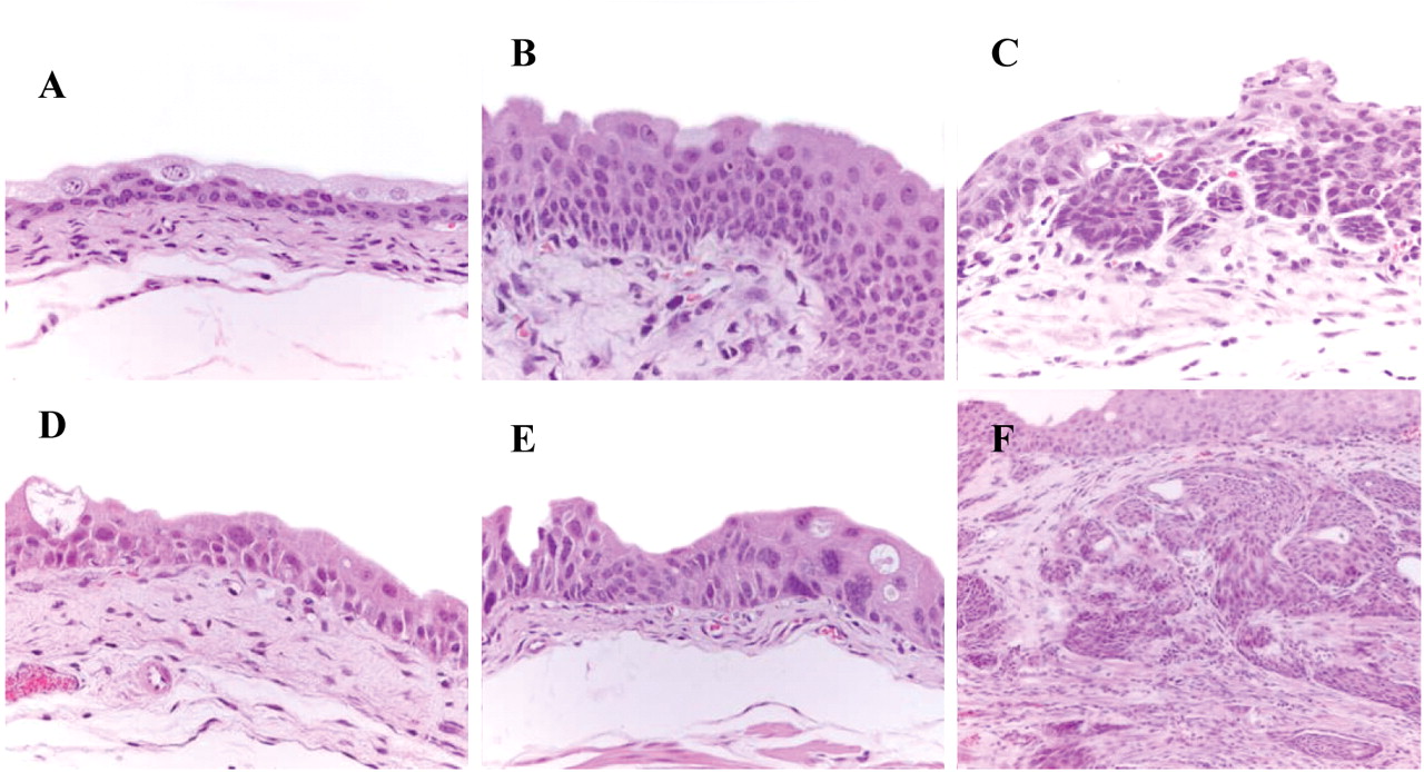
Histopathological evaluation of lesions identified in mouse urothelial carcinogenesis induced by BBN. A: Normal urothelium (control group, H&E, ×400). B: Simple hyperplasia (H&E, ×600). C: Nodular hyperplasia (H&E, ×400). D: Dysplasia (H&E, ×600). E: Carcinoma in situ (H&E, ×400). F: Invasive carcinoma with squamous differentiation (H&E, ×400).

Histopathological evaluation of lesions identified in rat urothelial carcinogenesis induced by BBN. A: Normal urothelium (control group, H&E, ×400). B: Simple hyperplasia (H&E, ×200). C: Nodular hyperplasia (H&E, ×200). D: Papilloma (H&E, ×100). E: Invasive carcinoma (H&E, ×200). F: Squamous metaplasia (H&E, ×100).

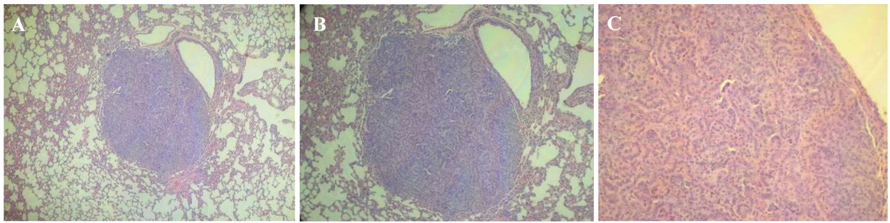
Macroscopic metastasis in the lymph node of a female Wistar rat exposed to N-butyl-N-(4-hydroxybutyl)nitrosamine.
Microscopic lesions are also different in mice and rats. For the past ten years, we have been using the bladder tumour model in which BBN was administered by gavage and in drinking water using mice (ICR) and rats (Wistar; Fisher 344). Further studies on the spectrum of urothelial lesions have been conducted by our team. As shown in Table II, the spectrum and incidence of urothelial lesions can be seen to increase over time.
Mice usually develop dysplasia, with or without hyperplasia, carcinoma in situ and invasive carcinoma (Figure 3). Microscopic lesions in rats usually start as simple hyperplasia, evolving into papillary and nodular hyperplasia, papilloma, papillary tumour of low malignant potential, low-grade papillary carcinoma, high-grade papillary carcinoma, and non-invasive carcinomas (Figure 4) (3, 23, 94-97). In contrast to low-grade papillary lesions in humans, in rats these lesions can eventually progress to higher grade, non-invasive carcinomas and finally to invasive neoplasms (3).

Lung metastasis in a male ICR mouse treated with N-butyl-N-(4-hydroxybutyl)nitrosamine with invasive urothelial carcinoma (H&E) (A: ×100; B: ×200; C: ×400).

Histopathological evaluation of lesions identified in mouse urethra after BBN treatment. A: Normal urethra (transverse section) (H&E, ×100). B: Dysplasia (H&E, ×600). C: Nodular hyperplasia (H&E, ×200). D: Invasive carcinoma (H&E, ×100). E: Invasive carcinoma (D) with higher magnification (H&E, ×400). F: Squamous metaplasia (H&E, ×100).
Rats are most susceptible to BBN, followed by mice, hamsters and guinea pigs. In hamsters, diffuse cell growth of the urothelium is observed in response to BBN, but no papillomatous growth is seen. Following treatment with BBN, the bladder urothelium of guinea pigs does not increase in thickness (6). In dogs, development of urinary bladder tumours induced by BBN required longer periods than needed in mice or rats, but the incidence and development of bladder tumours and morphological patterns were very similar. BBN-induced transitional cell carcinoma in dog urinary bladder was very similar to the one observed in human cases (98, 99).
Extra Urinary Bladder Tumours
In mice and rats, tumour development can lead to the emergence of considerably larger urothelial tumours, which in turn can cause obstruction of their ureters. This situation often leads to hydronephrosis and death (100), even before metastasis can arise. On the other hand, since experimental animals are euthanized early in life (at the end of the experimental period) it is not surprising that this would be too soon for metastases to develop (101). However, according to several authors, invasive carcinomas are highly malignant and frequently develop metastasis in mice, particularly to the lungs (3, 102) but also to regional lymph nodes and occasionally other organs (3). Throughout our years of research using BBN, we have only observed metastasis in one inguinal lymph node (Figure 5), in a female Wistar rat after 20 weeks' exposure to BBN and 8 weeks' of water, and in the uterine wall of another animal submitted to the same protocol.
In mice, we observed lung metastasis in one ICR male mouse submitted to 12 weeks of BBN followed by 8 weeks of water (Figure 6), and alterations in the urethra of seven mice subjected to the same protocol, consistent with the pre-neoplasic and neoplasic lesions induced by BBN (Figure 7). As yet, we have not been able to find reports demonstrating alterations of normal urethral cells as a direct effect of BBN. All metastases found were associated with invasive urothelial carcinomas.
During our experimental studies using BBN, the mortality rate in mice and rats has been very low. When death happens, it is generally associated with hydronephrosis, which is diagnosed at necropsy.
BBN-induced Urothelial Tumours: Applications
The similarity between bladder cancer in humans and rodents enables the investigation of several areas that cannot be studied under clinical conditions, such as pharmacokinetics and toxicity. Rodent models of bladder cancer chemically-induced by BBN are useful in the evaluation of new bladder cancer therapies. Such models can be used to determine the efficacy of therapeutic drugs that are intravesically instilled (6) or administered by other routes, such per os (103) or intraperitoneally (86, 87). However, before beginning treatment, it is absolutely necessary to confirm tumour uptake. Physical examination by means of bladder palpation and urine inspection are classically reported methods that can be used in mice and rats. However, haematuria and palpable mass might represent the latter stages of cancer. In vivo imaging systems that can monitor tumour development are useful. Non-invasive methods such as ultrasonography (104), endoscopy (105), cystoscopy (106) and magnetic resonance imaging (MRI) (107, 108), computerized axial tomography (CAT) (109) and bioluminescence (110, 111), can be applied with these aims in mind. The possibility of applying all these detection and measurement methods are very useful in animal models of human cancer, since they allow the reduction of the number of animals needed for such studies. If we are able to determine the presence of pre-neoplasic and neoplasic lesions without the need to sacrifice animals, we can progress to the treatment phase, and each animal will function as its own control (109).
Genetic Alterations in BBN-induced Urothelial Tumours
Bladder cancer in humans is associated with several genetic and chromosomal changes (112). In rats, chromosomal alterations associated with oral BBN exposure have been previously reported by our team (113) and others (114-117). In mice, there are some studies that describe several genetic and chromosomal changes (118-120). These genetic alterations are similar to those found in humans. BBN-induced rodent tumours, particularly murine tumours, have p53 mutations, or mutations in genes related to the p53 pathway, especially in high-grade tumours (3, 115). Numerous chromosomal abnormalities and mutations in other key growth control genes are frequently detected in these tumours, even at their early non-invasive stages (3). H-ras mutations are observed infrequently in mouse and rat models, although BBN-induced carcinogenesis occurs more efficiently in H-ras transgenic mice. BBN-induced tumours display elevated levels of Epidermal growth factor receptor (EGFR) (115, 120, 121). In rats, an up-regulation of H19, an imprinted maternally expressed oncofetal gene which is used as a tumour marker in human bladder cancer, has also been shown (116). Allelic losses within mouse chromosome 4 (syntenic to human 9p21-p22) are also common, mirroring the loss of 9p21-p22 that occurs in human bladder cancer (121).
These findings confirm the similarity of rodent models to human bladder cancer, and emphasize the applicability of such models for future therapeutic studies.
Other Chemicals
In the past, N-[4-(5-nitro-2-furyl)-2-thiazolyl]formamide (FANFT) and N-methyl-N-nitrosourea (MNU), both genotoxic carcinogenic compounds, were widely used to induce bladder cancer in laboratory animals. However, nowadays, their use is very limited, taking into consideration the fact that both compounds have very particular handling requirements.
FANFT is a nitrofurane derivative, administered in the animals' diet, with specific bladder carcinogenicity for mice, rats, dogs (122-124) and hamsters (124, 125). FANFT is a dangerous compound that must be carefully manipulated, otherwise carcinogenic and teratogenic lesions may arise in the experimenters (6).
MNU is a direct- and complete-acting carcinogen causing tumour initiation and/or promotion in several organs, including the urinary bladder, lung, liver, thyroid, pancreas, prostate, intestine, forestomach, glandular stomach, alimentary tract, kidney, nervous system and hematopoietic system (126-128). In order for it to induce bladder tumours, it must be administered by the intravesical route. It decomposes spontaneously in aqueous solution at a rate proportional to the pH (129, 130).
Conclusion
The BBN-induced rodent model is an orthotopic model widely used in urinary bladder cancer research, especially for the evaluation of the preventive and therapeutic effect of drugs for human urinary bladder cancer. Mouse and rat models simulate the natural environment of bladder cancer, with intact pathological and immunological responses.
Stable, reliable and reproducible orthotopic animal models are critical, as they provide the opportunity to study pathogenesis mechanisms and enable the research and development of novel therapeutic agents. Developing therapeutic agents for bladder cancer requires adequate in vivo models. The BBN model of urothelial tumours includes the vascular and stromal environment that is crucial to determine the efficacy of therapeutic drugs. Moreover, rodents have a lower urinary tract which is comparable to humans, and neoplasms in the bladder are morphologically very similar, with a similar phenotype to human urinary carcinoma with respect to tumourigenesis and gene expression. This experimental model of urinary bladder tumour in vivo enables the study of the biology and evolution of tumours subjected to different agents, using imaging, genetic, molecular and histological techniques.
Acknowledgements
This study was supported by a grant from the Fundação para a Ciência e Tecnologia (FCT), Ministério da Ciência e Ensino Superior, Portugal (SFRH/BD/40896/2007) and by the strategic research Project Pest-OE/AGR/UI0772/2011 also financed by FCT.
- Received January 24, 2012.
- Revision received March 1, 2012.
- Accepted March 6, 2012.
- Copyright © 2012 International Institute of Anticancer Research (Dr. John G. Delinassios), All rights reserved