


Abstract
Background/Aim: The correlation between the intestinal microbiome and endocrine disorders has recently been drawing attention as an important key for determining their pathology and clinical assessment. In this study, we evaluated the microbiome of dogs with insulin-dependent diabetes mellitus (IDDM) with respect to blood lactate. Materials and Methods: Fecal samples were obtained from 17 subjects and real-time quantitative polymerase chain reaction determinations were performed to quantify the gene expression levels of lactate-producing and dysbiosis index-related bacteria. Results: Expression levels of the lactate-producing bacteria Lactobacillus spp., Enterococcus spp., and Bifidobacterium spp., were confirmed in patients with high concentrations of lactate in the blood. The abundance of Enterococcus and Bifidobacterium was higher in diabetic dogs compared to that of non-diabetic dogs. When blood lactate concentrations were high, the abundance of Bifidobacterium also increased. Conclusion: Blood lactate levels influence the gut microbiome in dogs with IDDM. This study will help understand the gut microbiota in the context of diabetes in human and veterinary medicine.
Intestinal dysbiosis, an imbalance of the microbiome, is associated with metabolic disorders, obesity, insulin resistance, type 2 diabetes, inflammatory bowel disease, and impaired immune function (1). Recent research on the fecal microbiome has attracted the attention of scientists and healthcare workers worldwide, focusing on the interaction of diabetes with the gut microbiome (2).
Insulin-dependent diabetes mellitus (IDDM) or type 1 diabetes (T1D), is the most common type of diabetes mellitus in dogs (3). Persistent diabetes induces the loss of beta cells; hypoinsulinemia, which results in an insufficient supply of circulating glucose to most cells; and accelerated hepatic gluconeogenesis and glycogenolysis (4). Recent evidence suggests a correlation between dysbiosis and the pathogenesis of IDDM. However, these studies remain controversial, and further studies are needed to define a gut microbiome imbalance in IDDM and identify the causative factors (5).
Patients with IDDM, especially if poorly controlled, can store excess hepatic glycogen and exhibit increased plasma lactate levels (6). Elevated blood lactate levels can lead to emergency complications in human IDDM patients. Previous studies have reported a correlation between intestinal dysbiosis and lactate concentration (7). However, there has been no study focusing on the underlying mechanism between increased blood lactate concentrations and the fecal microbiome in patients with IDDM.
In this study, we assessed the microbial communities in non-diabetic and diabetic dogs and hypothesized that blood lactate concentrations in diabetic dogs are a major factor influencing the gut microbiota.
Materials and Methods
Animals. We categorized subjects into diabetic and healthy (non-diabetic) dogs. Dogs who had any diseases which could affect their blood lactate concentrations were not included in this study. The normal circulating lactic acid level in dogs is ≤2.5 mmol/l (8). Dogs with diabetes were divided into two groups according to their blood lactate concentration, within the normal range and above the normal range, according to the criteria.
Sample collection. Dogs with naturally occurring IDDM (n=11) and healthy dogs (n=6) were enrolled from the hospital population at the Veterinary Medical Teaching Hospital of Seoul National University. Also, dogs with IDDM were divided into two groups: those with normal blood lactate levels (N-DM, n=5) and those with levels above the normal range (L-DM, n=6). Dogs with DM were diagnosed based on urinalysis and clinical signs. All dogs participated in this study after informed consent was obtained from their guardians. All protocols were previously approved by Seoul National University Institutional Animal Care and Use Committee and adhered to the guidelines (SNU-200912-3).
Sample analysis. Blood was collected from both groups of dogs in a fasting state by routine venipuncture using the jugular or cephalic veins. Glucose, ketone (i-Sens, CareSens Dual, Seoul, Republic of Korea), hemoglobin A1c (Hb1ac; Biattic, Aniscan, Gyeonggi-do, Republic of Korea), and lactate (Nova biomedical, Stat Strip xpress lactate, Como, Italy) were measured in fresh whole blood, while fructosamine (IDEXX, Catalyst one, Jericho, NY, USA) was measured in serum. Serum was separated quickly within 15 min of collection. A sample for blood gas analysis (i-Sens, i-Smart, Seoul, Republic of Korea) was collected with a heparin-coated syringe. All dogs were fasted for at least 12 h before samples were obtained. Free catch urine was used. The presence of urine glucose was confirmed using urine test strips (Combur-Test®; Roche, Basel, Switzerland). The majority (n=8) of dogs with IDDM were fed a prescription diet for diabetes and three dogs with IDDM were fed commercial maintenance rations. All control dogs were fed commercial maintenance rations. No dogs had a history of antibiotic administration for at least six months prior to sample collection. Fecal samples were also free cached and stored at −80°C until all samples were collected.
RNA extraction, cDNA synthesis, and RT-qPCR. Fecal RNA was extracted using RNeasy Power Microbiome Kit following the manufacturer’s instruction (Qiagen, Hilden, Germany). cDNA was synthesized by isolating sample RNA using Cell Script cDNA MasterMix (Cell-Safe) according to the manufacturer’s instructions. A total of 10 μl of cDNA was synthesized and diluted with DEPC. In this experiment, a total of seven bacteria were targeted and quantified using RT-qPCR (StepOne™ Real-Time PCR System, ThermoFisher, Waltham, MA, USA). The target bacteria and primer sequences are shown in Table I. For comparison, universal cDNA was measured together.
Primer sequences used to detect gene expression of microbiota in this study.
Statistical analysis. All experiments were performed in triplicate and repeated three times. GraphPad prism (version 6.01) software (GraphPad, Inc., La Jolla, CA, USA) was used for statistical analysis. Comparisons of more than two groups were performed using one-way analysis of variance followed by Bonferroni’s multiple comparison test. The results are presented as the means±standard deviations. p-Values <0.05 were considered statistically significant.
Results
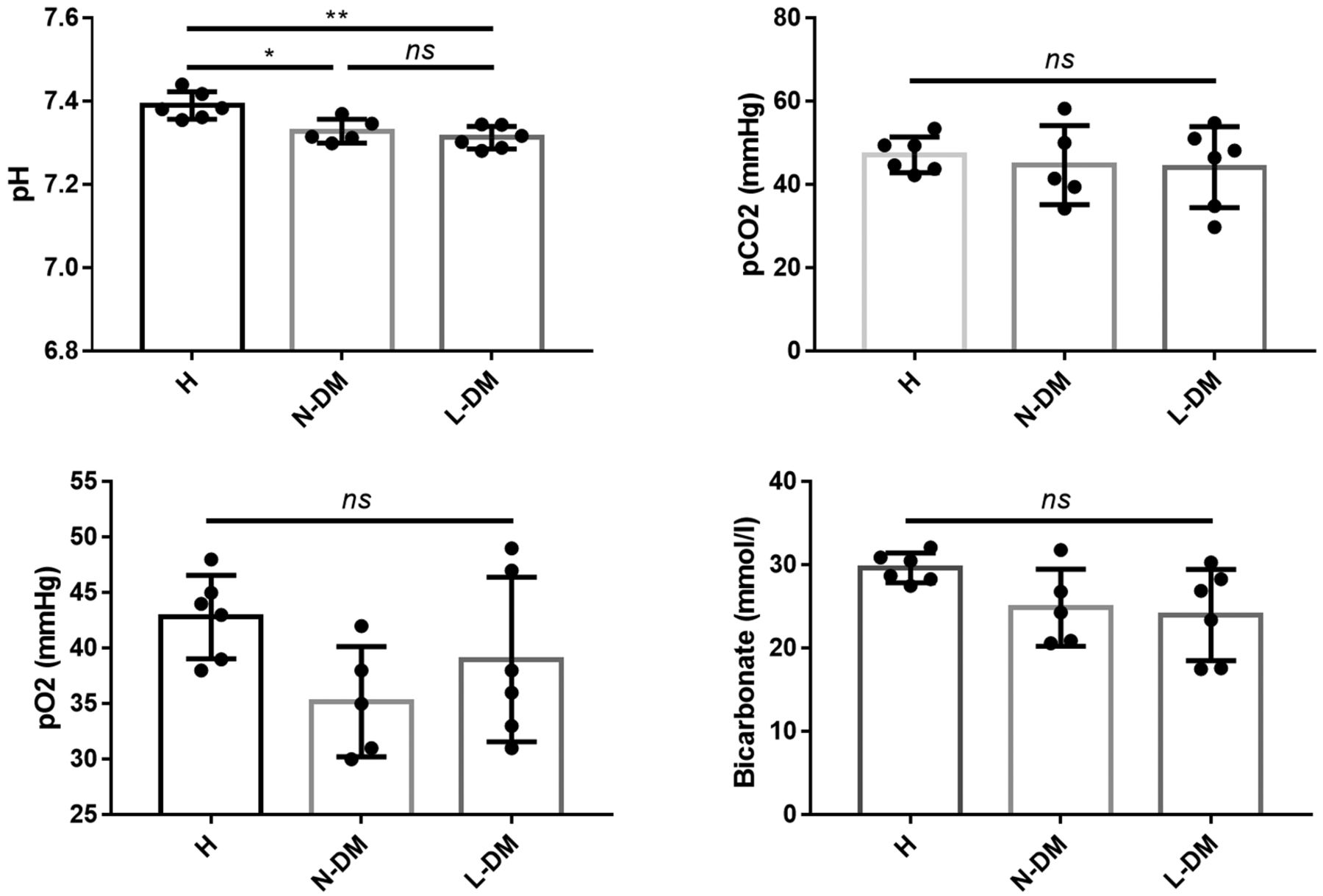
Characteristics of participants. Signalment is described in Table II. Healthy dogs had no specific abnormalities. Dogs with IDDM who had normal lactate levels did not have any diseases that affected their blood lactate concentration such as urolithiasis, tracheal collapse, chronic valvular heart disease, or mast cell tumor resection history. Two dogs with IDDM who had increased lactate levels had hyperadrenocorticism, while the others had no diseases. We evaluated differences in the blood lactate concentration between diabetic and healthy dogs. Except for two dogs, dogs with IDDM had higher than normal blood glucose levels. The blood glucose of healthy dogs was within the normal range. Diabetic dogs also exhibited higher lactate concentrations. Diabetic dogs were further divided into high and normal lactate groups. We measured three DM control indicators (fasting glucose, ketone, Hb1ac, and fructosamine) (Figure 1). The DM groups had higher lactate, blood glucose, ketone, Hb1ac, and fructosamine levels than those in the healthy group. The L-DM group had significantly higher ketone levels than those in the N-DM group. The DM groups had a higher blood pH compared to that of the healthy group, but there was no significant difference between the DM groups (Figure 2). There was no significant difference in pCO2 and pO2 between the DM and healthy groups. Bicarbonate was lower in the DM groups compared to that in the healthy group.
Characteristics, including breed, age, and sex, of the 17 dogs included in this study.
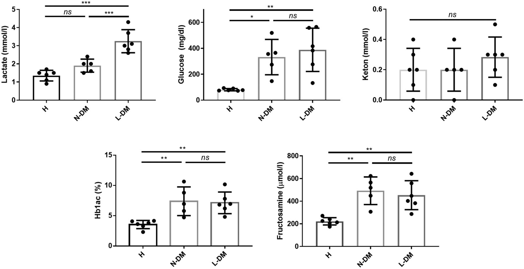
Concentration of blood lactate, glucose, ketone, fructosamine, and Hb1Ac. Groups were divided according to blood lactate levels, and results values were compared for each group. H: Healthy (non-diabetes mellitus) dog group; N-DM: normal blood lactate concentration diabetes mellitus dog group; L-DM: blood lactate concentration above the normal range diabetes mellitus dog group (*p<0.05, **p<0.01, ***p<0.001, ****p<0.0001, as determined by one-way ANOVA).

Venous blood gas analysis in diabetes dogs. The blood gas test result values were compared for each group. H: Healthy (non-diabetes mellitus) dog group; N-DM: normal blood lactate concentration in diabetes mellitus dog group; L-DM: blood lactate concentration above the normal range in diabetes mellitus dog group. (*p<0.05, **p<0.01, ***p<0.001, ****p<0.0001, as determined by one-way ANOVA).
Correlation between fecal bacteria and blood lactate concentration. We investigated lactate-producing bacteria in all groups. Bifidobacterium and Enterococcus were more abundant in both DM groups than in the healthy group. Especially Bifidobacterium, was higher in the L-DM group than in the N-DM group. Lactobacillus was lower in the DM groups than that in the healthy group (Figure 3).
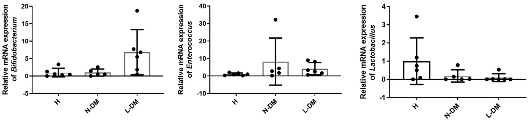
Relative mRNA expression of lactate-producing bacteria in feces of IDDM dogs. Relative mRNA expression of Bifidobacterium, Enterococcus, Lactobacillus in healthy and diabetics dogs. Bacterial mRNA for each group was compared between diseased and healthy controls. H: Healthy (non-diabetes mellitus) dog group; N-DM: normal blood lactate concentration in diabetes mellitus dog group; L-DM: blood lactate concentration above the normal range in diabetes mellitus dog group.
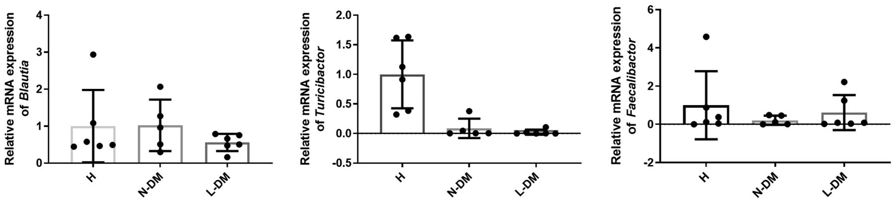
Fecal bacteria associated with dysbiosis. We evaluated three bacteria associated with dysbiosis. Blautia mRNA expression was lower in the DM groups than that in the healthy group, however, it was lower in the L-DM group than that in the N-DM group. Turicibactor mRNA expression was significantly lower in the DM groups. When the lactate levels increased, the Turicibactor mRNA expression tended to decrease. The Feacalibactor mRNA expression was lower in the DM groups than that in the healthy group (Figure 4).

Dysbiosis Index-related bacteria relative mRNA expression. Relative mRNA expression of Blautia, Turicibactor, and Faecalibactor. Bacterial mRNA for each group was compared between diseased and healthy controls. H: Healthy (non-diabetes mellitus) dog group; N-DM: normal blood lactate concentration in diabetes mellitus dog group, L-DM: blood lactate concentration above the normal range in diabetes mellitus dog group.
Discussion
In this study, we hypothesized that the blood concentration of lactic acid correlates with the changes in fecal microbiota of patients with IDDM. In order to investigate the changes in the microbiome according to blood lactate concentration, bacteria associated with lactate production, such as bifidobacterium spp., enterococcus spp., and lactobacillus spp., were examined (9). We confirmed that bifidobacterium spp. and enterococcus spp. tended to increase and lactobacillus spp. tended to decrease in diabetic patients compared to healthy counterparts. It is noteworthy that these findings are related to previously studied intestinal microbial flora.
Brugman et al. investigated the fecal microbiome and found that Lactobacillus and Bifidobacterium populations exhibited lower microbial counts in rats with T1DM than in rats without diabetes (10). Yang et al. found that the proportion of enterococci in the feces was higher in mice with T1DM (11). Interestingly, Paterson et al. studied the changes in intestinal microflora with diabetes onset and progression and found that T1DM progression coincided with an increase in lactate-producing bacteria (i.e., Lactobacillus and Bifidobacterium) (12). Barnett et al. proposed that diabetes-associated hyperlactatemia may be an early event in the time course of the disease (13). In addition, Brouwers et al. reported increased lactate levels in patients with poorly controlled T1DM and glycogenic hepatopathy, implying that enhanced plasma lactate concentrations are part of the clinical spectrum of these diseases (6). This study confirmed that even in diabetic dogs, the intestinal fecal cells changed according to the blood lactate concentration. Although further research is needed on the factors that affect the gut microbiota in diabetic patients, this is the first study to report changes in lactate-producing bacteria in diabetics with elevated lactate levels.
In addition, lactate-producing microbiomes are known to play a major role in the formation of microflora in the intestinal tract, and by identifying blautia, turicibacter, and faecalibactor related to dysbiosis, it was confirmed how it affects other flora (9). Our findings indicate that the intestinal flora of diabetic patients changed according to their blood lactate levels, which means that the blood lactate concentration could be a major indicator of intestinal dysbiosis in diabetic patients.
It is well-known that the intestinal microbiota plays an important role in health by regulating digestion and supporting the immune system (14). Therefore, various studies are being conducted to utilize intestinal bacteria in the treatment of chronic diseases such as metabolic diseases, cancer, inflammatory bowel disease, and autoimmune diseases (15). In particular, an imbalance of the intestinal microflora is known to affect the immune system of patients with T1DM, which has been reported to exacerbate intestinal inflammation (16). However, studies on the intestinal microflora in diabetic patients are still insufficient, and further research is necessary for the development of therapeutic agents that can target the intestinal microflora and their potential applications in diabetic patients.
While we have confirmed the presence of restrictive microbial bacteria, we have not been able to determine whether dysbiosis is present in the feces. In addition, further studies are needed to determine whether the changes in microbial flora observed in DM are a result of disease phenotype or have a causal relationship to pathophysiology. However, our findings are of great value as this was the first study to confirm changes in the fecal microbiota in terms of blood lactate concentration in diabetic dogs. Our results will provide important basic data to help further our understanding of the microbiome in diabetic diseases in both veterinary and human medicine.
We have confirmed that blood lactate levels are a major factor influencing changes in the fecal microflora of dogs with IDDM. These findings help further our understanding of the intestinal microflora of patients with IDDM.
Footnotes
Authors’ Contributions
JHK: Data curation, formal analysis, investigation, resources, writing-original draft. JHA: Conceptualization, data curation, formal analysis, investigation, methodology, resources, writing-original draft, writing-review & editing. JHL: data curation, writing-review & editing. SMP: data curation, writing-review & editing. GHL: data curation, writing-review & editing. YIO: data curation. KWS: data curation, writing-review & editing HYY: Conceptualization, data curation, formal analysis, funding acquisition, investigation, methodology, resources, software, supervision, validation, visualization, writing-review & editing.
Funding
This study was partially supported by the Research Institute for Veterinary Science, Seoul National University, Seoul, Republic of Korea.
Conflicts of Interest
The Authors have no conflicts of interest to report in relation to this study.
- Received December 27, 2022.
- Revision received January 12, 2023.
- Accepted January 13, 2023.
- Copyright © 2023, International Institute of Anticancer Research (Dr. George J. Delinasios), All rights reserved
This article is an open access article distributed under the terms and conditions of the Creative Commons Attribution (CC BY-NC-ND) 4.0 international license (https://creativecommons.org/licenses/by-nc-nd/4.0).
References
In this issue


Jump to section
Related Articles
Cited By...
- No citing articles found.