


Article Figures & Data
Figures
- Figure 1.
Computed tomography (CT) findings in the axial (left) and coronal and sagittal planes (right). A: CT showed the solid mass in the enlarged uterus (yellow arrows). B: The enlarged cystic solid mass in the ovary was shown by CT (orange arrows). C: CT showing the mass in left pelvic cavity (red arrows).
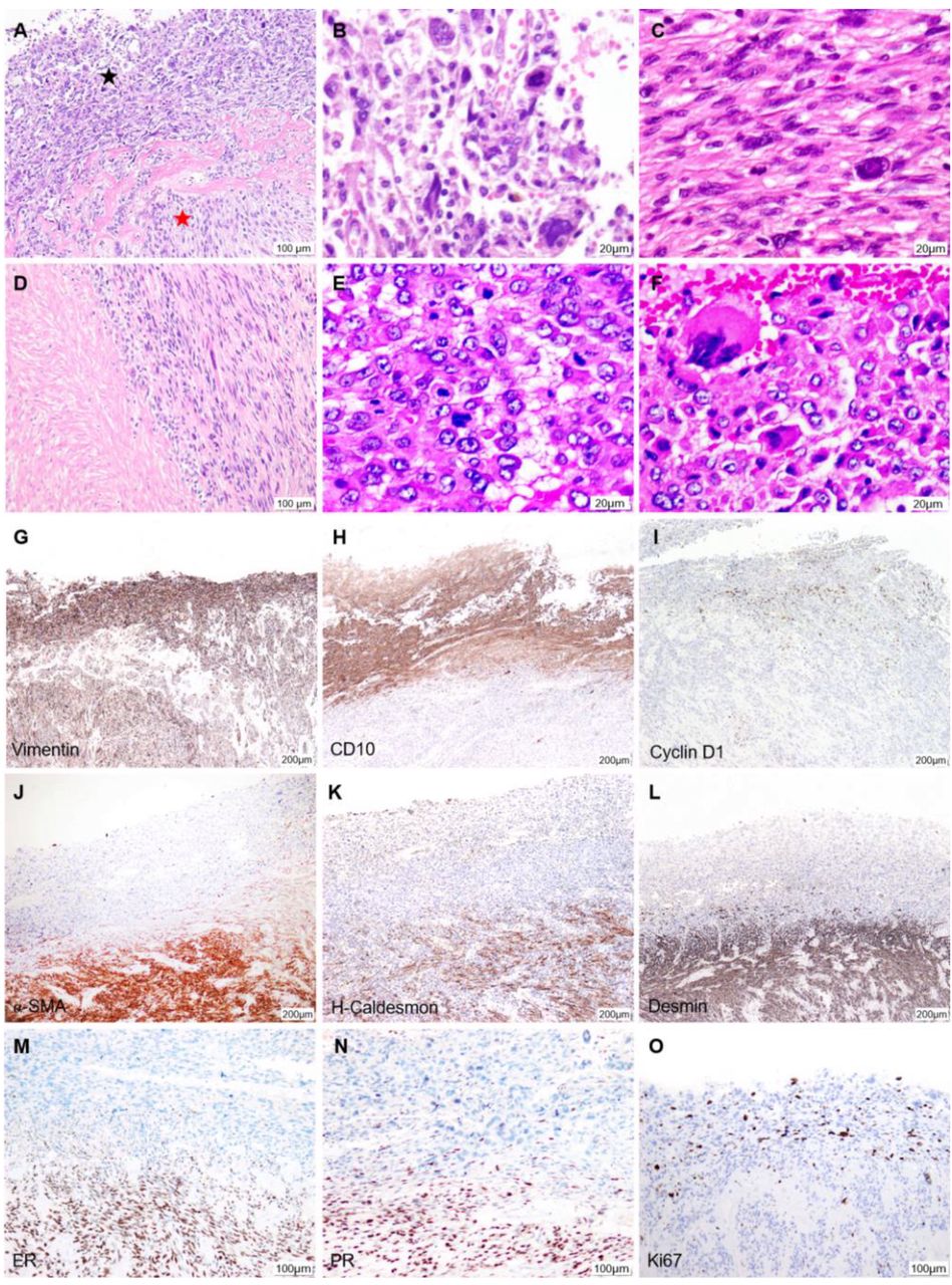
- Figure 2.
Hematoxylin and eosin staining (A-F) and immunohistochemical analysis (G-O). A: The two types of cells can be seen to be separated by hyalinized collagen, with a clear dividing line: Endometrial stromal sarcoma with round-like tumor cells (black star) and leiomyosarcoma with spindled tumor cells (red star), ×40. B: Endometrial stromal sarcoma, ×400. C: Leiomyosarcoma, ×400. D: Coagulative necrosis in leiomyosarcoma, ×100. E: Ovarian metastasis, ×400. F: Pelvic metastasis, ×400. G: Vimentin was diffusely positive in both tumor cells, ×40. H-N: Partial immunophenotyping (CD10, cyclin D1, α-smooth muscle actin, H-caldesmon, desmin, estrogen receptor and progesterone receptor) of the two cell types showed almost opposite results. H-L: ×40, M-N: ×100. O: The Ki-67 proliferation index was about 10% for leiomyosarcoma and 40% for endometrial stromal sarcoma, ×100.
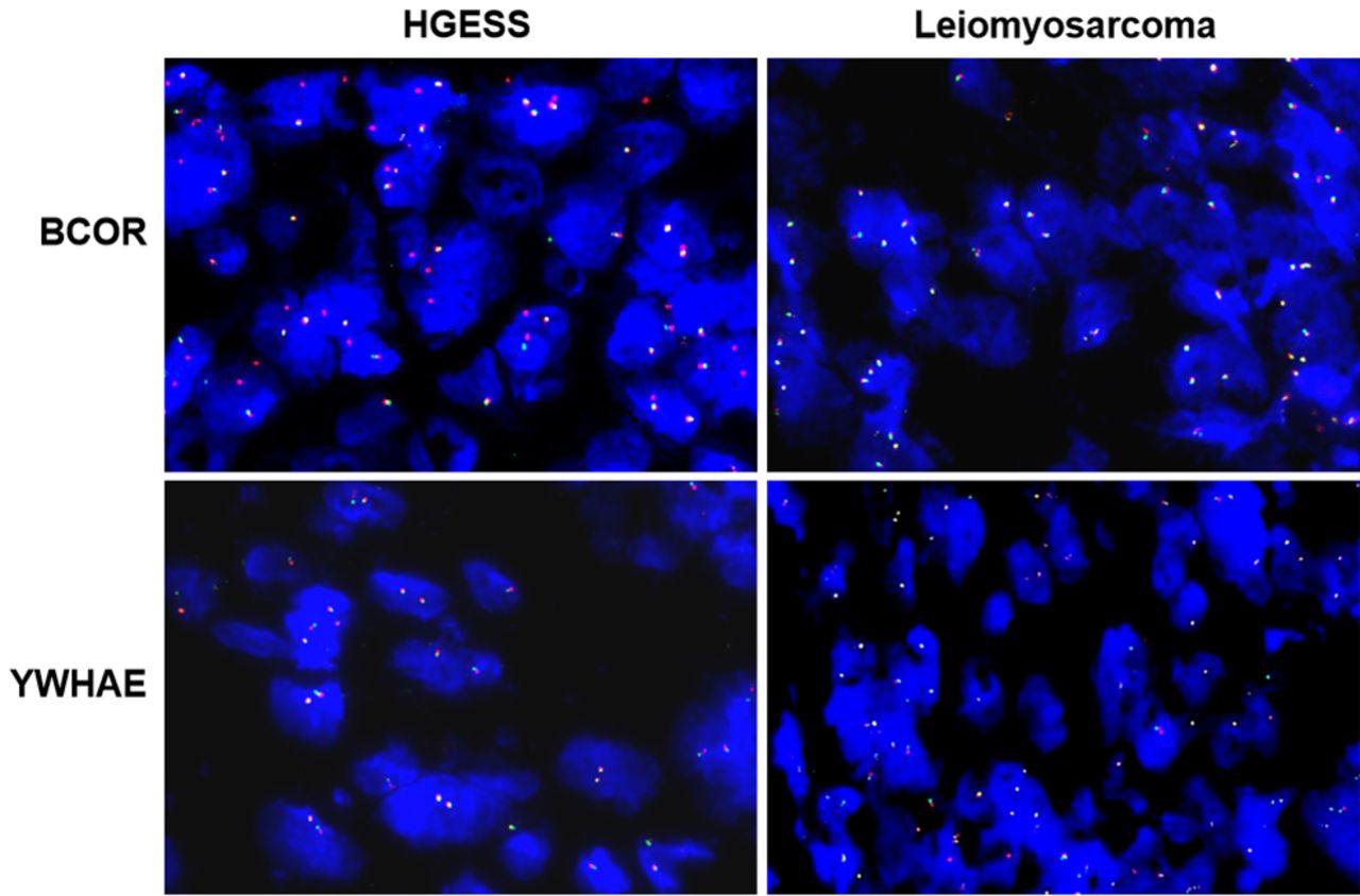
- Figure 3.
Fluorescence in situ hybridization (red probe centromeric, green probe telomeric). High-grade endometrial stromal sarcoma (HGESS) cells were confirmed to have BCL6 corepressor (BCOR) rearrangements but not tyrosine 3-monooxygenase/tryptophan 5-monooxygenase activation protein epsilon (YWHAE) rearrangements. BCOR or YWHAE rearrangements were not detected in leiomyosarcoma cells.

In this issue


Jump to section
Related Articles
Cited By...
- No citing articles found.