


Abstract
Background: The role of epigenetic alterations in the pathogenesis of retinal degenerative diseases, such as macular degeneration is not well established. This study aimed to evaluate whether treatments with gamma-mangostin can rescue the hydrogen peroxide (H2O2)-induced cytotoxicity in human retinal pigment epithelial (ARPE-19) cells. Materials and Methods: ARPE-19 cells were treated with H2O2 alone or with gamma-mangostin plus H2O2 to investigate changes relating to cell viability, appearance of sub-G1 cells, antioxidant enzymes, and apoptotic-related proteins. Results: The data showed that under H2O2 treatment of 400 μM, there was a significant decrease in cell viability and enhanced apoptosis, together with an increased expression of Bax, Bad, cleaved-caspase-3, -8, and -9 at the protein level. On the contrary, the protein expression levels of Bcl2 and Bcl-xl were decreased. Gamma-mangostin pre-treatments (2-16 μM) could effectively prevent all alterations. Conclusion: Gamma-mangostin may conduct its eye-protective effects against H2O2-induced oxidative damage via anti- apoptotic and antioxidant mechanisms in ARPE-19 cells.
Across the retinal degenerative diseases, there are two typical pathological features, the chronic oxidative and inflammatory status. Among these retinal degenerative diseases, the age-related macular degeneration (AMD) is the most common cause of blindness among elderly citizens (1). Clinically, AMD will lead to progressive neurosensory macular destruction, which may cover the areas of retinal pigment epithelium (RPE), Bruch’s membrane, and choroid (2). AMD is cut into two stages: the early stage of AMD is characterized by the aberrant pigmentation of the RPE and the accumulation of extracellular deposits of lipid, cellular debris, and proteins (i.e., drusen), while the late (advanced) stage may manifest as non-exudative or exudative AMD (3, 4). In literature, AMD is one of the most investigated multifactorial eye diseases, and there have been many factors involved in the chronic oxidative and inflammatory status of the micro-environment, which may include sociodemographic (age and race) (5), environmental (cigarette smoking, light exposure, and nutrient intake) (6-10), and genetic risk factors (11-14). All of these factors are involved in the oxidative stress and inflammation of AMD (15).
To mimic the oxidative stress and damage, treatment with hydrogen peroxide (H2O2) is one of the most commonly used models both in vitro and in vivo (16, 17). However, the effect of H2O2 is very acute, occurs randomly, is dosedependent and has multi-output effects, including cell proliferation, migration, survival, differentiation (18, 19) and cell death (20, 21). There have been few studies investigating the influence of H2O2 on cell proliferation and apoptosis, and the alterations of antioxidant enzymes, including glutathione peroxidase, superoxide dismutase and catalase (22, 23). However, the results remain inconclusive.
Mangostins belong to natural xanthonoid compounds, which can be isolated from multiple fragments of the mangosteen trees (Garcinia mangostana). Structurally, mangostins all have a core of xanthone. In the literature, mangostins have been reported to have several characters. In addition to anti-bacterial, anticancer and anti-inflammatory capacities, the antioxidant activities attract the most attention (24). In mice and rats, alpha-mangostin has been found to have a central nervous system depressing capacity, although the mechanisms are not clear and there is a lack of clinical investigation (25). To prevent potential adverse effects, we focused on examining the efficacy of gamma-mangostin (Man), which is a member of the class of xanthones and is isolated from the stems of Cratoxylum cochinchinense. Man has been reported to exhibit anticancer activity (26). Compared with alpha- and beta-mangostin, it is less frequently studied. As far as we know, the eye-protecting effects of Man and its mechanisms have not been previously studied.
Materials and Methods
Reagents. Dulbecco’s Modified Eagle’s Medium (DMEM)/F12, penicillin/streptomycin, and certified fetal bovine serum were purchased from Invitrogen (Carlsbad, CA, USA). 3-(4,5- dimethylthiazol-2-yl)-2,5-diphenyltetrazolium bromide (MTT) were purchased from Sigma–Aldrich, Inc (St. Louis, MO, USA). Primary antibodies including anti-Bax (Cat. sc-7480), anti-Bad (Cat. sc- 8044), anti-Bcl2 (Cat. sc-7382), anti-Bcl-xl (Cat. sc-8392), antiuncleaved caspase-3 (Cat. sc-7272), anti-uncleaved caspase-8 (Cat. sc-56070), anti-uncleaved caspase-9 (Cat. sc-56076), anti-cleaved caspase-3 (Cat. sc-56052), anti-cleaved caspase-8 (Cat. sc-81657), anti-cleaved caspase-9 (Cat. sc-56073), anti-β-actin (Cat. sc-47778), and horseradish peroxidase–conjugated secondary antibodies were purchased from Santa Cruz Biotechnology (Santa Cruz, CA, USA).
Cell culture conditions. Human retinal pigment epithelium (ARPE- 19) cells were purchased from ATCC and routinely maintained in DMEM/F-12 medium, supplemented with 10% fetal bovine serum, 100 U/ml penicillin plus 100 μg/ml streptomycin in a steady 37°C incubator under a humidified 5% CO2 supplement. These cells were passaged every 2 or 3 days and disposed after 15 passages.
MTT assay. Cell viability was evaluated using MTT assay as previously described (27-29). First, to establish the H2O2-induced oxidative stress system, ARPE-19 cells (2×10⁵ cells/well) were treated with 0, 100, 200, 400 and 800 μM of H2O2 for 24 h, and then investigated with MTT assay. To examine the influence of Man on the cell viability of ARPE-19 cells, cells was exposed to 0, 2, 4, 8 and 16 μM of for 24 h. To examine the effects of Man against H2O2-induced cytotoxicity, cultured ARPE-19 cells were pre-treated with indicated concentrations of Man for 24 h, followed by a 24-h exposure of H2O2. After any designed experiments, 10 μl of MTT solution (5 mg/ml) was added to each well and the cells were incubated at 37°C in the dark for another 4 h. The medium was then aspirated and 100 μl of DMSO was added and kept for exactly 10 min. Finally, the absorption was evaluated using a Multiskan MS ELISA reader (Labsystems, Helsinki, Finland).
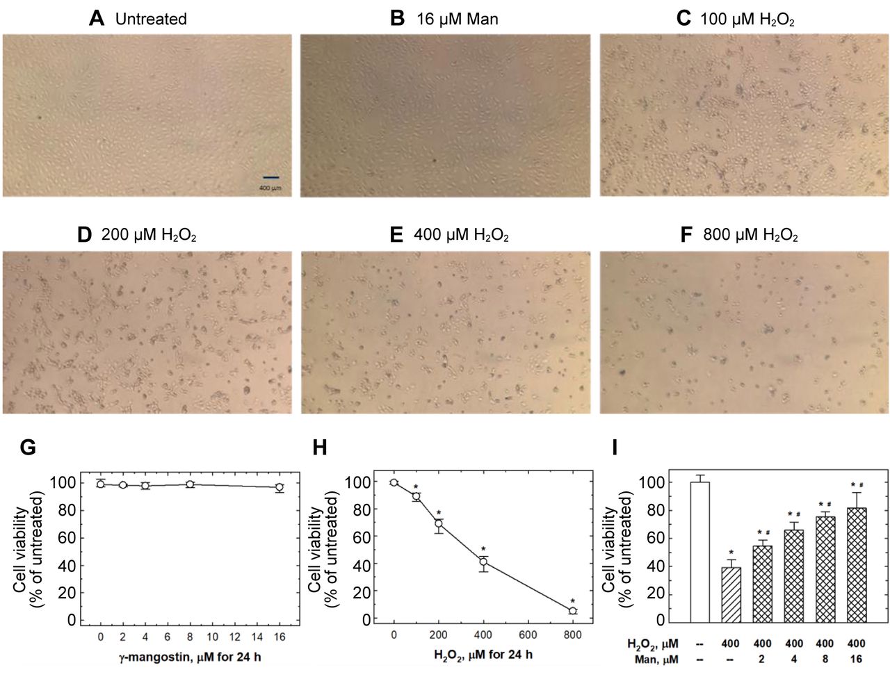
Flow cytometry analysis for cell apoptosis. Cell apoptosis was examined by Annexin V-FITC/PI methodology. ARPE-19 cells were grown in a six-well plate at a density of 2×10⁵ cells/well and treated with or without Man for 24 h, before treatment with H2O2 for 24 h. Then, the cells in all designed groups were washed twice with icecold PBS, resuspended in 300 μl of binding buffer, and stained with 10 μl of Annexin V-FITC plus 10 μl of PI in the dark for 20 min. After that, the stained cells were analyzed using the FACS Calibur instrument (BD Biosciences, San Jose, CA, USA). Morphological changes were photographed randomly as shown in Figure 1A-F.

The effects of H2O2 and Man on ARPE-19 cell viability. (A) ARPE-19 cells are observed under a microscope after 24 h treatment with 16 μM of Man (B), 0 (A), 100 (C), 200 (D), 400 (E), and 800 μM of H2O2 (F). (G) The quantification results of 0~16 μM Man treatment for 24 h in ARPE-19 cells. Data are presented as mean±SD of at least three experiments. (H) The quantification results of 0~800 μM H2O2 treatment for 24 h in ARPE-19 cells. Data are presented as mean±SD of at least three experiments. *Statistically significant (p<0.05) compared with the untreated group. (I) Pretreatments of 0~16 μM Man for 24 h to ARPE-19 cells before being challenged by 400 μM H2O2 for 24 h. Data are presented as mean±SD of at least three experiments. *Statistically significant (p<0.05) compared with the untreated group. #Statistically significant (p<0.05) compared with the 400 μM H2O2 alone group.
Antioxidant enzyme measurements. Following treatment, the ARPE- 19 cells were collected and three common antioxidant enzymes, superoxide dismutase (SOD), catalase (CAT) and glutathione peroxidase (GSH-PX), were measured. Generally, SOD (Cat. 706002), CAT (Cat. 707002) and GSH-PX (Cat. 703002) activity were detected using commercially available assay kits (Cayman Chemical Company, Milpitas, CA, USA), following the manufacturers’ instructions.
Glutathione measurement. Following treatment, the ARPE-19 cells were harvested, and glutathione were measured using a commercially available assay kit from Sigma–Aldrich, Inc. All the procedures were carried out following the manufacturer’s instructions.
Western blot analysis. Briefly, ARPE-19 cells were pre-treated with Man for 24 h and then challenged with H2O2 for 24 h. Cells were then lysed, and equal amount of cell lysates were separated on a 12% sodium dodecyl sulfate/polyacrylamide gel electrophoresis and transferred to a nitrocellulose membrane. The separated bands were then subjected to a panel of primary antibodies including anti-Bax, anti-Bad, anti-Bcl2, anti-Bcl-xl, anti-uncleaved caspase-3, antiuncleaved caspase-8, anti-uncleaved caspase-9, anti-cleaved caspase-3, anti-cleaved caspase-8, anti-cleaved caspase-9 and anti- β-actin, followed by incubation with the appropriate horseradish peroxidase-conjugated secondary antibodies. The western blotting process was performed as described previously (28, 29), and every experiment was conducted at least thrice.
Statistical analysis. All statistical comparisons were carried out using one-way ANOVA followed by Tukey’s post-hoc comparison, with p-Value less than 0.05 to be considered statistically significant. Each value is presented as mean±standard deviation (S.D.).
Results
Man protected ARPE-19 cells against H2O2-induced loss of cell viability. To establish an H2O2-induced ARPE-19 cytotoxicity model, the cells were challenged with 0, 100, 200, 400 and 800 μM of H2O2 for 24 h. As shown in Figure 1H, 100, 200, 400 and 800 μM of H2O2, treatments caused 11.6%, 33%, 60.7% and 95.7% loss of cell viability, respectively (Figure 1H). At the same time, the treatments of 2, 4, 8 and 16 μM of Man did not cause a significant loss of cell viability (Figure 1G). Simultaneously, ARPE-19 cells treated with 100, 200, 400, 800 μM of H2O2’ together with those treated with 16 μM of Man, were observed directly under the microscope. Consistently, H2O2 induced a loss of cell viability dose-dependently, while 16 μM of Man did not cause any obvious alteration in cell integrity or loss of viability (Figure 1A-F). Interestingly, the pre-treatment of Man before H2O2 challenge can prevent the consequent damage and loss of cell viability in a dose-dependent manner (Figure 1I).
Man protected ARPE-19 cells against H2O2-induced apoptosis. To examine H2O2-induced ARPE-19 apoptosis, the cells were challenged with 0, 100, 200, 400 and 800 μM of H2O2 for 24 h. As shown in Figure 2A, 100, 200, 400 and 800 μM of H2O2 treatments induced 16.7%, 22.7%, 39.3, 75.7% of ARPE-19 cells to undergo apoptosis, respectively. The treatments of 2, 4, 8 and 16 μM of Man did not induce any significant apoptosis of ARPE-19 cells (data not shown). Meanwhile, 2, 4, 8 and 16 μM of Man pre-treatments could reduce the percentages of apoptotic cells induced by 400 μM of H2O2 dose-dependently (Figure 2B).

The effects of H2O2 and Man on ARPE-19 cell apoptosis. (A) The effects of 24-h treatment of 0~800 μM H2O2 on ARPE-19 cells. Apoptotic cells were detected by flow cytometry with sub-G1. Data are presented as mean±SD for at least 3 experiments. *statistically significant (p<0.05) compared with the untreated group. (B) Pre-treatments of 0~16 μM Man for 24 h to ARPE-19 cells before being challenged by 400 μM H2O2 for 24 h. Data are presented as mean±SD for at least 3 experiments. *Statistically significant (p<0.05) compared with the untreated group. #Statistically significant (p<0.05) compared with the 400 μM H2O2 alone group.
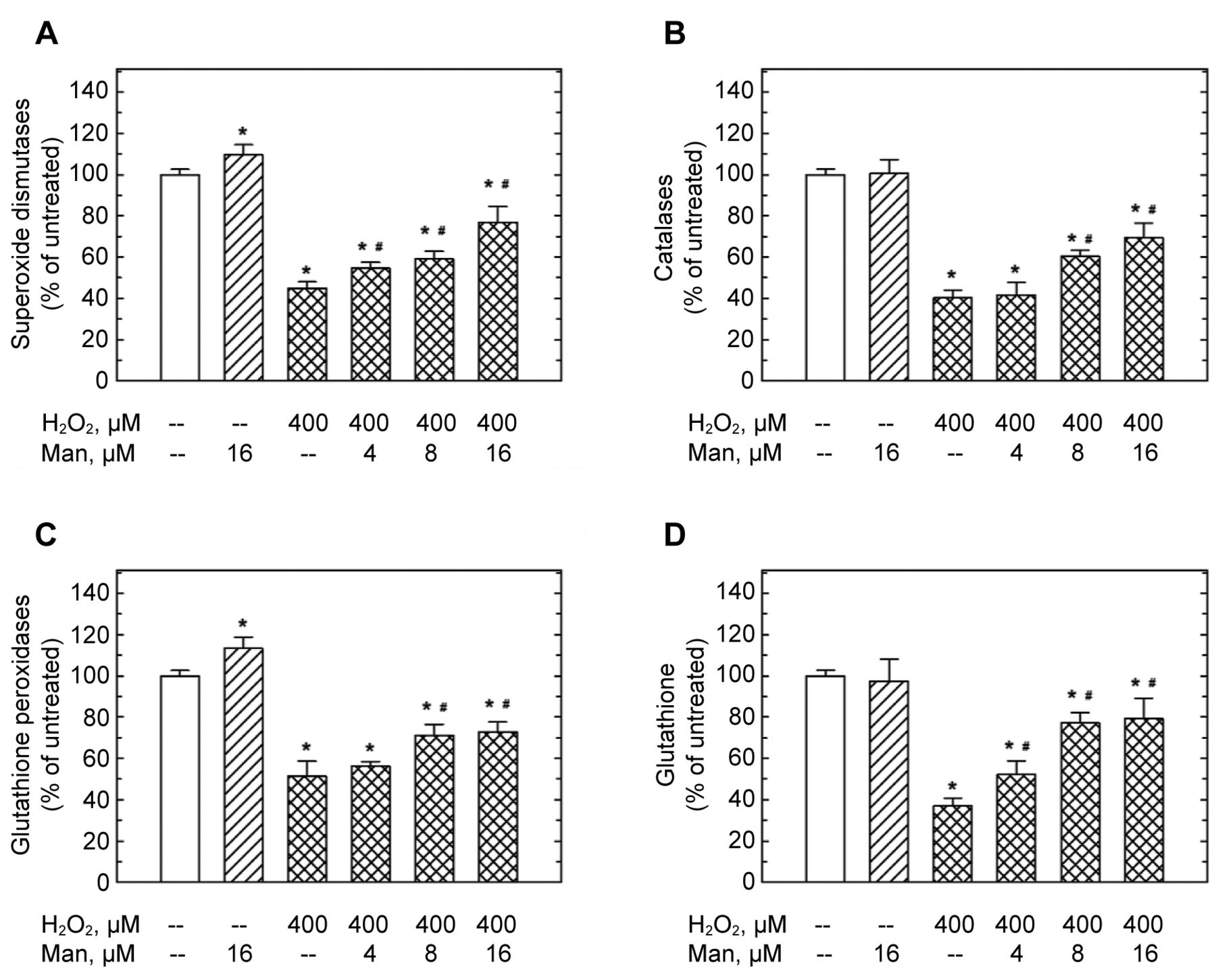
Man rescued H2O2-induced loss of antioxidant capacity in ARPE-19 cells. To explore the influence of H2O2 on antioxidant capacity in ARPE-19 cells, the cells were challenged with 400 μM of H2O2 for 24 h, and the status of 3 antioxidant enzymes, SOD, CAT and GSH-Px, together with GSH were measured. As shown in the figures, 400 μM of H2O2 treatment could decrease the activity of SOD, CAT and GSH-Px, in addition to the level of GSH (Figure 3A-D). At the same time, 16 μM of Man could enhance the activity of SOD and GSH-Px (Figure 3A and C), but not those of CAT or GSH (Figure 3B and D). Although to different degrees, 2, 4, 8 and 16 μM of Man were capable of rescuing the H2O2-induced loss of antioxidant status (Figure 3A-D).

The effects of H2O2 and Man on antioxidant status of ARPE-19 cells. Pre-treatments of 0~16 μM Man for 24 h to ARPE-19 cells before challenged by 400 μM H2O2for 24 h. Then the activity of SOD (A), CAT (B), GSH-Px (C), and the relative amounts of GSH (D) were measured. Data are presented as mean±SD of at least three experiments. *Statistically significant (p<0.05) compared with the untreated group. *Statistically significant (p<0.05) compared with the 400 μM H2O2 alone group.
Man reversed H2O2-induced alterations in apoptotic-related proteins of ARPE-19 cells. To confirm the influence of H2O2, and to check the reverse effects of Man on H2O2-induced alterations in ARPE-19 cells, the cells were pre-treated with various doses of Man, then challenged with 400 μM of H2O2 for 24 h, and the expression levels of apoptotic-related proteins were measured, including Bax, Bad, Bcl2, Bcl-xl, together with uncleaved and cleaved forms of casepase-3, - 8, and -9. As shown in Figure 4, 400 μM of H2O2 treatment could enhance the levels of Bax, Bad (Figure 4A), cleaved caspase-3, -8 and -9 (Figure 4B). On the contrary, the levels of Bcl2 and Bcl-xl were decreased by the H2O2 challenge (Figure 4A), while the uncleaved form of caspase-3, -8 and -9 were unaltered. The pre-treatments of 4 and 16 μM Man could reverse the influences of H2O2 on Bax, Bad, Bcl2, Bcl-xl, cleaved casepase-3, -8, and -9 (Figure 4A and B).
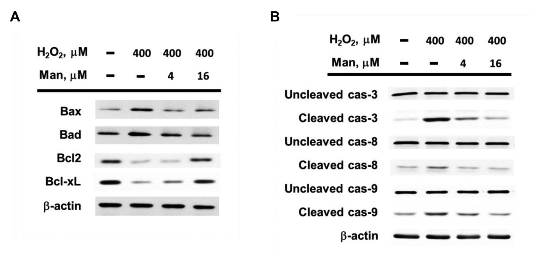
The effects of H2O2 and Man on the expression levels of apoptotic-relatedproteins in ARPE-19 cells. (A) The effects of H2O2 and Man on the expression levels of Bax, Bad, Bcl2 and Bcl-xL in ARPE-19 cells, accessed by Western blotting assay. Pre-treatments of 0~16 μM Man for 24 h to ARPE-19 cells before challenged by 400 μM H2O2 for 24 h. β-actin served as internal standard for loading control. (B) The effects of H2O2 and Man on the expression levels of uncleaved and cleaved forms of caspase-3, -8 and -9 in ARPE-19 cells, accessed by Western blotting assay. Pre-treatments of 0~16 μM Man for 24 h to ARPE-19 cells before being challenged by 400 μM H2O2 for 24 h. β-actin served as internal standard for loading control. Experiments were repeated at least thrice.
Discussion
Although the precise pathogenesis of age-related macular degeneration (AMD) remains largely unclear, oxidative damage-induced loss of function in retinal pigment epithelium cells is thought to be the pathological cause in the initial stage of the AMD progress (30, 31). In addition, several lines of evidence have shown that reactive oxygen species-induced damage to retinal pigment epithelium cells are closely related to AMD (32, 33). Thus, in order to reveal the etiology of AMD and provide eye-protective strategy, the models of reactive oxygen species-induced damage to retinal pigment epithelium cells should be established for drug discovery and screening. To fulfill that, we have set up an H2O2-damage cell model in ARPE-19 cells.
Mangostins have been found to have antioxidant capacity (34, 35). In recent decades, alpha-mangostin has attracted most of the attention, which has been focused on its apoptosisinducing capacity in colorectal cancer (36, 37). Following colorectal cell lines, a panel of cancer cell lines including breast and skin cancer cell lines, were also tested for validating its anti-cancer ability (38-42). In 2016, Fang and colleagues first found that in ARPE-19 cells, alpha-mangostin has protective effects on the cells in respect to oxidative-induced apoptosis (43). However, they did not investigate the effects of other mangostins (beta- and gamma-forms) and the effects of them on oxidative damage and apoptosis.
It is believed that reactive oxygen species-induced apoptotic cascades may play a critical role in AMD (44). From the same viewpoint, drugs which may improve the antioxidant capacity and do good to the mitochondrial integrity may have beneficial effects in fighting against AMD (45). Thus, we focused on the Bcl-2 family proteins which have anti-apoptotic (Bcl2 and Bcl-xl) and pro-apoptotic (Bax and Bad) properties, respectively (Figure 4A). In addition, the activation of caspase cascade is also critical; we have investigated the alterations about caspase-8, -9, and -3 (Figure 4B). Furthermore, we have checked the overall antioxidant status via measuring the alterations of SOD, CAT, GSH-Px, and GSH (Figure 3). We not only investigated the alterations after H2O2 treatment, but also the differences between with and without the pre-treatment of Man.
The highlights of the current study include that Man is effective in conducting its antioxidant capacity for the first time in ARPE-19 cells (Figure 1 and Figure 2). In addition, Man is capable of reversing H2O2-induced loss of antioxidant capacity of SOD, CAT, and GSH-Px (Figure 3). Furthermore, Man is also capable of reversing H2O2-induced alterations of Bax, Bad, Bcl2, Bcl-xl, cleaved casepase-3, -8, and -9 (Figure 4). The most important fact is that Man can be a strong antioxidant as alpha-mangostin was reported to protect ARPE-19 cells from oxidative stress (46). In our unpublished data, we have found that Man is more effective than alpha-mangostin in ARPE-19 cells (data not shown). Safety is another critical concern in clinical drugs. In the data of Chuang, 20 μM of alpha-mangostin caused a loss of cell viability by about 40% (46), while Man caused almost no loss of cell viability (Figure 1G) in the same ARPE-19 cell line. The oxidative stressor in our study is H2O2, while Chuang et al. used NaIO3 (46).
In conclusion, the study has a solid and systematic set of results showing that Man effectively protected ARPE-19 cells against H2O2-induced stress via re-activating anti-apoptotic and antioxidant mechanisms. These findings polish the potential of Man as a novel drug for AMD and other diseases. Further investigations, such as those in mice or rat models, are needed for a better understanding of the underlying mechanisms and possible clinical practice.
Acknowledgements
The study has been supported by Chang Bing Show Chwan Memorial Hospital (BRD-109011), China Medical University Hospital (DMR-110-117), Asia University plus China Medical University (CMU110-ASIA-05) and Changhua Christian Hospital (109-CCH-IRP-113).
Footnotes
Authors’ Contributions
Hu PS, Hsia NY and Chien WC conceived and designed the experiments. Mong MC, Wang YC and Chang WS performed the experiments. Hsia TC Chang HM and Tsai CW analyzed the data. Hu PS and Hsia NY contributed with reagents, materials and analysis tools. Tsai CW and Bau DT wrote and revised the article.
Conflicts of Interest
The Authors declare no conflicts of interest in regard to this study.
- Received April 2, 2022.
- Revision received April 26, 2022.
- Accepted April 27, 2022.
- Copyright © 2022, International Institute of Anticancer Research (Dr. George J. Delinasios), All rights reserved
This article is an open access article distributed under the terms and conditions of the Creative Commons Attribution (CC BY-NC-ND) 4.0 international license (https://creativecommons.org/licenses/by-nc-nd/4.0).
References
In this issue


Jump to section
Related Articles
Cited By...
- No citing articles found.