


Abstract
Background/Aim: Self-defense products that use high-intensity light are being developed. The intense light generated by the high-power light-emitting diodes (LEDs) of such self-defense products causes temporary blindness. However, few studies have been conducted on the visual safety of their devices. We, therefore, evaluated the effects of strong light of a short duration on the eyes of rabbits in this study. Materials and Methods: The right eyes of 15 rabbits were irradiated for 5 s with a lighting device (25 W, 150 lm/W at 700 mA LED) and four eyes of two rabbits were non-irradiated as controls. Changes in the eye structure and function were evaluated before, and immediately, 30 min, 1 h, 24 h, 7 days and 14 days after light irradiation by full-field electroretinogram (ERG), slit-lamp biomicroscopy, and retinal camera. The thickness of the outer nuclear layer of the retina tissue was measured, and histopathological signs of retinal damage were analyzed. Results: The ERG results showed that night vision was not affected. In day vision, the ERG waveform was temporarily affected immediately after light irradiation; however, it recovered within 24 h. No histopathological signs of damage were observed. Conclusion: Application of high-power LED light with short duration as used for self-defense was found to cause temporary phototoxicity, but safety was confirmed as vision recovered within 24 h.
Recently, self-defense products have been developed which use intense LED light to blind an attacker temporarily. Therefore, research on the safety of such devices also needs to be conducted. Several studies have reported retinal damage caused by various light sources, such as home lights using light-emitting diodes (LEDs) and ophthalmic surgical microscopes (1-4). However, no studies have been conducted to test the time needed for vision recovery and the extent of retinal damage after a short duration (5 s) of exposure to light bright enough to be used for self-defense.
The mechanisms of damage to the retina by light can be divided into mechanical, thermal, and chemical (5-7). Mechanical damage is caused by shock waves when the eye is exposed to light for a short duration, from nanoseconds to picoseconds, with intensity ranging from megawatts to terawatts per cm2. Thermal damage occurs when the eye is exposed to light with increasing kinetic energy that can cause an increase in the temperature around the retina by more than 10°C (6, 7). Sources of light that can cause mechanical and thermal damage are rarely used. However, chemical damage is a major result of light-induced phototoxicity. When incident radiation has a wavelength that falls within the high-energy portion of the visible spectrum, different interactions occur between the radiant energy and biological molecules. The excited electrons dissipate extra energy to return to the ground state. In the process, reactive oxygen species, such as hydrogen peroxide, singlet oxygen, hydroxyl radicals, and superoxide radicals, are formed in retinal molecules, resulting in photoreceptor apoptosis and damage to retinal tissue (8, 9).
There are various methods for evaluating phototoxicity. Krigel et al. conducted a study using electroretinography (ERG) as an evaluation method after light exposure (3). Lee et al. evaluated phototoxicity with ERG and transmission electron microscopy (4). ERG provides an objective method for separately assessing impairment in rod and cone functions in degenerative disorders and pharmacological studies. When testing dark-adapted eyes by ERG, rod cells mainly contribute to the results. Using various settings of ERG, different degrees of cone function can be assessed. Saenz-de-Viteri et al. also measured the thickness of the outer nuclear layer (ONL) of the retina and neurosensory retina using histological analysis (10). They counted the number of photoreceptors on hematoxylin-eosin (H&E)-stained sections after the induction of phototoxicity. A significant decrease in the thickness of the ONL and neurosensory retina was observed. The photoreceptor counts showed no significant differences, but an increase in vacuolization within the outer segments of the photoreceptors was observed.
This study used short-term bright light from a self-defense product designed to cause temporary blindness using a high-power LED light source. The changes within rabbit eyes were quantitatively evaluated for up to 2 weeks after irradiation. Structural damage was evaluated using slit-lamp biomicroscopy and a retinal camera. Functional damage was assessed with ERG, which investigated the recovery time, and color and contrast recognition. Histological examination was performed to determine light-induced histological changes within the retinal tissue. Through these examinations, ocular phototoxicity after exposure to light from a self-defense product was quantitatively evaluated. This study assessed how long a self-defense product affects normal vision and whether it causes any other damage after temporary blindness.
Materials and Methods
In vivo studies of the effects of light irradiation. This study used 17 male New Zealand White rabbits (weight, 2.6-3.3 kg; age, 13-16 weeks; Damool Science, Daejeon, Republic of Korea). The light-irradiated groups (with five animals each) were divided into three based on the day of sacrifice after irradiation: Groups were sacrificed 24 h, 7 and 14 days after light irradiation, respectively. The right eyes of 15 rabbits comprised the three light-irradiated groups, and a total of four eyes of two rabbits comprised the control group. All experimental procedures were submitted and approved by the Institutional Animal Care and Use Committee of Chonnam National University (Authorization number: CNU IACUC-YB-2020-34).
Light source. LUHERO® (Newseogwang Co., Gwangju, Korea), a self-defense product made with high-output light sources (four LEDs emitting a total of 25 W, 150 lm/W at 700 mA LED) and a narrow-angle lens that collects light was used. The lighting device was directed toward the rabbit cornea at 1 cm from the corneal surface, and the eye was irradiated for 5 s.
Ophthalmic evaluations. All animals were examined by slit-lamp biomicroscopy (SLM-3ER-E Digital Slit Lamp; Kanghua Ruiming Science & Technology Co., Ltd., Chongqing, PR China) and a retinal camera (Horus DEC-200; MiiS, Hsinchu, Taiwan, ROC) before light irradiation and at sacrifice. The slit-lamp was used to evaluate the conjunctiva, cornea, anterior chamber, light reflex, iris, and lens. The retinal camera was used to assess the fundus, including the optic disc and retinal blood vessels. Full-field ERG (RETevet™; LKC Technologies Inc., Gaithersburg, MD, USA) was performed before, and immediately, 30 min, 1 h, 24 h, 7 days, and 14 days after light irradiation. For the dark-adapted ERG test, dark adaption was induced at least 50 min before testing. The rabbits were anesthetized with an intramuscular injection of 5 mg/kg xylazine hydrochloride (Rompun®; Bayer Korea Ltd., Seoul, Republic of Korea) and 20 mg/kg ketamine hydrochloride (Yuhan Ketamine 50 Inj®; Yuhan Co., Seoul, Republic of Korea). The pupil was dilated with 0.5% tropicamide/phenylephrine eye drops (Mydrin®-P; Santen Pharmaceutical Co., Osaka, Japan). The cornea was anesthetized with 0.5% proparacaine HCL eye drops (Alcaine® eye drops 0.5%; Alcon Laboratories Inc., Fort Worth, TX, USA), and ERG electrodes were placed as follows: Jet contact lens-type electrode with 2% hydroxypropyl methylcellulose (Hycell®, Samil Pharm Co., Seoul, Republic of Korea) on the cornea (active electrode), needle electrode ipsilaterally between the caudal and the lateral canthi (reference electrode), and gold-dome electrode in the mouth (ground electrode). In the dark-adapted state, scotopic tests were performed, and then photopic tests were performed after 5 min of light adaptation. After removing the ERG lens-type active electrode, the eye was irradiated for 5 s with bright light from the self-defense device at 1 cm from the corneal surface. The ERG electrodes were replaced, and photopic tests were performed immediately, and 30 min, and 1 h after light irradiation. The amplitudes of a- and b-waves and the implicit durations of the a- and b-waves were analyzed as parameters. The results are presented in microvolts for amplitudes and milliseconds for implicit times. All the parameter values before and after light irradiation were compared.
ERG was performed according to the research protocols set for testing rabbits in a machine. The stimuli were 2.5-6.0 times brighter than that recommended in the dog, cat and primate protocol by LKC Technologies Inc. In this protocol, six responses are recorded. The set values were as follows: (a) Dark-adapted test 1: scotopic rod response, flash=0.060 cd·s/m2 at a frequency of 0.5 Hz, of ideal white light with Commission Internationale de l’Eclairage coordinates (0.33, 0.33) (hereinafter referred to as white light), background luminance=0.0 cd/m2; (b) dark-adapted test 2: scotopic rod and cone mixed response, flash=8.0 cd·s/m2 at a frequency of 0.1 Hz, white light (0.33, 0.33), background luminance=0.0 cd·s/m2; (c) dark-adapted test 3: oscillatory potentials were automatically calculated from the dark-adapted test 2 and up to five cursors were automatically placed on the peaks and troughs of the oscillatory potentials; (d) dark-adapted test 4: scotopic rod and cone mixed response to a higher intensity flash, flash=25 cd·s/m2 at a frequency of 0.05 Hz, white light (0.33, 0.33), background luminance=0.0 cd/m2; (e) light-adapted test 1: photopic rod and cone mixed response, flash=8.0 cd·s/m2 at a frequency of 2 Hz, white light (0.33, 0.33), background luminance=30 cd/m2; (f) light-adapted test 2: photopic cone response, flash=8.0 cd·s/m2 at a frequency of 28.3 Hz white light (0.33, 0.33), background luminance=30 cd/m2.
Histological analysis. The animals were sacrificed using intravenous potassium chloride (KCl-40® inj.; Daihan Pharmacy, Seoul, Republic of Korea) after general anesthesia, and the eyes were enucleated. The eyes were immersed in Davidson’s Fixative solution (BBC Biochemical Co., Mount Vernon, WA, USA). The tissues were processed and stained with H&E following the conventional procedure. ONL thickness measurements were performed on the H&E-stained sections. The ONL thickness was randomly measured at four points 2,000 μm from the optic nerve, and the mean values were compared.
Statistical analysis. ERG analysis was performed to compare the effects pre light irradiation with those post light irradiation quantitatively, and the ONL thickness measurements of the control and light-irradiated groups were compared. The values are presented as the mean±standard deviation (SD). Student’s t-test was used to evaluate statistical significance; differences with p<0.05 were considered significant.
Results
Ophthalmic evaluations. The light-irradiated eyes did not have corneal edema, corneal vascularization, corneal melanosis, uveitis, or iritis (Figure 1). One animal had conjunctival hyperemia and edema in the light-irradiated eye. No structural damage to the general fundus, including the optic disc and the retinal and choroidal vasculature, was observed after light irradiation (Figure 2).
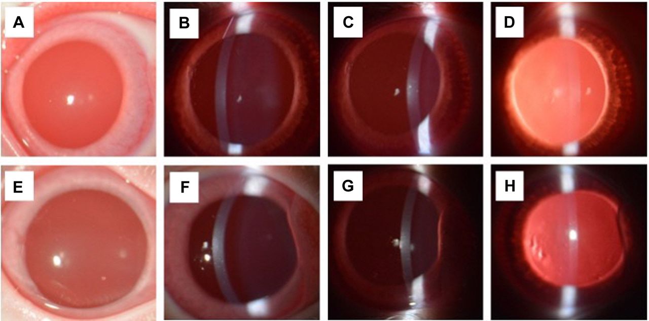
Slit-lamp biomicroscopy of the control and light-irradiated groups. Broad-beam illumination before (A) and after light irradiation (E). Narrow-beam (2 mm) optic section before (B, C) and after light irradiation (F, G). Retroillumination from the fundus before (D) and after light irradiation (H). The cornea, iris, and anterior chamber (left side, temporal; right side, nasal) showed no abnormal findings.

Photographs of the fundus in control and light-irradiated groups. Control (A), Group 1 (24 h after light irradiation) (B), Group 2 (7 days after light irradiation) (C), and Group 3 (14 days after light irradiation) (D). No structural damage of the general fundus, including the optic disc and retinal and choroidal vasculatures, was observed after light irradiation (left side: temporal, right side: nasal).
Electroretinography (ERG). The results of ERG are summarized in Table I and Table II. In the light-adapted test 1, which provided an overall assessment of the outer and inner retina during day vision. The light irradiation caused significant (p<0.001) reductions in both a- and b-wave amplitudes to 77.6% and 65.6% of the pre-irradiation value immediately after light irradiation, respectively (Figure 3A and B). Significant (p<0.001) increases in both a- and b-wave amplitudes to 158.2% and 127.6% of the pre-irradiation values were measured 30 min after light irradiation. After that, they recovered to the pre-irradiation values within 24 h. In the light-adapted test 2 (light-adapted 8.0 flicker, 28.3 Hz), the amplitudes of the b-waves were 82.78 μV before light irradiation. A significant (p<0.05) reduction to 88.11% of the pre-irradiation value was measured immediately after light irradiation (Figure 3C); after 30 min, there was no significant difference from the pre-irradiation value. The implicit durations shown by the light-adapted ERG tended to decrease immediately after light irradiation and recovered over time, but it was not always statistically significant.
Results of light-adapted electroretinography before and after light exposure.
Results of dark-adapted electroretinography before and after light exposure.

Light-adapted electroretinography amplitudes in rabbits before and after light exposure. (A) The a-wave amplitude in light-adapted test 1. (B) The b-wave amplitude in light-adapted test 1. (C) The b-wave amplitude in light-adapted test 2. Statistically significant decrements were measured immediately after light irradiation. Pre-: Pre-irradiation. Results are expressed as the mean±SD. Significantly different at *p<0.05 and **p<0.001 versus pre-irradiation.
In the dark-adapted test 2, which provided a mixed assessment of rods and cones during night vision, the amplitudes of the a- and b-waves tended to increase 24 h after light irradiation; however, the change was not statistically significant.
Histological analysis. No histopathological signs of retinal damage were observed during the histological study of the controls and experimental eyes at 1, 7, and 14 days after irradiation (Figure 4). No pyknotic nuclei, apoptotic bodies of photoreceptors, retinal detachment from retinal pigment epithelium, and phagocytosis of apoptotic bodies by retinal pigment epithelium cells (RPE cells) were observed. Other signs, including gliosis, activation of RPE cells, glial scar formation, newly formed canals filled with edematous fluid, muller glial activation and hypertrophy, and inflammatory cells between the photoreceptor layer and RPE, were also not observed. There were no significant differences in the thickness of the ONL between the control and light-irradiated groups (Figure 5).

Histopathological evaluations of the light-irradiated retina on retinal cross-sections. No histopathological signs of retinal damage were observed during the histological study of the controls and groups at 1, 7 and 14 days after irradiation (Scale bar: 25 μm). ONF: Optic nerve fiber layer; GC: ganglion cell layer; IP: inner plexiform layer; IN: inner nuclear layer; OP: outer plexiform layer; ON: outer nuclear layer; P: photoreceptor layer.
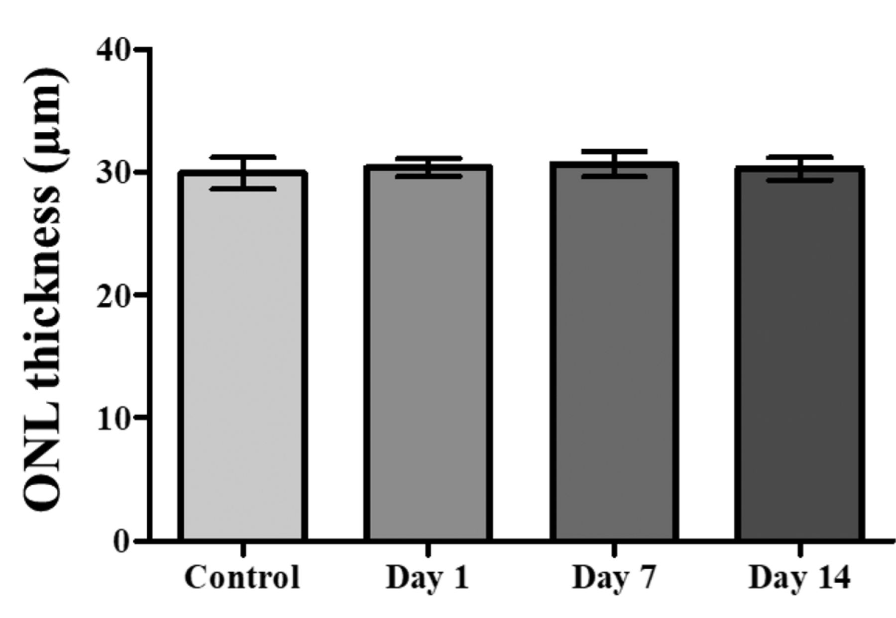
Comparison of the thickness of the outer nuclear layer (ONL) of the retina of the controls and light-irradiated groups. Results are expressed as the mean±SD. There were no significant differences between the irradiated groups and the controls.
Discussion
Several experiments have been conducted to study retinal toxicity using various light sources (2, 11-14). Various clinical findings and histological results have been reported, ranging from reversible damage to extensive and permanent retinal damage, depending on the intensity and the duration of exposure to the light source (2, 11-14). Therefore, the light source’s intensity and duration of exposure associated with visual paralysis were selected as the parameters for irradiation in assessing retinal damage. The experimental results showed no abnormal findings in the cornea, iris, conjunctiva, and aqueous humor on slit-lamp biomicroscopy before and 1, 7, and 14 days after light irradiation. Conjunctival hyperemia and swelling were observed in one animal 1 day after light irradiation, but they were attributed to the stimulation by a contact lens type electrode when the ERG test device was placed or the hydroxypropyl methylcellulose gel used during installation (15).
The retinal ERG was carried out after dark adaptation and light adaptation. The functioning of rod cells, predominantly responsible for night vision, was mainly evaluated using dark-adapted ERG. The cone cell function, mostly responsible for daytime vision, was assessed using light-adapted ERG. However, the contributions of rods and cones to vision depend on the intensity of light and its frequency, and the functions of the two types of cell cannot be completely separated and evaluated. Therefore, the evaluation was performed comprehensively using ERG under various conditions (16, 17).
The light-adapted ERG showed a significant decrease in amplitudes of both waveforms immediately after irradiation. During the light-adapted test 1, the amplitude decreased immediately after irradiation, and a significant increase was observed 30 min and 1 h after irradiation. Moreover, amplitudes recovered 24 h after irradiation. As the dark-adapted state of the retina is not maintained after light irradiation, the dark-adapted ERG was performed immediately, and 1, 7, and 14 days after irradiation. All the tests showed no significant amplitude reduction or implicit time delay which was indicative of retinal damage. Instead, during the dark-adapted tests 2, 3, and 4, the amplitude increased immediately through 14 days after irradiation. In other words, it was confirmed that short-term exposure to bright light did not affect normal night vision based on the ERG results. The visual function was temporarily affected immediately after irradiation for day vision, but color recognition recovered within 30 min. It was also confirmed that both contrast and color recognition were restored within 24 h.
The increase in the ERG wave amplitude after light irradiation can be regarded as the temporary activation of photoreceptors. The mechanism by which amplitude temporarily decreases and increases is unknown, and it cannot be considered that this change leads to activation of visual function. As a result, no complete ERG waveform was lost enough to affect vision after light irradiation; after approximately 10-30% reduction, the amplitude was restored within 24 h. Thus, it may be concluded that the self-defense product temporarily affects vision but vision recovers within 24 h.
Animal models for retinal phototoxicity have been used in several studies. Light-induced damage in nocturnal rats was shown to induce damage of retinal cells and rod cells, mostly limited to the ONL (3, 18-20). Even in rabbits, the thickness of the ONL decreases when there is damage to the retina by light. The initial process of retinal damage is a rapid increase in lipid peroxidation, which destroys cells in the retina and photoreceptors via apoptosis. If oxidative damage is not severe, the cells remain alive but damaged, with vacuolated outer segments. Antioxidant efforts are made in the cell itself, but the damage continues its course, and gradual destruction causes a large reduction in ONL thickness (7, 10, 19). In this experiment, after irradiating the rabbit’s eye with short-term bright light, the thickness of the ONL did not show any significant change over time compared with the control group. According to several studies, various characteristics of retinal degeneration can be observed in retinal tissues after exposure to light in experimental animals (21-27). In this study, the histological analysis showed no specific findings within 1, 7 and 14 days after exposure to short-term bright light.
This study confirmed that when short-term bright light from a self-defense product was irradiated into the rabbit’s eye, the visual function recovered within 24 h, and no structural damage or histological changes occurred. No further changes in the ERG waveform and histopathological results were observed up to the second week after visual function had recovered (within 24 h). This indicates that no further damage was caused after temporary visual paralysis. However, as the degree of damage varies with the exposure conditions, it may change depending on the exposure time, source distance, or the intensity of the light source.
Acknowledgements
This research was supported by the Basic Science Research Program through the National Research Foundation of Korea (NRF) funded by the Ministry of Education (NRF-2020R1C1C1009798). The Authors thank the Ophthalmic Optic Medical Device Globalization Team and Newseogwang Co. for their assistance.
Footnotes
↵* These Authors contributed equally to the study.
Authors’ Contributions
H Park, K Jang, KM Shim and SE Kim designed the study concept. H Park and Y Jo collected and analyzed the data. H Park and K Jang wrote the whole article. KM Shim and C Bae critically revised the article. SE Kim and SS Kang supervised the study. All Authors read and approved the final version of the article.
Conflicts of Interest
The Authors have no financial conflicts of interest.
- Received October 22, 2021.
- Revision received November 17, 2021.
- Accepted December 1, 2021.
- Copyright © 2022 International Institute of Anticancer Research (Dr. George J. Delinasios), All rights reserved
References
In this issue


Jump to section
Related Articles
Cited By...
- No citing articles found.