
Main menu

  • Home
  • Current Issue
  • Archive
  • Info for
    • Authors
    • Editorial Policies
    • Advertisers
    • Editorial Board
    • Special Issues 2025
  • Journal Metrics
  • Other Publications
    • Anticancer Research
    • Cancer Genomics & Proteomics
    • Cancer Diagnosis & Prognosis
  • More
    • IIAR
    • Conferences
  • About Us
    • General Policy
    • Contact
  • Other Publications
    • In Vivo
    • Anticancer Research
    • Cancer Genomics & Proteomics

User menu

  • Register
  • Subscribe
  • My alerts
  • Log in
  • My Cart

Search

  • Advanced search
In Vivo
  • Other Publications
    • In Vivo
    • Anticancer Research
    • Cancer Genomics & Proteomics
  • Register
  • Subscribe
  • My alerts
  • Log in
  • My Cart
In Vivo

Advanced Search

  • Home
  • Current Issue
  • Archive
  • Info for
    • Authors
    • Editorial Policies
    • Advertisers
    • Editorial Board
    • Special Issues 2025
  • Journal Metrics
  • Other Publications
    • Anticancer Research
    • Cancer Genomics & Proteomics
    • Cancer Diagnosis & Prognosis
  • More
    • IIAR
    • Conferences
  • About Us
    • General Policy
    • Contact
  • Visit iiar on Facebook
  • Follow us on Linkedin
Research ArticleExperimental Studies

Curcumin Derivative MTH-3 Regulates Palmitate-induced Insulin Resistance in Mouse Myoblast C2C12 Cells

YU-JEN CHIU, YU-HSIANG LO, JAI-SING YANG, SHENG-CHU KUO and SHIH-CHANG TSAI
In Vivo November 2021, 35 (6) 3181-3191; DOI: https://doi.org/10.21873/invivo.12613
YU-JEN CHIU
1Division of Plastic and Reconstructive Surgery, Department of Surgery, Taipei Veterans General Hospital, Taipei, Taiwan, R.O.C.;
2Department of Surgery, School of Medicine, National Yang Ming Chiao Tung University, Taipei, Taiwan, R.O.C.;
3Institute of Clinical Medicine, National Yang Ming Chiao Tung University, Taipei, Taiwan, R.O.C.;
  • Find this author on Google Scholar
  • Find this author on PubMed
  • Search for this author on this site
YU-HSIANG LO
4Department of Biological Science and Technology, China Medical University, Taichung, Taiwan, R.O.C.;
5Department of Medical Research, China Medical University Hospital, China Medical University, Taichung, Taiwan, R.O.C.;
  • Find this author on Google Scholar
  • Find this author on PubMed
  • Search for this author on this site
JAI-SING YANG
5Department of Medical Research, China Medical University Hospital, China Medical University, Taichung, Taiwan, R.O.C.;
  • Find this author on Google Scholar
  • Find this author on PubMed
  • Search for this author on this site
SHENG-CHU KUO
6Chinese Medicinal Research and Development Center, China Medical University Hospital, School of Pharmacy, China Medical University, Taichung, Taiwan, R.O.C.
  • Find this author on Google Scholar
  • Find this author on PubMed
  • Search for this author on this site
SHIH-CHANG TSAI
4Department of Biological Science and Technology, China Medical University, Taichung, Taiwan, R.O.C.;
  • Find this author on Google Scholar
  • Find this author on PubMed
  • Search for this author on this site
  • For correspondence: sctsai{at}mail.cmu.edu.tw
  • Article
  • Figures & Data
  • Info & Metrics
  • PDF
Loading

Abstract

Background/Aim: At present, there are no effective drugs for the treatment of insulin resistance. MTH-3, a curcumin derivative, exerts potent anti-cancer effects. The aim of the present study was to explore whether MTH-3 is capable of regulating palmitic acid (PA)-induced insulin resistance in C2C12 cells. Materials and Methods: Cell viability was examined using the MTT assay. C2C12 cells were treated with PA and evaluated for the production of oil droplets using an Oil Red O assay. Glucose uptake was analysed by the 2-NBDG assay. Results: Treatment of cells with up to 500 μM PA for 24 h or with 5 or 10 μM MTH-3 had no effect on cell viability. PA induced production of oil droplets in C2C12 cells. After adding MTH-3, the quantity of oil droplets decreased significantly and glucose uptake recovered. Conclusion: MTH-3 may become an efficient drug for the treatment of insulin resistance and associated diseases.

  • Curcumin derivative
  • MTH-3
  • palmitic acid
  • insulin resistance
  • mouse myoblast cells

As the prevalence rate of obesity is increasing on a yearly basis, the incidence of obesity-related complications has also risen sharply. Obesity is regarded as a chronic disease and requires long-term observation and management (1). Thus, there is an urgent need to study potential treatments for obesity-related diseases. Obesity refers to a physical condition that has a negative impact on health due to an excessive increase and accumulation in body fat. The primary causes of obesity are excessive energy intake and a sedentary lifestyle; in a small number of cases, the cause is genetic and/or psychological or due to physical diseases. The current treatment methods are behavioral therapy, drug treatment, and bariatric surgery. Behavior therapy is currently the most important treatment method, including diet control and increased exercise (2). The effects of obesity can be roughly divided into two groups. The first effect of obesity is the mental illness/awareness it can cause due to social obstacles/perceptions (3). The second effect of obesity is the metabolic changes including increased risk of diabetes, insulin resistance, metabolic syndrome, gallbladder disease, hypertension, cardiovascular disease, and cancer (4).

Insulin serves a role in regulating glucose metabolism in tissues such as the fat, liver, heart, kidney and in the central nervous system. It can also inhibit glucose production and increase the speed of glucose absorption by muscle and fat cells. Insulin resistance refers to a situation in which liver cells, fat cells, and muscle cells do not respond to normal concentrations of insulin, thus requiring increased production of insulin (5). In individuals with insulin resistance, a physiological concentration of insulin cannot induce absorption of glucose by muscle and fat cells. To compensate for this situation, the pancreas secretes increased quantities of insulin to stimulate these cells to absorb glucose, which results in hyperinsulinemia. Excessive levels of insulin in the blood triggers obesity, as insulin binds receptors on fat cells to inhibit the activity of lipase, which would normally breakdown triglycerides, resulting in the formation of larger fat cells (6).

Palmitic acid (PA) is one of the most commonly found fatty acids in animals and plants. Butter, cheese, milk, and meat all possess this fatty acid, which is the only fatty acid synthesized in the human body. The liver, adipose tissue, and small intestines in the human body are the primary organs involved in PA synthesis, of which the liver possesses the greatest capacity. Studies have shown that excess lipids affect the levels of free fatty acids, and causes insulin resistance in muscle and liver cells (7, 8). PA is one of the most abundant free fatty acids, accounting for 30-35% of the total free fatty acids in the human blood (9). PA affects the delivery of insulin to hepatocytes and muscle cells in culture (10, 11).

Curcumin is extracted and purified from the root of turmeric. In the past, it was often used in traditional pharmacology to treat biliary diseases, loss of appetite, and cough. Curcumin possesses anti-inflammatory, anti-oxidant, and anti-tumor activities (12-14). Studies have shown that curcumin significantly reduces blood sugar and blood lipid levels in diabetic rats (15, 16), and significantly improves the symptoms of disordered fat metabolism in diabetic rats (17). Although curcumin has been shown to possess a favorable therapeutic effect on several diseases (18), its shortcomings are the poor hydrophilicity and low bioavailability. The curcumin derivative, MTH-3, has thus been developed and studied with the aim of improving hydrophilicity (19). MTH-3 has also been shown to be more effective with regards to anti-cancer effects against triple-negative breast cancer in animal experiments compared with curcumin (20). However, the mechanism by which MTH-3 regulates insulin resistance caused by free fatty acid metabolism remains unclear. The aim of the present study was to explore the effects and mechanism of MTH-3 on insulin resistance in C2C12 cells.

Materials and Methods

Cell differentiation. Cell differentiation was assayed as described previously by Bajaj et al. (21). Briefly, cells were plated at a density of 2×10⁵ cells/ml in each well of a 6-well plate for 24 h. Cells were cultured in DMEM supplemented with 2% horse serum, and the medium was changed every day for 5 days. The formation of a muscular tube represented the completion of differentiation.

MTT assay. Cell viability was assessed using an MTT assay, as previously described (22). Briefly, cells were seeded at a density of 2×10⁵ cells/ml in each well of a 96-well plate (Thermo Fisher Scientific, Inc., Waltham, MA, USA), cultured for 24 h, and then treated with different concentrations (0, 5 and 10 μM) of MTH-3 or different concentrations of PA (0, 100, 200, 300, 400 or 500 μM) for 24 h. MTT was added to each well at a final concentration of 0.5 mg/ml, and incubated at 37°C for 3 h. The supernatant was discarded and DMSO was added to dissolve the formazan crystals. The absorbance at 595 nm was recorded using a microplate reader, and control absorbance was normalized to 100%. A total of six wells per condition were used, across at least four independent experiments.

Oil Red O staining. Cells were cultured in DMEM containing 2% horse serum, and medium was changed every day for 5 days. After 5 days, the culture medium was aspirated, and different concentrations (0, 100, 200, 300, 400 or 500 μM) of PA were added for 24 h. Cells were washed with PBS and then 1 ml of 10% formalin was added to each well for 1 h. Cells were washed with ddH2O and then 1 ml 60% isopropanol was added to each well. After isopropanol was aspirated, cells were stained with Oil-Red-O at room temperature for 2 h in the dark and then observed using an inverted microscope at 100× magnification (23).

Glucose uptake assay. The C2C12 myotubes were treated with 0.4 mM PA and MTH-3 (5 and 10 μM) simultaneously for 24 h. The cells were washed three times with PBS, and subsequently incubated in the presence or absence of 100 nM insulin for 10 min before the addition of 20 μl 2-NBDG (5 mM) for 2 h at 37°C. Subsequently, 30 μl cell suspension was immediately loaded into a 2-chamber slide (NC-Slide A2; ChemoMetec A/S, Allerod, Denmark) for analysis on the NucleoCounter NC-3000 using the built-in caspase assay program (24).

RNA extract. Total RNA was isolated using TRIzol® reagent (Invitrogen; Thermo Fisher Scientific, Inc.) according to the manufacturer’s protocol, as described previously (25). Briefly, 2×10⁶ cells for each treatment were harvested and mixed with 1 ml TRIzol® reagent at room temperature for 5 min. To each tube, 0.2 ml chloroform was added, and centrifuged at 12,000×g for 15 min at 4°C. The supernatant was collected and mixed with 0.5 ml isopropanol for 10 min. This was further centrifuged at 12,000×g for 15 min at 4°C. RNA pellets were dissolved in 100 μl DEPC-treated H2O.

Quantitative (q)PCR. qPCR was performed using a TaKaRa One Step SYBR Ex Taq™ qRT-PCR kit (Takara Bio, Inc., Shiga, Japan) as described previously (25) in a QuantStudio™ 3 Real-Time PCR system (Thermo Fisher Scientific, Inc.). Relative expression levels of target genes were normalized to β-actin. The relative expression levels of mRNA are presented as the mean±standard deviation of duplicates. The sequences of the primers used for qPCR were: GAPDH, forward, 5’-gagtgtttcctcgtcccgtaga-3’ and reverse, 5’-gtgaccaggcgcccaata-3’; glucose transporter 4 (GLUT4) forward, 5’-caatggttgggaaggaaaagg-3’ and reverse, 5’-ggcgccccccaaca-3’; Myogenin forward, 5’-accctacagacgcccacaatc-3’ and reverse, 5’-acatatcctccaccgtgatgct-3’; transcription factor EB (TFEB) forward, 5’-gcggcagaagaaagacaatcac-3’ and reverse, 5’-tgatccggtcattgatgttgaa-3’; peroxisome proliferator-activated receptor-γ (PPAR-γ) coactivator 1-α (PGC1α) forward, 5’-ggcacgcagccctattca-3’ and reverse, 5’-tctcgacacggagagttaaagga-3’; and PPARγ forward, 5’-accacagttgatttctccagcatt-3’ and reverse, 5’-ccattgggtcagctcttgtga-3’.

Western blotting. Western blotting was performed as described previously (26). Briefly, 100 μg protein prepared form each sample was used for western blot analysis. The primary antibodies used were: Anti-GLUT4 (cat. no. GTX79317; GeneTex Inc., Irvine, CA, USA) and anti-β-Actin (cat. no. GTX629630; GeneTex Inc.). Protein bands were semi-quantified using ImageJ 1.4 (National Institutes of Health, Bethesda, MD, USA).

Statistical analysis. The experimental data are presented as the mean±standard deviation. A one-way ANOVA was used for multiple comparisons. p<0.05 was considered to indicate a statistically significant difference. Data were analyzed using GraphPad Prism version 7 (GraphPad Software, Inc., San Diego, CA, USA).

Results

Effect of PA on C2C12 cell survival. To establish a cell model for evaluating C2C12 differentiation, C2C12 cells were cultured in a differentiation medium. C2C12 cells differentiated after cells were cultured for 5 days in the differentiation medium (Figure 1A). Myotubes were formed that were observed under an inverted microscope. Myogenin expression, a biological indicator of C2C12 differentiation, was evaluated by reverse transcription (RT)-qPCR (Figure 1B). Myogenin expression was significantly increased after cells were cultured for 5 days in the differentiation medium. To examine whether PA affected cell viability in C2C12 cells, an MTT assay was performed. C2C12 myotubes cells were treated with 0-500 μM PA (Figure 2A) for 24 h to examine cell viability (Figure 2B). Cell viability was decreased when cells were treated with 500 μM PA. PA at a concentration of 100-400 μM had no effect on C2C12 myotube cells. Therefore, PA concentrations <400 μM were used in the present study.

Figure 1.
  • Download figure
  • Open in new tab
  • Download powerpoint
Figure 1.

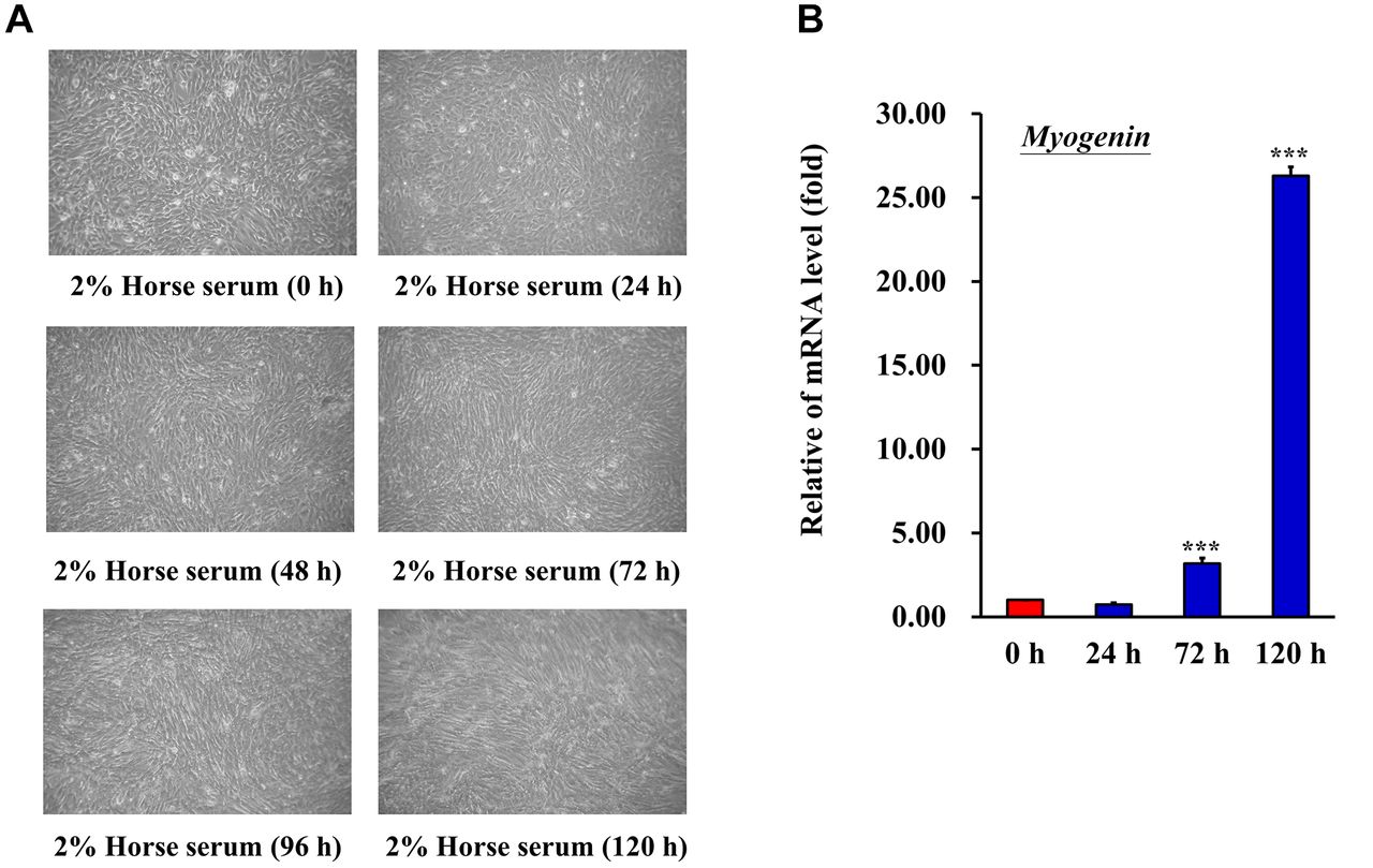
Differentiation of C2C12 cells. (A) Changes in morphology of C2C12 cells. C2C12 were cultured in DMEM supplemented with 2% horse serum to induce differentiation. Culture medium was changed daily. Formation of myotubes was observed under an inverted microscope daily. (B) mRNA expression of myogenin, a biological indicator of C2C12 differentiation, was evaluated. ***p<0.001 vs. control.

Figure 2.
  • Download figure
  • Open in new tab
  • Download powerpoint
Figure 2.

Effect of palmitic acid (PA) on C2C12 cell survival. (A) Chemical structure of PA. (B) C2C12 cells were treated with 0, 100, 200, 300, 400, or 500 μM PA for 24 h. Cell viability was determined using an MTT assay. ***p<0.001 vs. control. PA: Palmitic acid.

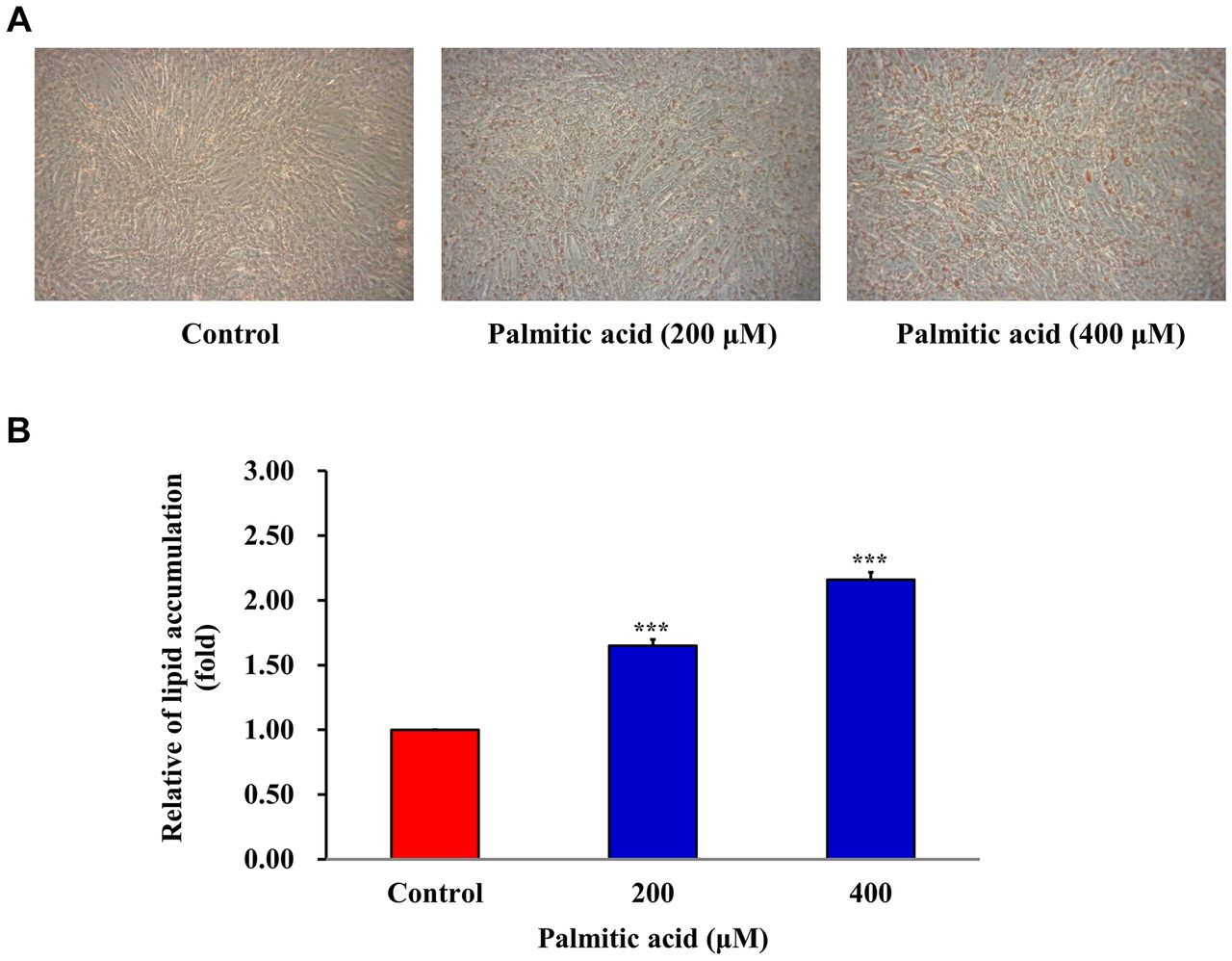
PA induces the production of oil droplets in C2C12 cells. Lipid droplets contain neutral lipids, primarily triglycerides, and cholesterol esters. Excessive accumulation of lipid droplets in cells may be an indicator of metabolic defects or pathogenesis. Oil Red O binds to triglycerides, lipids and lipoproteins, and dyes them red. The quantity of oil droplets was determined using Oil Red O. C2C12 cells were treated with 200 and 400 μM PA for 24 h. PA treated C2C12 cells produced more oil droplets than the control group (Figure 3A and B).

Figure 3.
  • Download figure
  • Open in new tab
  • Download powerpoint
Figure 3.

Palmitic acid (PA) induces the production of oil droplets in C2C12 cells. (A) C2C12 cells were treated with 0, 200 or 400 μM PA for 24 h. Production of oil droplets was assayed using Oil Red O staining. (B) Fold change in the treated group compared with the control group was calculated. ***p<0.001 vs. control.

PA down-regulates GLUT4 expression in C2C12 cells. The primary glucose absorption receptor in muscle cells is GLUT4. To examine whether PA affected the function of GLUT4 to alter insulin resistance, the protein and mRNA expression levels of GLUT4 were analyzed using western blotting and RT-qPCR, respectively. The protein and mRNA expression levels of GLUT4 were significantly reduced in PA-treated C2C12 cells (Figure 4A and B). These results highlight a potential mechanism for the insulin resistance in PA-treated C2C12 cells.

Figure 4.
  • Download figure
  • Open in new tab
  • Download powerpoint
Figure 4.

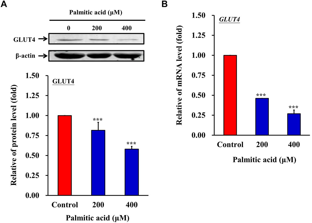
PA down-regulates GLUT4 expression in C2C12 cells. C2C12 cells were treated with 0, 200 or 400 μM PA for 24 h. (A) Protein and (B) RNA expression levels of GLUT4 were analyzed by western blotting and reverse transcription-quantitative PCR, respectively. ***p<0.001 vs. control. GLUT4: Glucose transporter 4.

MTH-3 reduces the effect of PA on the production of oil droplets in C2C12 cells. C2C12 myotubes cells were treated with 5 or 10 μM MTH-3 (Figure 5A) for 24 h to examine cell viability using an MTT assay (Figure 5B). MTH-3 at a concentration of 5 or 10 μM had no effect on C2C12 cells. To investigate whether MTH-3 reduced the production of oil droplets in PA-stimulated C2C12 cells, Oil Red O staining was performed. C2C12 cells were treated with MTH-3 (0, 5 or 10 μM) in the absence or presence of PA (400 μM) for 24 h. The results showed that MTH-3 reduced the production of oil droplets in PA-treated C2C12 cells (Figure 6).

Figure 5.
  • Download figure
  • Open in new tab
  • Download powerpoint
Figure 5.

Effect of MTH-3 on C2C12 cell survival. (A) Chemical structure of MTH-3. (B) C2C12 cells were treated with 0, 5 or 10 μM MTH-3 for 24 h. Cell viability was determined using an MTT assay. MTH-3 at a concentration of 5 and 10 μM had no effect on the viability of C2C12 cells.

Figure 6.
  • Download figure
  • Open in new tab
  • Download powerpoint
Figure 6.

MTH-3 reduces the effect of palmitic acid (PA) on the production of oil droplets in C2C12 cells. C2C12 cells were treated with MTH-3 (0, 5 and 10 μM) in the absence or presence of PA (400 μM) for 24 h. The production of oil droplets was assayed using Oil Red O staining. Fold change was calculated in the treated groups compared with the control group. ***p<0.001 vs. untreated group.

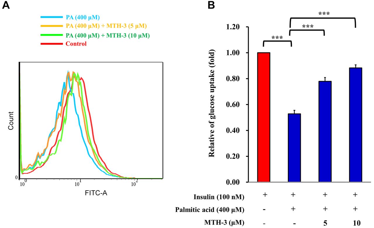
MTH-3 ameliorates glucose uptake in PA-treated C2C12 cells. To investigate whether MTH-3 caused insulin resistance, 2-NBDG was used to detect glucose uptake in PA-stimulated C2C12 cells. C2C12 cells were pre-treated with 100 nM insulin for 10 min, and subsequently treated with MTH-3 (0, 5 or 10 μM) in the absence or presence of PA (400 μM) for 24 h. 2-NBDG was added to cells to analyze glucose uptake. As shown in Figure 7, glucose uptake was significantly increased in the MTH-3 treated C2C12 cells.

Figure 7.
  • Download figure
  • Open in new tab
  • Download powerpoint
Figure 7.

MTH-3 ameliorates glucose uptake in palmitic acid (PA)-treated C2C12 cells. (A) C2C12 cells were pre-treated with 100 nM insulin for 10 min. C2C12 cells were subsequently treated with MTH-3 (0, 5 and 10 μM) in the absence or presence of PA (400 μM) for 24 h. 2-NBDG was added to cells to assess glucose uptake. (B) Relative changes were calculated in the treated group compared with the untreated group. ***p<0.001 vs. untreated group.

MTH-3 reverses the expression of TFEB and PPAR-γ in PA-treated C2C12 cells. To characterize the signaling pathways involved, the expression of TFEB and PPAR-γ were determined. TFEB is the primary transcription factor in lysosomal biosynthesis. In a state of starvation, TFEB increases autophagy, and thus increases fat metabolism. In the liver, TFEB regulates genes involved in lipid catabolism, fatty acid oxidation, and ketone synthesis (27). TFEB also plays a key role in mediating insulin sensitivity, glucose stabilization, lipid oxidation, and mitochondrial function (28). C2C12 cells were treated with 5 and 10 μM MTH-3 in the absence or presence of PA (400 μM) for 24 h, and RNA was subsequently extracted. Expression of TFEB was determined by RT-qPCR. TFEB expression was significantly increased in the MTH-3-treated C2C12 cells (Figure 8A). PPARs are a group of nuclear proteins that function as transcription factors to regulate gene expression. PPARs serve a key role in cell differentiation, metabolism and cancer (29). There are three types of PPARs, namely PPARα, β and γ. These three types are expressed in different tissues and reflect different physiological functions. Amongst these, PPAR-γ plays an important regulatory role in insulin sensitivity (30). C2C12 cells were treated with 5 and 10 μM MTH-3 in the absence or presence of PA (400 μM) for 24 h and RNA was subsequently extracted. Expression of PPAR-γ was determined by RT-qPCR. PPAR-γ expression was significantly increased in the MTH-3-treated C2C12 cells (Figure 8B). Taken together, TFEB and PPAR-γ expression was significantly increased in the MTH-3-treated C2C12 cells.

Figure 8.
  • Download figure
  • Open in new tab
  • Download powerpoint
Figure 8.

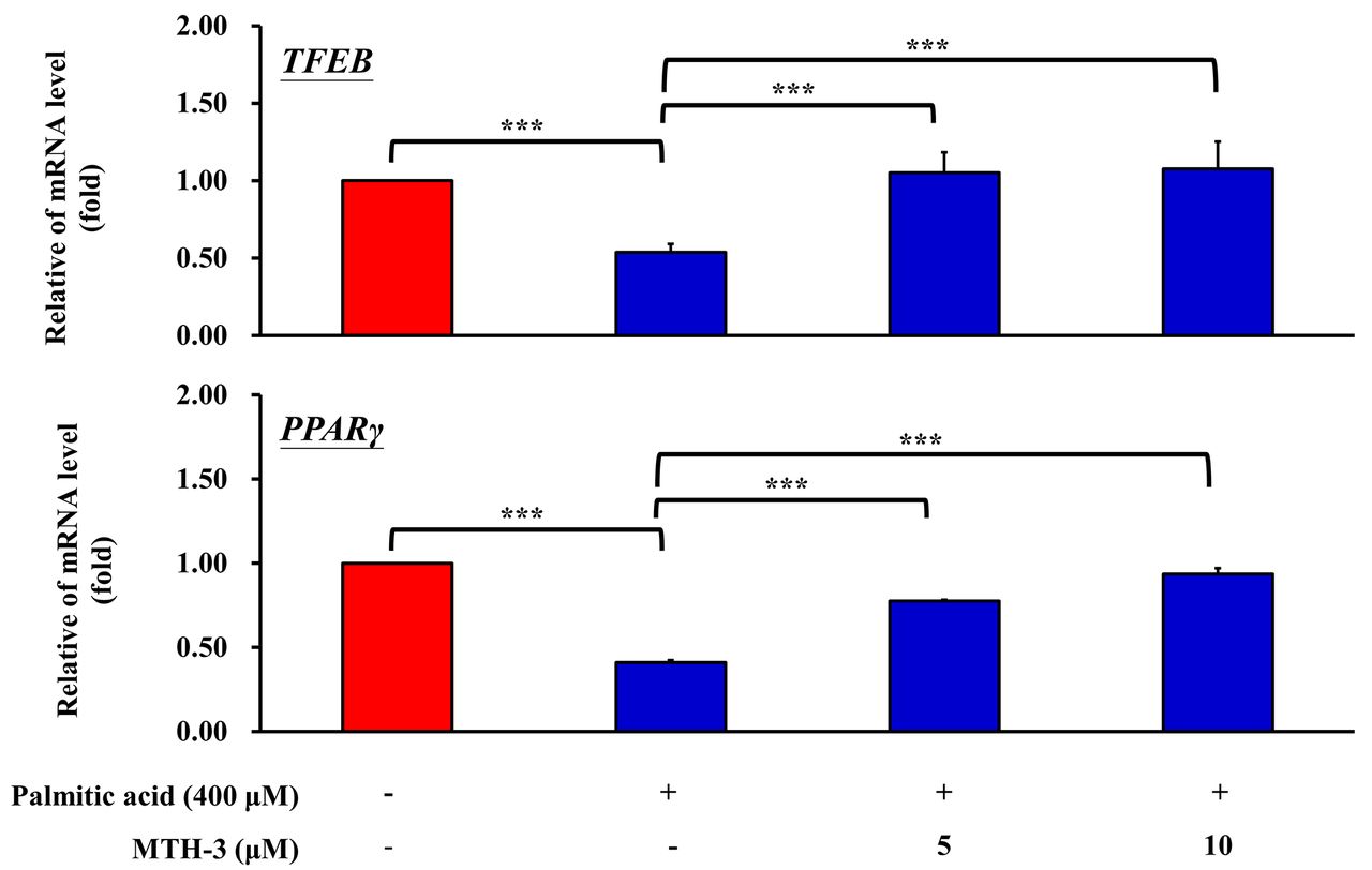
MTH-3 decreases the expression of TFEB and PPAR-γ in PA-treated C2C12 cells. C2C12 cells were treated with MTH-3 (0, 5 and 10 μM) in the absence or presence of PA (400 μM) for 24 h, and RNA was subsequently extracted. Expression of (A) TFEB and (B) PPAR-γ was determined by reverse transcription-quantitative PCR. Relative changes were calculated in the treated group compared with the untreated group. ***p<0.001 vs. control. TFEB: Transcription factor EB; PPARγ: peroxisome proliferator-activated receptor-γ; PA: palmitic acid.

Discussion

Skeletal myoblasts are the optimal cells for studying muscle cell proliferation and differentiation in vitro (31, 32). When skeletal myoblasts are cultured in medium supplemented with 10% fetal calf serum, they continue to proliferate. However, when they are cultured in medium supplemented with 2% horse serum, the cells readily fuse and form myotubes (31, 33). In the present study, the C2C12 cells were an immortalized mouse myoblast cell line, and a well-established in vitro model for studying muscle regeneration and differentiation. In the present study, C2C12 cells were used to study myoblast proliferation and differentiation. During C2C12 cell differentiation, myoblasts undergo remodeling to form mature myotubes, and exhibit increased myogenin mRNA expression (34, 35). As shown in Figure 1A, 2% horse serum medium triggered myogenic differentiation after 72, 96, and 120 h of induction, and the cells that formed the myotubes presented considerable numbers of fused myotubes. Myogenin expression at the mRNA level was increased after 72 and 120 h of culture with 2% horse serum. Thus, it was demonstrated that 2% horse serum induced myogenic differentiation of C2C12 cells.

Insulin resistance is one of the major physiological characteristics of a type II diabetes mellitus (T2D) phenotype (36-38). High levels of free fatty acids in the plasma are associated with insulin resistance in T2D (39, 40). PA is a saturated fatty acid that has been implicated in the development of T2D (41). Excess transpiration of PA to the skeletal muscle is implicated in the development of insulin resistance, resulting from lipid oversupply to skeletal muscles (42-44). In the present study, 100, 200, 300, 400, and 500 μM PA was used to treat C2C12 cells cultured with 2% horse serum supplemented medium for up to 120 h. Treatment for 24 h with 100-400 μM PA did not affect cell viability. PA-induced intracellular lipid accumulation as determined using Oil Red O staining when cells were treated with 200 and 400 μM PA. It has been reported that GLUT4 protein is highly expressed in C2C12 cells, which can be used in the investigation of glucose uptake and insulin resistance mechanisms (45, 46). In the present study, it was shown that GLUT4 mRNA and protein expression levels were reduced in cells treated with 200 and 400 μM PA. It was hypothesized that PA induced intracellular lipid droplet deposition and increased insulin resistance in C2C12 myotubes. Thus, an in vitro model of insulin resistance using C2C12 myotubes was successfully established.

Curcumin, a constituent compound of Curcuma longa plants, has various pharmacological properties, including anti-oxidant, anti-inflammatory, anti-bacterial, anti-viral, anti-cancer, and anti-diabetic properties (47, 48). A clinical study showed that curcumin was safe even at doses of up to 8 g per day (20). However, the pharmacokinetic studies of curcumin on animal and human models have demonstrated poor absorption in the gastrointestinal tract (49, 50). Low systemic bioavailability of curcumin prevents a suitable concentration reaching the target tissues to achieve its pharmacological effects (20). To overcome its poor bioavailability and increase curcumin absorption in the gastrointestinal tract, a novel curcumin derivative, (1E,3Z,6E)-3-hydroxy-5-oxohepta-1,3,6-triene-1,7-diyl)-bis(2-methoxy-4,1-phenylene)-bis(3-hydroxy-2-hydroxymethyl)-2-methyl propanoate, referred to as MTH-3 in the present study, was established. A previous study demonstrated that MTH-3 inhibits proliferation of triple-negative breast adenocarcinoma cells and induces cell death in vitro and in vivo (20). In our previous study, it was also demonstrated that MTH-3 inhibits triple-negative breast adenocarcinoma cell metastasis via the MAPK/ERK/AKT signaling pathway (51). MTH-3 was effective against several different cancer cell lines and exerted only limited toxicity on physiologically healthy cells (19). To the best of our knowledge, the present study is the first to demonstrate that MTH-3 regulates PA-induced insulin resistance in mouse myoblast C2C12 cells. The results showed that 5 and 10 μM MTH-3 treatment for 24 h did not affect cell viability. MTH-3 reduced intracellular lipid accumulation and glucose uptake when 5 and 10 μM MTH-3 was administered in C2C12 cells treated with PA to induce insulin resistance.

TFEB is a transcription factor, and acts as a primary regulator of autophagy, lysosomal genesis, lipid and energy metabolism, and the immune response (52-55). Previous studies demonstrated that TFEB can advance neurodegenerative diseases, such as Parkinson’s disease and Huntington’s disease (56, 57). TFEB also serves an important role in the metabolic response to starvation (58-60). Absence of TFEB results in impairment of lipid catabolism, and TFEB over-expression rescues obesity and the associated metabolic syndrome. It was reported that TFEB regulated insulin resistance and a PPARα agonist (Fenofibrate) reduces hepatic fat accumulation through the upregulation of TFEB-mediated lipophagy (28, 61, 62). In addition, Verma et al. (63) demonstrated that PPARγ expression also regulated insulin resistance in C2C12 skeletal muscle cells. MTH-3 induced TFEB and PPARγ mRNA expression when cells were treated with 5 or 10 μM MTH-3 in PA-induced insulin resistant C2C12 cells. These results suggested that MTH-3 relieves PA-induced intracellular lipid droplet deposition and increases insulin resistance in C2C12 myotubes through upregulating TFEB and PPARγ gene expression.

In conclusion, it was shown that PA induced intracellular lipid droplet deposition and increased insulin resistance in C2C12 myotubes. MTH-3 improved PA-induced-insulin resistance in C2C12 cells through increased TFEB and PPARγ expression at the mRNA level. Based on these results, MTH-3 may be considered a promising therapeutic strategy for the treatment of T2D.

Acknowledgements

The Authors would like to thank the Office of Research & Development at China Medical University, Taichung, Taiwan, R.O.C. for allowing the use of the Medical Research Core Facilities to perform the experiments and data analysis.

Footnotes

  • This article is freely accessible online.

  • Authors’ Contributions

    YJC, YSL, JSY and SCT conceived and designed the study. YJC, YSL and SCT performed the experiments. YSL, JSY and SCT analyzed the data. YJC, JSY SCK and SCT wrote and modified the manuscript. All the Authors read and approved the manuscript. All Authors confirm the authenticity of all the raw data.

  • Funding

    The present study was supported in part by the China Medical University Hospital (grant no. DMR-110-155), China Medical University (grant no. CMU103-S-16 and MOST 106-2314-B-039-046), Taipei Veterans General hospital (grant no. V110B-038), Yen Tjing Ling Medical Foundation (grant no. CI-110-6) and the Chinese Medicine Research Center, China Medical University from The Featured Areas Research Center Program within the framework of the Higher Education Sprout Project by the Ministry of Education (MOE) in Taiwan, R.O.C.

  • Conflicts of Interest

    All the Authors have declared no conflicts of interest in relation to this study.

  • Received July 7, 2021.
  • Revision received July 31, 2021.
  • Accepted August 5, 2021.
  • Copyright © 2021 The Author(s). Published by the International Institute of Anticancer Research.

References

  1. ↵
    1. Brown JC,
    2. Carson TL,
    3. Thompson HJ and
    4. Agurs-Collins T
    : The triple health threat of diabetes, obesity, and cancer-epidemiology, disparities, mechanisms, and interventions. Obesity (Silver Spring) 29(6): 954-959, 2021. PMID: 34029445. DOI: 10.1002/oby.23161
    OpenUrlCrossRefPubMed
  2. ↵
    1. Pan XF,
    2. Wang L and
    3. Pan A
    : Epidemiology and determinants of obesity in China. Lancet Diabetes Endocrinol 9(6): 373-392, 2021. PMID: 34022156. DOI: 10.1016/S2213-8587(21)00045-0
    OpenUrlCrossRefPubMed
  3. ↵
    1. Fontaine KR,
    2. Cheskin LJ and
    3. Barofsky I
    : Health-related quality of life in obese persons seeking treatment. J Fam Pract 43(3): 265-270, 1996. PMID: 8797754.
    OpenUrlPubMed
  4. ↵
    1. Sivasubramanian R,
    2. Malhotra S,
    3. Fitch AK and
    4. Singhal V
    : Obesity and metabolic care of children of South Asian ethnicity in western society. Children (Basel) 8(6): 447, 2021. PMID: 34070381. DOI: 10.3390/children8060447
    OpenUrlCrossRefPubMed
  5. ↵
    1. Bratanova-Tochkova TK,
    2. Cheng H,
    3. Daniel S,
    4. Gunawardana S,
    5. Liu YJ,
    6. Mulvaney-Musa J,
    7. Schermerhorn T,
    8. Straub SG,
    9. Yajima H and
    10. Sharp GW
    : Triggering and augmentation mechanisms, granule pools, and biphasic insulin secretion. Diabetes 51 Suppl 1: S83-S90, 2002. PMID: 11815463. DOI: 10.2337/diabetes.51.2007.s83
    OpenUrlAbstract/FREE Full Text
  6. ↵
    1. DeFronzo RA
    : Lilly lecture 1987. The triumvirate: beta-cell, muscle, liver. A collusion responsible for NIDDM. Diabetes 37(6): 667-687, 1988. PMID: 3289989. DOI: 10.2337/diab.37.6.667
    OpenUrlFREE Full Text
  7. ↵
    1. Kahn BB and
    2. Flier JS
    : Obesity and insulin resistance. J Clin Invest 106(4): 473-481, 2000. PMID: 10953022. DOI: 10.1172/JCI10842
    OpenUrlCrossRefPubMed
  8. ↵
    1. Perry RJ,
    2. Samuel VT,
    3. Petersen KF and
    4. Shulman GI
    : The role of hepatic lipids in hepatic insulin resistance and type 2 diabetes. Nature 510(7503): 84-91, 2014. PMID: 24899308. DOI: 10.1038/nature13478
    OpenUrlCrossRefPubMed
  9. ↵
    1. Wilding JP
    : The importance of free fatty acids in the development of Type 2 diabetes. Diabet Med 24(9): 934-945, 2007. PMID: 17725706. DOI: 10.1111/j.1464-5491.2007.02186.x
    OpenUrlCrossRefPubMed
  10. ↵
    1. Gao D,
    2. Nong S,
    3. Huang X,
    4. Lu Y,
    5. Zhao H,
    6. Lin Y,
    7. Man Y,
    8. Wang S,
    9. Yang J and
    10. Li J
    : The effects of palmitate on hepatic insulin resistance are mediated by NADPH Oxidase 3-derived reactive oxygen species through JNK and p38MAPK pathways. J Biol Chem 285(39): 29965-29973, 2010. PMID: 20647313. DOI: 10.1074/jbc.M110.128694
    OpenUrlAbstract/FREE Full Text
  11. ↵
    1. Yang M,
    2. Wei D,
    3. Mo C,
    4. Zhang J,
    5. Wang X,
    6. Han X,
    7. Wang Z and
    8. Xiao H
    : Saturated fatty acid palmitate-induced insulin resistance is accompanied with myotube loss and the impaired expression of health benefit myokine genes in C2C12 myotubes. Lipids Health Dis 12: 104, 2013. PMID: 23866690. DOI: 10.1186/1476-511X-12-104
    OpenUrlCrossRefPubMed
  12. ↵
    1. Hatcher H,
    2. Planalp R,
    3. Cho J,
    4. Torti FM and
    5. Torti SV
    : Curcumin: from ancient medicine to current clinical trials. Cell Mol Life Sci 65(11): 1631-1652, 2008. PMID: 18324353. DOI: 10.1007/s00018-008-7452-4
    OpenUrlCrossRefPubMed
    1. Rahmani AH,
    2. Alsahli MA,
    3. Aly SM,
    4. Khan MA and
    5. Aldebasi YH
    : Role of curcumin in disease prevention and treatment. Adv Biomed Res 7: 38, 2018. PMID: 29629341. DOI: 10.4103/abr.abr_147_16
    OpenUrlCrossRefPubMed
  13. ↵
    1. Vallianou NG,
    2. Evangelopoulos A,
    3. Schizas N and
    4. Kazazis C
    : Potential anticancer properties and mechanisms of action of curcumin. Anticancer Res 35(2): 645-651, 2015. PMID: 25667441.
    OpenUrlAbstract/FREE Full Text
  14. ↵
    1. Arun N and
    2. Nalini N
    : Efficacy of turmeric on blood sugar and polyol pathway in diabetic albino rats. Plant Foods Hum Nutr 57(1): 41-52, 2002. PMID: 11855620. DOI: 10.1023/a:1013106527829
    OpenUrlCrossRefPubMed
  15. ↵
    1. Babu PS and
    2. Srinivasan K
    : Hypolipidemic action of curcumin, the active principle of turmeric (Curcuma longa) in streptozotocin induced diabetic rats. Mol Cell Biochem 166(1-2): 169-175, 1997. PMID: 9046034. DOI: 10.1023/a:1006819605211
    OpenUrlCrossRefPubMed
  16. ↵
    1. Pari L and
    2. Murugan P
    : Antihyperlipidemic effect of curcumin and tetrahydrocurcumin in experimental type 2 diabetic rats. Ren Fail 29(7): 881-889, 2007. PMID: 17994458. DOI: 10.1080/08860220701540326
    OpenUrlCrossRefPubMed
  17. ↵
    1. Kostrzewa T,
    2. Przychodzen P,
    3. Gorska-Ponikowska M and
    4. Kuban-Jankowska A
    : Curcumin and cinnamaldehyde as PTP1B inhibitors with antidiabetic and anticancer potential. Anticancer Res 39(2): 745-749, 2019. PMID: 30711953. DOI: 10.21873/anticanres.13171
    OpenUrlAbstract/FREE Full Text
  18. ↵
    1. Hsieh MT,
    2. Chang LC,
    3. Hung HY,
    4. Lin HY,
    5. Shih MH,
    6. Tsai CH,
    7. Kuo SC and
    8. Lee KH
    : New bis(hydroxymethyl) alkanoate curcuminoid derivatives exhibit activity against triple-negative breast cancer in vitro and in vivo. Eur J Med Chem 131: 141-151, 2017. PMID: 28319780. DOI: 10.1016/j.ejmech.2017.03.006
    OpenUrlCrossRefPubMed
  19. ↵
    1. Chang LC,
    2. Hsieh MT,
    3. Yang JS,
    4. Lu CC,
    5. Tsai FJ,
    6. Tsao JW,
    7. Chiu YJ,
    8. Kuo SC and
    9. Lee KH
    : Effect of bis(hydroxymethyl) alkanoate curcuminoid derivative MTH-3 on cell cycle arrest, apoptotic and autophagic pathway in triple-negative breast adenocarcinoma MDA-MB-231 cells: An in vitro study. Int J Oncol 52(1): 67-76, 2018. PMID: 29138806. DOI: 10.3892/ijo.2017.4204
    OpenUrlCrossRefPubMed
  20. ↵
    1. Bajaj P,
    2. Reddy B Jr.,
    3. Millet L,
    4. Wei C,
    5. Zorlutuna P,
    6. Bao G and
    7. Bashir R
    : Patterning the differentiation of C2C12 skeletal myoblasts. Integr Biol (Camb) 3(9): 897-909, 2011. PMID: 21842084. DOI: 10.1039/c1ib00058f
    OpenUrlCrossRefPubMed
  21. ↵
    1. Yang JS,
    2. Lin CA,
    3. Lu CC,
    4. Wen YF,
    5. Tsai FJ and
    6. Tsai SC
    : Carboxamide analog ITR-284 evokes apoptosis and inhibits migration ability in human lung adenocarcinoma A549 cells. Oncol Rep 37(3): 1786-1792, 2017. PMID: 28098885. DOI: 10.3892/or.2017.5374
    OpenUrlCrossRefPubMed
  22. ↵
    1. Mehlem A,
    2. Hagberg CE,
    3. Muhl L,
    4. Eriksson U and
    5. Falkevall A
    : Imaging of neutral lipids by oil red O for analyzing the metabolic status in health and disease. Nat Protoc 8(6): 1149-1154, 2013. PMID: 23702831. DOI: 10.1038/nprot.2013.055
    OpenUrlCrossRefPubMed
  23. ↵
    1. Zou C,
    2. Wang Y and
    3. Shen Z
    : 2-NBDG as a fluorescent indicator for direct glucose uptake measurement. J Biochem Biophys Methods 64(3): 207-215, 2005. PMID: 16182371. DOI: 10.1016/j.jbbm.2005.08.001
    OpenUrlCrossRefPubMed
  24. ↵
    1. Yang JS,
    2. Lee CY,
    3. Cho HC,
    4. Lu CC,
    5. Kuo SC,
    6. Wen YF,
    7. Tsai FJ,
    8. Lee MR and
    9. Tsai SC
    : ITR 284 modulates cell differentiation in human chronic myelogenous leukemia K562 cells. Oncol Rep 39(1): 383-391, 2018. PMID: 29138846. DOI: 10.3892/or.2017.6090
    OpenUrlCrossRefPubMed
  25. ↵
    1. Tsai SC,
    2. Tsai MH,
    3. Chiu CF,
    4. Lu CC,
    5. Kuo SC,
    6. Chang NW and
    7. Yang JS
    : AMPK-dependent signaling modulates the suppression of invasion and migration by fenofibrate in CAL 27 oral cancer cells through NF-κB pathway. Environ Toxicol 31(7): 866-876, 2016. PMID: 25545733. DOI: 10.1002/tox.22097
    OpenUrlCrossRefPubMed
  26. ↵
    1. Settembre C,
    2. De Cegli R,
    3. Mansueto G,
    4. Saha PK,
    5. Vetrini F,
    6. Visvikis O,
    7. Huynh T,
    8. Carissimo A,
    9. Palmer D,
    10. Klisch TJ,
    11. Wollenberg AC,
    12. Di Bernardo D,
    13. Chan L,
    14. Irazoqui JE and
    15. Ballabio A
    : TFEB controls cellular lipid metabolism through a starvation-induced autoregulatory loop. Nat Cell Biol 15(6): 647-658, 2013. PMID: 23604321. DOI: 10.1038/ncb2718
    OpenUrlCrossRefPubMed
  27. ↵
    1. Sun J,
    2. Lu H,
    3. Liang W,
    4. Zhao G,
    5. Ren L,
    6. Hu D,
    7. Chang Z,
    8. Liu Y,
    9. Garcia-Barrio MT,
    10. Zhang J,
    11. Chen YE and
    12. Fan Y
    : Endothelial TFEB (Transcription Factor EB) improves glucose tolerance via upregulation of IRS (Insulin Receptor Substrate) 1 and IRS2. Arterioscler Thromb Vasc Biol 41(2): 783-795, 2021. PMID: 33297755. DOI: 10.1161/ATVBAHA.120.315310
    OpenUrlCrossRefPubMed
  28. ↵
    1. Berger J and
    2. Moller DE
    : The mechanisms of action of PPARs. Annu Rev Med 53: 409-435, 2002. PMID: 11818483. DOI: 10.1146/annurev.med.53.082901.104018
    OpenUrlCrossRefPubMed
  29. ↵
    1. Kintscher U and
    2. Law RE
    : PPARgamma-mediated insulin sensitization: the importance of fat versus muscle. Am J Physiol Endocrinol Metab 288(2): E287-E291, 2005. PMID: 15637349. DOI: 10.1152/ajpendo.00440.2004
    OpenUrlCrossRefPubMed
  30. ↵
    1. Fujita H,
    2. Shimizu K and
    3. Nagamori E
    : Novel method for fabrication of skeletal muscle construct from the C2C12 myoblast cell line using serum-free medium AIM-V. Biotechnol Bioeng 103(5): 1034-1041, 2009. PMID: 19350625. DOI: 10.1002/bit.22318
    OpenUrlCrossRefPubMed
  31. ↵
    1. Lawson MA and
    2. Purslow PP
    : Differentiation of myoblasts in serum-free media: effects of modified media are cell line-specific. Cells Tissues Organs 167(2-3): 130-137, 2000. PMID: 10971037. DOI: 10.1159/000016776
    OpenUrlCrossRefPubMed
  32. ↵
    1. Chaturvedi V,
    2. Dye DE,
    3. Kinnear BF,
    4. van Kuppevelt TH,
    5. Grounds MD and
    6. Coombe DR
    : Interactions between skeletal muscle myoblasts and their extracellular matrix revealed by a serum free culture system. PLoS One 10(6): e0127675, 2015. PMID: 26030912. DOI: 10.1371/journal.pone.0127675
    OpenUrlCrossRefPubMed
  33. ↵
    1. Rimington RP,
    2. Capel AJ,
    3. Chaplin KF,
    4. Fleming JW,
    5. Bandulasena HCH,
    6. Bibb RJ,
    7. Christie SDR and
    8. Lewis MP
    : Differentiation of bioengineered skeletal muscle within a 3D printed perfusion bioreactor reduces atrophic and inflammatory gene expression. ACS Biomater Sci Eng 5(10): 5525-5538, 2019. PMID: 33464072. DOI: 10.1021/acsbiomaterials.9b00975
    OpenUrlCrossRefPubMed
  34. ↵
    1. Yamaguchi A
    : Regulation of differentiation pathway of skeletal mesenchymal cells in cell lines by transforming growth factor-beta superfamily. Semin Cell Biol 6(3): 165-173, 1995. PMID: 7548856. DOI: 10.1006/scel.1995.0023
    OpenUrlCrossRefPubMed
  35. ↵
    1. Hoca M,
    2. Becer E and
    3. Vatansever HS
    : The role of resveratrol in diabetes and obesity associated with insulin resistance. Arch Physiol Biochem: 1-7, 2021. PMID: 33719825. DOI: 10.1080/13813455.2021.1893338
    OpenUrlCrossRefPubMed
    1. Yaribeygi H,
    2. Maleki M,
    3. Sathyapalan T,
    4. Jamialahmadi T and
    5. Sahebkar A
    : Obesity and insulin resistance: a review of molecular interactions. Curr Mol Med 21(3): 182-193, 2021. PMID: 32787760. DOI: 10.2174/1566524020666200812221527
    OpenUrlCrossRefPubMed
  36. ↵
    1. Swarup S,
    2. Goyal A,
    3. Grigorova Y and
    4. Zeltser R
    : Metabolic syndrome. Treasure Island, FL, USA, Statpearls, 2021.
  37. ↵
    1. Stuttgen GM and
    2. Sahoo D
    : FFAR4: A new player in cardiometabolic disease? Endocrinology 162(8): bqab111, 2021. PMID: 34043793. DOI: 10.1210/endocr/bqab111
    OpenUrlCrossRefPubMed
  38. ↵
    1. Marušić M,
    2. Paić M,
    3. Knobloch M and
    4. Liberati Pršo AM
    : NAFLD, insulin resistance, and diabetes Mellitus Type 2. Can J Gastroenterol Hepatol 2021: 6613827, 2021. PMID: 33681089. DOI: 10.1155/2021/6613827
    OpenUrlCrossRefPubMed
  39. ↵
    1. Desoye G and
    2. Herrera E
    : Adipose tissue development and lipid metabolism in the human fetus: The 2020 perspective focusing on maternal diabetes and obesity. Prog Lipid Res 81: 101082, 2021. PMID: 33383022. DOI: 10.1016/j.plipres.2020.101082
    OpenUrlCrossRefPubMed
  40. ↵
    1. da Paixão AO,
    2. Bolin AP,
    3. Silvestre JG and
    4. Rodrigues AC
    : Palmitic acid impairs myogenesis and alters temporal expression of miR-133a and miR-206 in C2C12 myoblasts. Int J Mol Sci 22(5): 2748, 2021. PMID: 33803124. DOI: 10.3390/ijms22052748
    OpenUrlCrossRefPubMed
    1. Kauffman EC,
    2. Ricketts CJ,
    3. Rais-Bahrami S,
    4. Yang Y,
    5. Merino MJ,
    6. Bottaro DP,
    7. Srinivasan R and
    8. Linehan WM
    : Molecular genetics and cellular features of TFE3 and TFEB fusion kidney cancers. Nat Rev Urol 11(8): 465-475, 2014. PMID: 25048860. DOI: 10.1038/nrurol.2014.162
    OpenUrlCrossRefPubMed
  41. ↵
    1. Junkin KA,
    2. Dyck DJ,
    3. Mullen KL,
    4. Chabowski A and
    5. Thrush AB
    : Resistin acutely impairs insulin-stimulated glucose transport in rodent muscle in the presence, but not absence, of palmitate. Am J Physiol Regul Integr Comp Physiol 296(4): R944-R951, 2009. PMID: 19193939. DOI: 10.1152/ajpregu.90971.2008
    OpenUrlCrossRefPubMed
  42. ↵
    1. Nieuwoudt S,
    2. Mulya A,
    3. Fealy CE,
    4. Martelli E,
    5. Dasarathy S,
    6. Naga Prasad SV and
    7. Kirwan JP
    : In vitro contraction protects against palmitate-induced insulin resistance in C2C12 myotubes. Am J Physiol Cell Physiol 313(5): C575-C583, 2017. PMID: 28835436. DOI: 10.1152/ajpcell.00123.2017
    OpenUrlCrossRefPubMed
  43. ↵
    1. Cheema U,
    2. Yang SY,
    3. Mudera V,
    4. Goldspink GG and
    5. Brown RA
    : 3-D in vitro model of early skeletal muscle development. Cell Motil Cytoskeleton 54(3): 226-236, 2003. PMID: 12589681. DOI: 10.1002/cm.10095
    OpenUrlCrossRefPubMed
  44. ↵
    1. Berry A,
    2. Collacchi B,
    3. Masella R,
    4. Varì R and
    5. Cirulli F
    : Curcuma Longa, the “Golden Spice” to counteract neuroinflammaging and cognitive decline-what have we learned and what needs to be done. Nutrients 13(5): 1519, 2021. PMID: 33946356. DOI: 10.3390/nu13051519
    OpenUrlCrossRefPubMed
  45. ↵
    1. Fallahi F,
    2. Borran S,
    3. Ashrafizadeh M,
    4. Zarrabi A,
    5. Pourhanifeh MH,
    6. Khaksary Mahabady M,
    7. Sahebkar A and
    8. Mirzaei H
    : Curcumin and inflammatory bowel diseases: From in vitro studies to clinical trials. Mol Immunol 130: 20-30, 2021. PMID: 33348246. DOI: 10.1016/j.molimm.2020.11.016
    OpenUrlCrossRefPubMed
  46. ↵
    1. Zhang S,
    2. Asghar S,
    3. Yu F,
    4. Hu Z,
    5. Ping Q,
    6. Chen Z,
    7. Shao F and
    8. Xiao Y
    : The enhancement of N-acetylcysteine on intestinal absorption and oral bioavailability of hydrophobic curcumin. Eur J Pharm Sci 154: 105506, 2020. PMID: 32763460. DOI: 10.1016/j.ejps.2020.105506
    OpenUrlCrossRefPubMed
  47. ↵
    1. Feng J,
    2. Huang M,
    3. Chai Z,
    4. Li C,
    5. Huang W,
    6. Cui L and
    7. Li Y
    : The influence of oil composition on the transformation, bioaccessibility, and intestinal absorption of curcumin in nanostructured lipid carriers. Food Funct 11(6): 5223-5239, 2020. PMID: 32458895. DOI: 10.1039/d0fo00473a
    OpenUrlCrossRefPubMed
  48. ↵
    1. Chiu YJ,
    2. Tsai FJ,
    3. Bau DT,
    4. Chang LC,
    5. Hsieh MT,
    6. Lu CC,
    7. Kuo SC and
    8. Yang JS
    : Next generation sequencing analysis reveals that MTH 3, a novel curcuminoid derivative, suppresses the invasion of MDA MB 231 triple negative breast adenocarcinoma cells. Oncol Rep 46(1): 133, 2021. PMID: 34013378. DOI: 10.3892/or.2021.8084
    OpenUrlCrossRefPubMed
  49. ↵
    1. Sardiello M
    : Transcription factor EB: from master coordinator of lysosomal pathways to candidate therapeutic target in degenerative storage diseases. Ann N Y Acad Sci 1371(1): 3-14, 2016. PMID: 27299292. DOI: 10.1111/nyas.13131
    OpenUrlCrossRefPubMed
    1. Napolitano G and
    2. Ballabio A
    : TFEB at a glance. J Cell Sci 129(13): 2475-2481, 2016. PMID: 27252382. DOI: 10.1242/jcs.146365
    OpenUrlAbstract/FREE Full Text
    1. Medina DL and
    2. Ballabio A
    : Lysosomal calcium regulates autophagy. Autophagy 11(6): 970-971, 2015. PMID: 26000950. DOI: 10.1080/15548627.2015.1047130
    OpenUrlCrossRefPubMed
  50. ↵
    1. Decressac M and
    2. Björklund A
    : TFEB: Pathogenic role and therapeutic target in Parkinson disease. Autophagy 9(8): 1244-1246, 2013. PMID: 23715007. DOI: 10.4161/auto.25044
    OpenUrlCrossRefPubMed
  51. ↵
    1. Song JX,
    2. Liu J,
    3. Jiang Y,
    4. Wang ZY and
    5. Li M
    : Transcription factor EB: an emerging drug target for neurodegenerative disorders. Drug Discov Today 26(1): 164-172, 2021. PMID: 33099023. DOI: 10.1016/j.drudis.2020.10.013
    OpenUrlCrossRefPubMed
  52. ↵
    1. Xu J,
    2. Zhang XQ and
    3. Zhang Z
    : Transcription factor EB agonists from natural products for treating human diseases with impaired autophagy-lysosome pathway. Chin Med 15(1): 123, 2020. PMID: 33292395. DOI: 10.1186/s13020-020-00402-1
    OpenUrlCrossRefPubMed
  53. ↵
    1. Ikari S,
    2. Lu SL,
    3. Hao F,
    4. Imai K,
    5. Araki Y,
    6. Yamamoto YH,
    7. Tsai CY,
    8. Nishiyama Y,
    9. Shitan N,
    10. Yoshimori T,
    11. Otomo T and
    12. Noda T
    : Starvation-induced autophagy via calcium-dependent TFEB dephosphorylation is suppressed by Shigyakusan. PLoS One 15(3): e0230156, 2020. PMID: 32134989. DOI: 10.1371/journal.pone.0230156
    OpenUrlCrossRefPubMed
    1. Soukas AA and
    2. Zhou B
    : Surviving starvation simply without TFEB. PLoS Biol 17(5): e3000285, 2019. PMID: 31136567. DOI: 10.1371/journal.pbio.3000285
    OpenUrlCrossRefPubMed
  54. ↵
    1. Settembre C and
    2. Ballabio A
    : TFEB regulates autophagy: an integrated coordination of cellular degradation and recycling processes. Autophagy 7(11): 1379-1381, 2011. PMID: 21785263. DOI: 10.4161/auto.7.11.17166
    OpenUrlCrossRefPubMed
  55. ↵
    1. Yoo J,
    2. Jeong IK,
    3. Ahn KJ,
    4. Chung HY and
    5. Hwang YC
    : Fenofibrate, a PPARα agonist, reduces hepatic fat accumulation through the upregulation of TFEB-mediated lipophagy. Metabolism 120: 154798, 2021. PMID: 33984335. DOI: 10.1016/j.metabol.2021.154798
    OpenUrlCrossRefPubMed
  56. ↵
    1. Evans TD,
    2. Zhang X,
    3. Jeong SJ,
    4. He A,
    5. Song E,
    6. Bhattacharya S,
    7. Holloway KB,
    8. Lodhi IJ and
    9. Razani B
    : TFEB drives PGC-1α expression in adipocytes to protect against diet-induced metabolic dysfunction. Sci Signal 12(606): eaau2281, 2019. PMID: 31690633. DOI: 10.1126/scisignal.aau2281
    OpenUrlAbstract/FREE Full Text
  57. ↵
    1. Verma NK,
    2. Singh J and
    3. Dey CS
    : PPAR-gamma expression modulates insulin sensitivity in C2C12 skeletal muscle cells. Br J Pharmacol 143(8): 1006-1013, 2004. PMID: 15504754. DOI: 10.1038/sj.bjp.0706002
    OpenUrlCrossRefPubMed
PreviousNext
Back to top

In this issue

In Vivo
Vol. 35, Issue 6
November-December 2021
  • Table of Contents
  • Table of Contents (PDF)
  • Index by author
  • Back Matter (PDF)
  • Ed Board (PDF)
  • Front Matter (PDF)
Print
Download PDF
Article Alerts
Sign In to Email Alerts with your Email Address
Email Article

Thank you for your interest in spreading the word on In Vivo.

NOTE: We only request your email address so that the person you are recommending the page to knows that you wanted them to see it, and that it is not junk mail. We do not capture any email address.

Enter multiple addresses on separate lines or separate them with commas.
Curcumin Derivative MTH-3 Regulates Palmitate-induced Insulin Resistance in Mouse Myoblast C2C12 Cells
(Your Name) has sent you a message from In Vivo
(Your Name) thought you would like to see the In Vivo web site.
CAPTCHA
This question is for testing whether or not you are a human visitor and to prevent automated spam submissions.
14 + 5 =
Solve this simple math problem and enter the result. E.g. for 1+3, enter 4.
Citation Tools
Curcumin Derivative MTH-3 Regulates Palmitate-induced Insulin Resistance in Mouse Myoblast C2C12 Cells
YU-JEN CHIU, YU-HSIANG LO, JAI-SING YANG, SHENG-CHU KUO, SHIH-CHANG TSAI
In Vivo Nov 2021, 35 (6) 3181-3191; DOI: 10.21873/invivo.12613

Citation Manager Formats

  • BibTeX
  • Bookends
  • EasyBib
  • EndNote (tagged)
  • EndNote 8 (xml)
  • Medlars
  • Mendeley
  • Papers
  • RefWorks Tagged
  • Ref Manager
  • RIS
  • Zotero
Reprints and Permissions
Share
Curcumin Derivative MTH-3 Regulates Palmitate-induced Insulin Resistance in Mouse Myoblast C2C12 Cells
YU-JEN CHIU, YU-HSIANG LO, JAI-SING YANG, SHENG-CHU KUO, SHIH-CHANG TSAI
In Vivo Nov 2021, 35 (6) 3181-3191; DOI: 10.21873/invivo.12613
Twitter logo Facebook logo Mendeley logo
  • Tweet Widget
  • Facebook Like
  • Google Plus One

Jump to section

  • Article
    • Abstract
    • Materials and Methods
    • Results
    • Discussion
    • Acknowledgements
    • Footnotes
    • References
  • Figures & Data
  • Info & Metrics
  • PDF

Related Articles

  • No related articles found.
  • PubMed
  • Google Scholar

Cited By...

  • Anti-cancer Effects of a Novel Curcumin Derivative, 4,4-Diallyl Curcumin Bis(2,2-Hydroxymethyl)Propanoate (35e), on Radio-resistant Colorectal Cancer Cells
  • Google Scholar

More in this TOC Section

  • Effect of Decay-accelerating Factor on LPS-induced Acute Lung Injury in the Rat
  • Early and Late Effects of Difluorodeoxycytidine on Murine Cell Radiosensitivity In Vivo
  • Reduced Pain Responses With a Digital Automatic Syringe: Behavioral and Molecular Evidence in a Rodent Formalin Model
Show more Experimental Studies

Similar Articles

Keywords

  • Curcumin derivative
  • MTH-3
  • palmitic acid
  • insulin resistance
  • mouse myoblast cells
In Vivo

© 2025 In Vivo

Powered by HighWire