
Main menu

  • Home
  • Current Issue
  • Archive
  • Info for
    • Authors
    • Editorial Policies
    • Advertisers
    • Editorial Board
  • Other Publications
    • Anticancer Research
    • Cancer Genomics & Proteomics
    • Cancer Diagnosis & Prognosis
  • More
    • IIAR
    • Conferences
  • About Us
    • General Policy
    • Contact
  • Other Publications
    • In Vivo
    • Anticancer Research
    • Cancer Genomics & Proteomics

User menu

  • Register
  • Subscribe
  • My alerts
  • Log in
  • My Cart

Search

  • Advanced search
In Vivo
  • Other Publications
    • In Vivo
    • Anticancer Research
    • Cancer Genomics & Proteomics
  • Register
  • Subscribe
  • My alerts
  • Log in
  • My Cart
In Vivo

Advanced Search

  • Home
  • Current Issue
  • Archive
  • Info for
    • Authors
    • Editorial Policies
    • Advertisers
    • Editorial Board
  • Other Publications
    • Anticancer Research
    • Cancer Genomics & Proteomics
    • Cancer Diagnosis & Prognosis
  • More
    • IIAR
    • Conferences
  • About Us
    • General Policy
    • Contact
  • Visit iiar on Facebook
  • Follow us on Linkedin
Research ArticleExperimental Studies

Suppression of Tumor Growth in a Rabbit Hepatic Cancer Model by Boron Neutron Capture Therapy With Liposomal Boron Delivery Systems

HIRONOBU YANAGIE, MASASHI YANAGAWA, YASUYUKI MORISHITA, ATSUKO SHINOHARA, NOVRIANA DEWI, YASUMASA NONAKA, YOSHITAKA FURUYA, RYOUJI MIZUMACHI, YUUJI MURATA, HIROYUKI NAKAMURA, MINORU SUZUKI, YOSHINORI SAKURAI, HIROKI TANAKA, SHINICHIRO MASUNAGA, KOJI ONO, TAKUMICHI SUGIHARA, MASAYUKI NASHIMOTO, HARUO YAMAUCHI, MINORU ONO, JUN NAKAJIMA and HIROYUKI TAKAHASHI
In Vivo November 2021, 35 (6) 3125-3135; DOI: https://doi.org/10.21873/invivo.12607
HIRONOBU YANAGIE
1Institute of Engineering Innovation, School of Engineering, The University of Tokyo, Tokyo, Japan;
2Cooperative Unit of Medicine and Engineering, The University of Tokyo Hospital, Tokyo, Japan;
3Research Institute of Healthy Living, Niigata University of Pharmacy and Applied Life Sciences, Niigata, Japan;
  • Find this author on Google Scholar
  • Find this author on PubMed
  • Search for this author on this site
  • For correspondence: h.yanagie@gmail.com
MASASHI YANAGAWA
4Veterinary Medical Center, Obihiro University of Agriculture and Veterinary Medicine, Obihiro, Japan;
  • Find this author on Google Scholar
  • Find this author on PubMed
  • Search for this author on this site
YASUYUKI MORISHITA
5Department of Human and Molecular Pathology, Graduate School of Medicine, The University of Tokyo, Tokyo, Japan;
  • Find this author on Google Scholar
  • Find this author on PubMed
  • Search for this author on this site
ATSUKO SHINOHARA
6Graduate School of Humanities, Seisen University, Tokyo, Japan;
  • Find this author on Google Scholar
  • Find this author on PubMed
  • Search for this author on this site
NOVRIANA DEWI
7Laboratory of Pharmaceutical Chemistry, Faculty of Pharmaceutical Sciences, Niigata University of Pharmacy and Applied Life Sciences, Niigata, Japan;
  • Find this author on Google Scholar
  • Find this author on PubMed
  • Search for this author on this site
YASUMASA NONAKA
8Department of Surgery, Keiai-kai Houyou Hospital, Hanamaki, Japan;
  • Find this author on Google Scholar
  • Find this author on PubMed
  • Search for this author on this site
YOSHITAKA FURUYA
9Department of Surgery, Sodegaura Satukidai Hospital, Sodegaura, Japan;
  • Find this author on Google Scholar
  • Find this author on PubMed
  • Search for this author on this site
RYOUJI MIZUMACHI
10Department of Pharmacology, Kumamoto Institute Branch, LSI Medience Ltd. Co., Uto, Japan;
  • Find this author on Google Scholar
  • Find this author on PubMed
  • Search for this author on this site
YUUJI MURATA
10Department of Pharmacology, Kumamoto Institute Branch, LSI Medience Ltd. Co., Uto, Japan;
  • Find this author on Google Scholar
  • Find this author on PubMed
  • Search for this author on this site
HIROYUKI NAKAMURA
11Laboratory for Chemistry and Life Science, Institute of Innovative Research, Tokyo Institute of Technology, Yokohama, Japan;
  • Find this author on Google Scholar
  • Find this author on PubMed
  • Search for this author on this site
MINORU SUZUKI
12Institute for Integrated Radiation and Nuclear Science, Kyoto University, Osaka, Japan;
  • Find this author on Google Scholar
  • Find this author on PubMed
  • Search for this author on this site
YOSHINORI SAKURAI
12Institute for Integrated Radiation and Nuclear Science, Kyoto University, Osaka, Japan;
  • Find this author on Google Scholar
  • Find this author on PubMed
  • Search for this author on this site
HIROKI TANAKA
12Institute for Integrated Radiation and Nuclear Science, Kyoto University, Osaka, Japan;
  • Find this author on Google Scholar
  • Find this author on PubMed
  • Search for this author on this site
SHINICHIRO MASUNAGA
12Institute for Integrated Radiation and Nuclear Science, Kyoto University, Osaka, Japan;
  • Find this author on Google Scholar
  • Find this author on PubMed
  • Search for this author on this site
KOJI ONO
13BNCT Joint Clinical Institute, Osaka Medical Pharmaceutical University, Osaka, Japan;
  • Find this author on Google Scholar
  • Find this author on PubMed
  • Search for this author on this site
TAKUMICHI SUGIHARA
7Laboratory of Pharmaceutical Chemistry, Faculty of Pharmaceutical Sciences, Niigata University of Pharmacy and Applied Life Sciences, Niigata, Japan;
  • Find this author on Google Scholar
  • Find this author on PubMed
  • Search for this author on this site
MASAYUKI NASHIMOTO
3Research Institute of Healthy Living, Niigata University of Pharmacy and Applied Life Sciences, Niigata, Japan;
  • Find this author on Google Scholar
  • Find this author on PubMed
  • Search for this author on this site
HARUO YAMAUCHI
2Cooperative Unit of Medicine and Engineering, The University of Tokyo Hospital, Tokyo, Japan;
14Department of Cardiovascular Surgery, The University of Tokyo Hospital, Tokyo, Japan;
  • Find this author on Google Scholar
  • Find this author on PubMed
  • Search for this author on this site
MINORU ONO
2Cooperative Unit of Medicine and Engineering, The University of Tokyo Hospital, Tokyo, Japan;
14Department of Cardiovascular Surgery, The University of Tokyo Hospital, Tokyo, Japan;
  • Find this author on Google Scholar
  • Find this author on PubMed
  • Search for this author on this site
JUN NAKAJIMA
2Cooperative Unit of Medicine and Engineering, The University of Tokyo Hospital, Tokyo, Japan;
15Department of Thoracic Surgery, The University of Tokyo Hospital, Tokyo, Japan
  • Find this author on Google Scholar
  • Find this author on PubMed
  • Search for this author on this site
HIROYUKI TAKAHASHI
1Institute of Engineering Innovation, School of Engineering, The University of Tokyo, Tokyo, Japan;
2Cooperative Unit of Medicine and Engineering, The University of Tokyo Hospital, Tokyo, Japan;
  • Find this author on Google Scholar
  • Find this author on PubMed
  • Search for this author on this site
  • Article
  • Figures & Data
  • Info & Metrics
  • PDF
Loading

Abstract

Background/Aim: Tumor cell destruction by boron neutron capture therapy (BNCT) is attributed to the nuclear reaction between 10B and thermal neutrons. The accumulation of 10B atoms in tumor cells without affecting adjacent healthy cells is crucial for effective BNCT. We previously reported that several types of liposomal boron delivery systems (BDS) delivered effective numbers of boron atoms to cancer tissues, and showed tumor-growth suppression after thermal neutron irradiation. In the present study, we examined the effects of BNCT after intra-arterial infusion of 10B-borono-dodecaborate (10BSH) by liposomal BDS in rabbit hepatic cancer models. Materials and Methods: We prepared 10BSH-entrapped transferrin-conjugated polyethylene glycol liposomes constructed with distearoyl-boron lipid (TF-PEG-DSBL), and performed thermal neutron irradiation at the Kyoto University Institute for Integrated Radiation and Nuclear Science after intra-arterial infusion into rabbit VX-2 hepatic tumors. Results: Concentrations of 10B in VX-2 tumors on delivery with TF-PEG-DSBL liposomes reached 25 ppm on day 3 after the injection. Tumor growth was suppressed by thermal neutron irradiation after intra-arterial injection of this 10BSH-containing liposomal BDS, without damage to normal cells. Conclusion: The present results demonstrate the applicability of 10B-containing TF-PEG-DSBL liposomes as a novel intra-arterial boron carrier in BNCT for cancer.

  • Boron neutron capture therapy
  • BNCT
  • transferrin-conjugated polyethylene glycol
  • PEG
  • liposome
  • 10B-borono-dodecaborate 10BSH
  • distearoylboron lipid
  • DSBL
  • hepatocellular carcinoma
  • HCC

Hepatocellular carcinoma (HCC) is one of the most difficult types of tumor to cure by surgery, chemotherapy, or radiotherapy (1, 2). Surgery is only indicated for 30% of patients with HCC due to the complications of liver cirrhosis and multiple intrahepatic tumors. In clinical settings, anticancer agents are generally administered intra-arterially after mixing with iodized poppy-seed oil (3). However, since anticancer agents easily separate from iodized poppy-seed oil within a short time period, they do not effectively accumulate in tumor cells. Higashi and co-workers previously reported the preparation of a long-term inseparable water-in-oil-in-water (WOW) emulsion containing 8-60 mg of epirubicin for use in arterial injection therapy for patients with HCC, and showed reductions in tumor sizes in patients with HCC (4-6).

Nanoscale liposomes have been extensively investigated as carriers for anticancer drugs in attempts to direct active agents to tumors or protect sensitive tissues from toxicity. Since the size of the WOW emulsion is in the micron (μm) scale, liposomes are able to deliver drugs to cancer cells in tumors via the enhanced permeability and retention effect.

Boron neutron capture therapy (BNCT) has been used to inhibit the growth of various cancer types, such as malignant brain tumors, melanoma, and head and neck tumors (7-9). The cytotoxic effects of BNCT are due to a nuclear reaction between 10B and thermal neutrons, which induces high linear-energy transfer of α particles and lithium recoil. These particles (α and 7Li) destroy cells within an approximately 10-μm path length from the site of the capture reaction. Therefore, the development of selective boron delivery systems for effective BNCT is of importance (10-15). As one of these drug delivery systems, we reported that immunoliposomes carrying 10B-borono-dodecaborate (10BSH) exerted cytotoxic effects against human pancreatic carcinoma cells in vitro by BNCT (16), and an intratumoral injection of boronated immunoliposomes suppressed tumor growth in vivo by BNCT (17). We also previously prepared polyethylene glycol (PEG) liposomes as an effective 10B carrier (18, 19).

The accumulation of 10B atoms in tumor cells without affecting adjacent healthy cells is crucial for effective BNCT. Transferrin (TF)-conjugated PEG liposomes may be useful for in vivo cytoplasmic targeting by chemotherapeutic agents or plasmid DNAs that target cells. TF-PEG liposomes readily bind to cancer cells and are internalized by receptor-mediated endocytosis, which enhances the extravasation of liposomes into solid tumor tissue (19, 20).

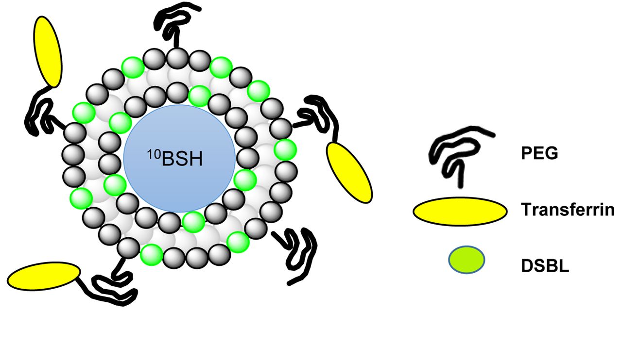
We are interested in applying BNCT to the treatment of HCC. Therefore, in the present study, we developed 10BSH-entrapped TF–PEG liposomes with 10B-distearoyl-boron lipid (DSBL) (21, 22). These liposomes were used as a selective boron delivery system to cancer tissues in a VX-2 hepatic tumor model in order to investigate the application of BNCT to the treatment of HCC with the aim of increasing the selection of therapies available for patients (Figure 1).

Figure 1.
  • Download figure
  • Open in new tab
  • Download powerpoint
Figure 1.

We propose the application of 10B-borono-dodecaborate-entrapped transferrin-conjugated polyethyleneglycol-distearoylboron lipid-coated (10BSH-TF-PEG-DSBL) liposomes as a delivery carrier of boron compounds for intra-arterial injection in boron neutron capture therapy: As TF-PEG has an endocytic role, it can deliver 10B atoms into cancer cells. Use of DSBL increases the 10B concentration in cancer cells.

Materials and Methods

Preparation of PEG and TF-PEG liposomes containing 10B-compound. Boron-entrapped liposomes were prepared by the reverse-phase evaporation method and extrusion method (18, 19).

The composition ratio of liposomes in uniting-type 25% DSBL stealth liposomes (PEG-DSBL) was as follows: distearoylphosphatidylcholine (DSPC):DSBL:cholesterol:PEG-2000=0.75:0.25:1.0: 0.11 (mol ratio). The concentration of boron entrapped in PEG-DSBL liposomes was 2, 700 ppm.

The composition element ratio of entrapping-type 10BSH-entrapped stealth (PEG) liposomes was as follows: DSPC: cholesterol:PEG-2000=1.0:1.0:0.11 (mol ratio) + 10BSH (125 mmol/l). The enclosed boron concentration entrapped in PEG liposomes was 4700 ppm.

10BSH-entrapped TF-PEG-DSBL liposomes were prepared by the coupling of TF to the PEG-COOH moieties of PEG-DSBL liposomes according to the protocol described by Ishida et al. [DSPC:DSBL:cholesterol:PEG-2000=2840:336:1546:928 (/mg)] (20) (Figure 2).

Figure 2.
  • Download figure
  • Open in new tab
  • Download powerpoint
Figure 2.

Schema of 10B-borono-dodecaborate (10BSH)-entrapped transferrin-conjugated polyethyleneglycol (PEG)-distearoylboron lipid (DSBL)-coated liposomes.

Boron concentrations in prepared liposomes were measured using inductively coupled plasma atomic emission spectroscopy (ICP-AES) (24).

Target tumor cells and rabbits. Rabbit VX-2 cells (a Shope virus-derived squamous cell carcinoma cell line) were cultured in vitro and supplemented with 10% fetal bovine serum and 500 μg streptomycin/penicillin under 5% CO2 conditions. Female New Zealand white rabbits with VX-2 cells inoculated into the left lobe of the liver were obtained from Nihon SLC Ltd. (Shizuoka, Japan) and used at a mean body weight of 2 kg (23). The procedures for tumor implantation and animal sacrifice were in accordance with the approved guidelines of the Institution’s Animal Ethics Committee and with the Declaration of Helsinki (approval number; LSI Medience: P090818 & P100455, KUR: 2010-18 & 25).

Evaluation of tumor-growth suppression by experimental BNCT. Rabbit VX-2 cells were inoculated into the left lobe of the liver, and 2 weeks after tumor cell inoculation, 10BSH-TF-PEG-DSBL liposomes were administered by an intra-arterial injection via the proper hepatic artery into the VX-2 rabbit hepatic tumor model. Tumor-bearing rabbits were irradiated with thermal neutrons (fluence: 2×10¹² n/cm–2) at the Kyoto University Institute for Integrated Radiation and Nuclear Science 48 h after the intra-arterial injection of liposomes. Neutron fluence was measured by gold foil at two points on the frontal side of the abdomen and rear side of the rabbit holder, while the gamma ray dose was assessed using a thermoluminescent dosimeter at the same points. After irradiation, the size of the tumor and the status of the abdomen were investigated on day 14 after BNCT by sacrificing rabbits for morphological observations. Pathological analyses were also performed on tumor samples resected after neutron irradiation, fixed in Optimal Cutting Temperature compound, and frozen at ‒80°C. Harvested tumor samples were sliced into 6-μm-thick sections using a cryostat and deposited on glass slides before being stained with hematoxylin and eosin.

Pathological findings of tumors after intra-arterial injection of 10BSH–PEG–DSBL liposomes. Histological and electron microscopic observations were performed. Rabbits were sacrificed 24 and 48 h after the injection of 10BSH–PEG–DSBL liposomes. Livers were resected and stored in Optimal Cutting Temperature compound frozen at ‒80°C. Six-micrometer-thick sections of the same specimens were stained with hematoxylin and eosin for light microscopy to assess the induction of necrosis, hyalinization by BNCT.

Measurement of 10B accumulated in each organ in vivo. We administered 10BSH-containing liposomes (15 mg/kg) to tumor-bearing rabbits. Regarding the hepatic artery injection technique, the abdomen was opened under general anesthesia, the proper hepatic artery facing the liver was identified, the catheter or injection needle was inserted, and the hepatic artery injection of 10BSH liposomes was performed. The administered volumes were 5.6 ml for 10BSH-PEG-DSBL liposomes and 3.3 ml for 10BSH-PEG liposomes. Boron concentrations in blood, VX-2 liver tumors, normal liver tissues, and other organs were measured using ICP-AES at 24 or 48 h after the intra-hepatic arterial injection of 10BSH-PEG-DSBL or 10BSH-PEG loposomes under general anesthesia in the VX-2 rabbit liver tumor model. Mean values and standard deviations were calculated. The number of rabbits in each group was three.

We also measured 10B concentrations in blood, VX-2 liver tumors, and normal liver tissues using ICP-AES 24, 48, 72, or 120 h after the intra-hepatic arterial injection of 10BSH-TF-PEG-DSBL liposomes. Tissues were weighed immediately after dissection and recorded as wet weights. Results were expressed as μg 10B per g tissue. Mean values and standard deviations were calculated.

Results

Characterization of liposomes. Mean 10B concentrations in liposomes were measured using ICP-AES. The concentration of 10B entrapped in 10BSH-PEG-DSBL liposomes was 2700 ppm, and that in 10BSH-PEG liposomes was 4,700 ppm. The 10B concentration entrapped in 10BSH-TF-PEG-DSBL liposomes was 3,200 ppm.

Comparison of 10B accumulation in each organ and biodistribution of 10BSH-PEG and 10BSH-PEG-DSBL liposomes after intra-hepatic arterial injection. The 10B concentrations in the VX-2 tumor using 10BSH-PEG-DSBL and 10BSH-PEG liposomes were 52.14±12.07 and 31.92±14.23 μg/g, respectively, 24 h after the intra-arterial injection, and were 60.62±23.89 and 23.02±3.47 μg/g, respectively, 48 h after (Figure 3). The 10B concentration in the tumor using DSBL liposomes was two-fold higher than that of 10BSH-PEG liposomes. The amount of boron that accumulated was also two-fold higher in hepatic tumors than in normal hepatic tissue (31 ppm on average) with 10BSH-PEG liposomes at 24 h after the intra-arterial injection (Figure 3).

Figure 3.
  • Download figure
  • Open in new tab
  • Download powerpoint
Figure 3.

Measurement of boron concentrations in tumors and each organ after the intra-arterial injection of boron-entrapped polyethyleneglycol (PEG) liposomes in the VX-2 hepatic cancer-bearing rabbit model. Two types of 10B-borono-dodecaborate (10BSH)-PEG liposomes were prepared: Distearoylboron lipid (DSBL) type and simple entrapping PEG type. Concentrations of 10B were measured by inductively coupled plasma atomic emission spectroscopy at Juntendo University. Mean values and standard deviations were calculated (n=3).

Histological and electron microscopic findings of tumors after intra-arterial injection of 10BSH-PEG-DSBL liposomes. Pathological findings revealed no damage to normal hepatic tissue with this dosage injection of 10BSH-PEG-DSBL liposomes (Figure 4).

Figure 4.
  • Download figure
  • Open in new tab
  • Download powerpoint
Figure 4.

Pathological and histological examinations. No damage was observed in normal liver tissue under light (original magnification, ×200) and electron microscopy (original magnification, ×600) after intra-hepatic arterial injection of 10B-borono-dodecaborate-entrapped polyethyleneglycol distearoyl boron lipid-coated (10BSH-PEG-DSBL) liposomes. A: Fat droplets were detected at the boundary of the tumor and normal hepatic tissue 24 h after administration. Degeneration and hyalinization were not observed in hepatocytes. B: Fat droplets were only noted in the liver vein surrounding the hepatic lobulus after 48 h. This phenomenon may have been due to the drainage of lipids constructed of liposomes from the liver. C: More fatty vesicles were detected in VX-2 tumor tissues 24 h after the intra-arterial injection of 10BSH-PEG-DSBL liposomes than in non-treated control groups by electron microscopy.

Hepatic biodistribution of 10BSH-TF-PEG-DSBL liposomes after intra-hepatic arterial injection. Modifications to the surface of PEG liposomes by conjugation with TF increased the 10B concentration in tumors to around 25 μg/g at 72 h after the intra-hepatic arterial injection of 10BSH-TF-PEG-DSBL liposomes into the VX-2 rabbit hepatic tumor model. The concentration of boron in normal hepatic tissue was 10-15 μg/g 72 h after the intra-hepatic arterial injection (Figure 5). The tumor/normal liver 10B concentration ratio was at least 1.5-fold higher until 120 h after intra-hepatic arterial injection (Figure 5). When 10BSH-TF-PEG-DSBL liposomes were administered intra-arterially at a dose of 6.4 mg 10B/kg, we observed the selective uptake of 10B by cancer tissues compared with that by normal hepatocytes in the VX-2 rabbit hepatic cancer model. TF-PEG-liposomes maintained a high 10B concentration in tumors, with concentrations higher than 25 μg/g for at least 72 h after intra-arterial injection. This high retention of 10B in tumor tissue indicates that the binding and concomitant cellular uptake of extravasated TF-PEG liposomes occurred by TF receptor and receptor-mediated endocytosis, with the concentration of 10B in hepatocytes eventually decreasing, resulting in a tumor/normal tissue ratio of 2.0 at 48 h after liposomal injection.

Figure 5.
  • Download figure
  • Open in new tab
  • Download powerpoint
Figure 5.

Boron concentrations in tumors and normal hepatic tissue in the VX-2 rabbit hepatic cancer model after intra-hepatic arterial injection of 10B-borono-dodecaborate -entrapped transferrin-conjugated polyethyleneglycol distearoyl-boron lipid liposomes. Liver-1: Normal liver near tumor in left lobe of liver; Liver-2: normal liver edge in left lobe of liver; Liver-3: normal liver central area in right lobe of liver; Liver-4: normal liver edge in right lobe of liver.

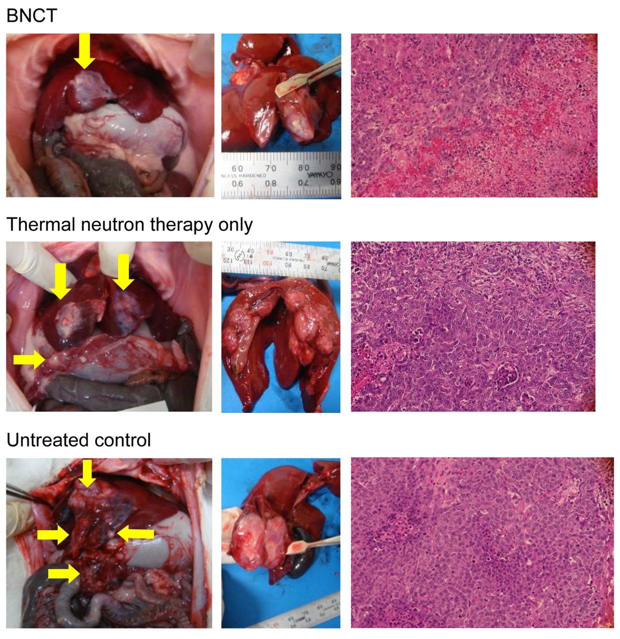
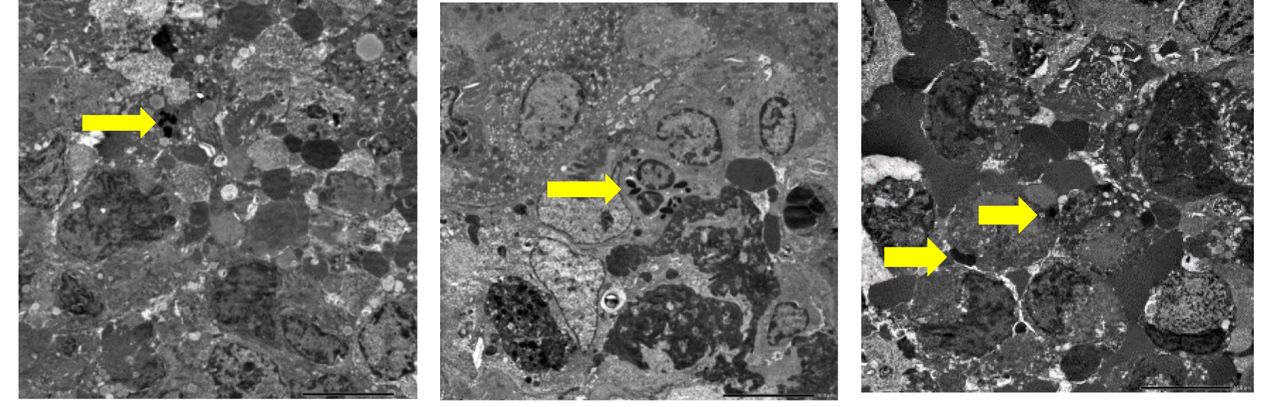
Morphological and pathological analyses after BNCT. In order to examine the tumor growth-suppressing effects of BNCT, the VX-2 rabbit hepatic cancer model was subjected under general anesthesia to thermal neutron irradiation 48 h after the intra-arterial injection of 10BSH-TF-PEG-DSBL liposomes at Kyoto University Institute for Integrated Radiation and Nuclear Science. The fluence of thermal neutrons was 2×10¹² n/cm–2 on the surface of the beam port, and details of the physical dose from each neutron energy range, including measurement results on the rear side, are shown in Table I. Rabbits were observed for 2 weeks, sacrificed, and liver tumors and the intra-abdominal state were then examined. After BNCT, the suppression of tumor growth was noted with thermal neutron irradiation after the intra-arterial injection of 10BSH-TF-PEG-DSBL liposomes, with smaller tumor sizes in the group treated with these liposomes than in the untreated group and the group treated only with thermal neutron irradiation (Figure 6; Table II). Many tumor nodules were detected in the liver, abdominal cavity, and peritoneum in the control group. Hematoxylin and eosin staining results shown in Figure 5 indicate the effectiveness of cancer cell killing by BNCT. Electron microscopic findings revealed apoptotic bodies and nuclear degradation in cancer cells (Figure 7).

View this table:
  • View inline
  • View popup
  • Download powerpoint
Table I.

Irradiation mode at the Kyoto University Institute for Integrated Radiation and Nuclear Science for boron neutron capture therapy for the hepatic cancer model.

Figure 6.
  • Download figure
  • Open in new tab
  • Download powerpoint
Figure 6.

Tumor growth suppression by boron neutron capture therapy (BNCT). Morphological and pathological findings of VX-2 tumors after BNCT with intra-arterial injection of 10B-borono-dodecaborate-entrapped transferrin-conjugated polyethyleneglycol-distearoylboron lipid-coated liposomes.

View this table:
  • View inline
  • View popup
  • Download powerpoint
Table II.

Tumor growth suppression by boron neutron capture therapy (BNCT) after an intra-arterial injection of 10B-borono-dodecaborate-entrapped transferrin-conjugated distearoyl-boron lipid-coated (10BSH-TF-PEG-DSBL) liposomes into the VX-2 hepatic tumor model. VX-2 cells were inoculated into the left lobe of the liver of female New Zealand White rabbits. Two weeks after the inoculation, tumor-bearing rabbits were irradiated with thermal neutrons (fluence: 2×10¹² n/cm–2) 48 h after intra-arterial injection of 10BSH-TF-PEG-DSBL liposomes. After irradiation, the effects of BNCT were evaluated based on the tumor volume after 14 days (calculated as 1/2× length × width2). Values are the means±standard deviation.

Figure 7.
  • Download figure
  • Open in new tab
  • Download powerpoint
Figure 7.

Electron microscopy findings (×600) of tumors after boron neutron capture therapy using intra-hepatic arterial injection of 10B-borono-dodecaborate-entrapped transferrin-conjugated polyethyleneglycol-distearoylboron lipid-coated liposomes. Nuclear deformation and apoptotic bodies were detected in tumor cells.

Discussion

PEG liposomes have a long circulation time and biodistribution patterns and pharmacokinetics that enhance their systemic therapeutic effects. The PEG liposome formulation prevents rapid uptake by the reticuloendothelial system and, thus, liposomes remain in the circulation for long periods. ‘Stealth’ liposomes in tumors combine slower plasma clearance and higher vascular permeability compared with conventional liposomes, and form depots of drugs in the perivascular space after extravasation which are available to neighboring cells within tumors for a few days (19, 20). Ishida et al. previously reported that PEG liposomes with an average diameter of 100-200 nm had the longest circulation time and the greatest accumulation in solid tumors in vivo (20). Maruyama et al. developed TF-binding PEG liposomes as an intracellular drug delivery system by receptor-mediated endocytosis (25).

The presence of a ligand facilitates the entry of 10B-containing compounds into cells through receptor-mediated endocytosis (11). We showed the suppression of tumor growth in a human pancreatic cancer model by BNCT after repeated intravenous injections of 10BSH-entrapped PEG liposomes (18). Furthermore, 10BSH-TF-PEG liposomes achieved superior tumor growth suppression by BNCT (19), and using neutron capture autoradiography, we demonstrated that 10B atoms more selectively accumulated in tumors using 10BSH-entrapped TF-PEG liposomes than conventional liposomes (26).

Nakamura et al. reported several boron lipids constructed of liposomes, which increased the uptake of boron-10 atoms in cancer cells, and termed these liposomal Boron Delivery Systems (27). 10BSH-DSBL-PEG liposomes have been developed, and their significant antitumor effects were observed in mice injected with these liposomes (15 mg 10B/kg) after thermal neutron irradiation (22). Furthermore, liposomes composed of the closo-dodecaborate lipids DSBL and dipalmitoyl boron lipid exhibited strong cytotoxicity with thermal neutron irradiation, and these lipid liposomes were taken up into the cytoplasm by endocytosis without degradation (21). TF-loaded nido-carborane liposomes also achieved a higher survival rate with BNCT in tumor-bearing mice (28).

Boron compounds are currently being developed (29-35). Previous studies showed that tumor uptake after the administration of a sulfhydryl borane dimer (Na4B24H22S2) was approximately two-fold that after the administration of equal amounts of boron as a monomer (36, 37). Feakes et al. encapsulated the apical-equatorial isomer of polyhedral borane [B20H17NH3]3-ion in liposomes prepared with 5% PEG-2000-distearoyl phosphatidyl-ethanolamine and found that the circulation time of these liposomes was prolonged, resulting in the continued accumulation of boron in tumors (38-41).

Cemazar et al. performed an electric pulse technique to enhance the accumulation of 10B into cancer cells after the intravenous injection of 10B-borono-phenylalanine (42). BNCT with an intravenous injection of TF-PEG liposomes with a high content of 10BSH has the ability to destroy malignant cells at the edge of the tumor mass, which is a hypervascular area. Experiments with these new 10B delivery systems that combine 10BSH-TF-PEG liposomes using the electric pulse technique to enhance the uptake of 10B will hopefully soon proceed to clinical BNCT trials.

The development of boron lipids for the construction of liposomes is very important because liposomes with boron atoms will increase the concentration of boron in cancer cells by liposomal endocytosis (21, 22, 28). The density of boron was previously shown to be higher in tumors than in normal hepatic tissue following modifications to the surface of PEG liposomes with TF, and tumor growth-suppressing effects were confirmed by thermal neutron irradiation (19, 21, 22, 28, 31). Intelligent targeting prevents accumulation in normal hepatic tissue and tumor selectivity in targeting is expected. In our study, the intra-arterial injection of 10BSH-TF-PEG-DSBL liposomes increased tumor retention of 10B atoms and also suppressed tumor growth in vivo upon thermal neutron irradiation. Pathological damage was not observed in normal hepatocytes after BNCT. The present results demonstrated the potential of 10BSH TF PEG-DSBL Lip as a novel intra-arterial boron carrier in BNCT for cancer.

Suzuki et al. reported preclinical studies and the treatment of patients with HCC by BNCT (43, 44). We also demonstrated that VX-2 tumor growth was suppressed by BNCT after an intra-hepatic arterial injection of 10BSH-entrapped WOW emulsion, and performed a clinical study on BNCT using 10BSH-entrapped WOW emulsion for patients with HCC (45, 46).

There were limitations to intra-arterial injection of drugs in these experiments as the proper hepatic artery of the rabbit is very thin, and it is very difficult to apply an injectional catheter to branches of hepatic arteries super-selectively. We hope to develop the boron-entrapped delivery systems for selective accumulation in tumors using this rabbit model, then proceed to a pilot clinical study of BNCT in the near future.

Conclusion

We prepared 10BSH-entrapped TF-PEG liposomes consisting of boron lipids (DSBL) as boron delivery systems in BNCT. The results demonstrated that 10BSH-TF-PEG-DSBL liposomes deliver and facilitate the retention of boron atoms in cancer cells of tumor tissues in a rabbit hepatic tumor model. Tumor growth was suppressed by thermal neutron irradiation after an intra-arterial injection of 10BSH-TF-PEG-DSBL liposomes without damage to normal cells. These results demonstrate the potential of 10BSH-TF-PEG-DSBL liposomes as a novel boron delivery carrier in BNCT for cancer using intra-arterial techniques. We intend to proceed to preclinical safety and clinical studies on patients with HCC using 10BSH-TF-PEG-DSBL liposomes in the near future.

Acknowledgements

This work was supported in part by Grants-in-Aid from the Ministry of Education, Science and Culture of Japan (No. 11691202 and No. 11557092 to Hironobu Yanagie), and a Grant-in-Aid from the Ministry of Health, Labour and Welfare of Japan (No. 2008-Nano-004 to Hiroyuki Nakamura). We express our appreciation to Mrs. Yuriko Sakurai and Mrs. Kikue Mouri for their preparation of pathological samples.

Footnotes

  • This article is freely accessible online.

  • Authors’ Contributions

    Conceived and designed the experiments; HY, HN, MS, YS, HT, SM, KO, and HT. Performed the experiments; HY, MY, YM, AS, RM, YM, MS, YS, HT, SM, and KO. Analyzed and interpreted the data; HY, YM, AS, HN, MS, YS, ND, YN, YF, and HT. Contributed reagents, materials, analysis tools or data; HY, YM, AS, HN, MS, YS, TS, MN, HY, MO, JN, and HT. Wrote the article; HY, ND, HN, MS, and HT.

  • Conflicts of Interest

    None declared.

  • Received July 2, 2021.
  • Revision received September 23, 2021.
  • Accepted September 24, 2021.
  • Copyright © 2021 International Institute of Anticancer Research (Dr. George J. Delinasios), All rights reserved

References

  1. ↵
    1. Chen Z,
    2. Xie H,
    3. Hu M,
    4. Huang T,
    5. Hu Y,
    6. Sang N and
    7. Zhao Y
    : Recent progress in treatment of hepatocellular carcinoma. Am J Cancer Res 10(9): 2993-3036, 2020. PMID: 33042631.
    OpenUrlPubMed
  2. ↵
    1. Yang JD,
    2. Hainaut P,
    3. Gores GJ,
    4. Amadou A,
    5. Plymoth A and
    6. Roberts LR
    : A global view of hepatocellular carcinoma: trends, risk, prevention and management. Nat Rev Gastroenterol Hepatol 16(10): 589-604, 2019. PMID: 31439937. DOI: 10.1038/s41575-019-0186-y
    OpenUrlCrossRefPubMed
  3. ↵
    1. Kanematsu T,
    2. Inokuchi K,
    3. Sugimachi K,
    4. Furuta T,
    5. Sonoda T,
    6. Tamura S and
    7. Hasuo K
    : Selective effects of Lipiodolized antitumor agents. J Surg Oncol 25(3): 218-226, 1984. PMID: 6199624. DOI: 10.1002/jso.2930250317
    OpenUrlCrossRefPubMed
  4. ↵
    1. Higashi S,
    2. Shimizu M,
    3. Nakashima T,
    4. Iwata K,
    5. Uchiyama F,
    6. Tateno S,
    7. Tamura S and
    8. Setoguchi T
    : Arterial-injection chemotherapy for hepatocellular carcinoma using monodispersed poppy-seed oil microdroplets containing fine aqueous vesicles of epirubicin. Initial medical application of a membrane-emulsification technique. Cancer 75(6): 1245-1254, 1995. PMID: 7882276. DOI: 10.1002/1097-0142(19950315)75:6<1245::aid-cncr2820750606>3.0.co;2-u
    OpenUrlCrossRefPubMed
    1. Higashi S,
    2. Tabata N,
    3. Kondo KH,
    4. Maeda Y,
    5. Shimizu M,
    6. Nakashima T and
    7. Setoguchi T
    : Size of lipid microdroplets effects results of hepatic arterial chemotherapy with an anticancer agent in water-in-oil-in-water emulsion to hepatocellular carcinoma. J Pharmacol Exp Ther 289(2): 816-819, 1999. PMID: 10215657.
    OpenUrlAbstract/FREE Full Text
  5. ↵
    1. Ikushima I,
    2. Higashi S,
    3. Ishii A,
    4. Seguchi K,
    5. Iryo Y and
    6. Yamashita Y
    : Ultraselective transcatheter infusion of epirubicin in water-in-oil-in-water emulsion for small hepatocellular carcinoma. Br J Radiol 85(1017): e584-e589, 2012. PMID: 22422389. DOI: 10.1259/bjr/16376960
    OpenUrlAbstract/FREE Full Text
  6. ↵
    1. Hatanaka H and
    2. Nakagawa Y
    : Clinical results of long-surviving brain tumor patients who underwent boron neutron capture therapy. Int J Radiat Oncol Biol Phys 28(5): 1061-1066, 1994. PMID: 8175390. DOI: 10.1016/0360-3016(94)90479-0
    OpenUrlCrossRefPubMed
    1. Mishima Y,
    2. Honda C,
    3. Ichihashi M,
    4. Obara H,
    5. Hiratsuka J,
    6. Fukuda H,
    7. Karashima H,
    8. Kobayashi T,
    9. Kanda K and
    10. Yoshino K
    : Treatment of malignant melanoma by single thermal neutron capture therapy with melanoma-seeking 10B-compound. Lancet 2(8659): 388-389, 1989. PMID: 2569577. DOI: 10.1016/s0140-6736(89)90567-9
    OpenUrlCrossRefPubMed
  7. ↵
    1. Larsson B,
    2. Crawford J and
    3. Weinreich R
    1. Ono K,
    2. Ueda S,
    3. Oda Y,
    4. Nakagawa Y,
    5. Miyatake S,
    6. Takagaki M,
    7. Osawa M and
    8. Kobayashi T
    : Boron neutron-capture therapy for malignant glioma at Kyoto University reactor. In: Advances in Neutron-Capture Therapy, Volume I, Medicine and Physics. Larsson B, Crawford J and Weinreich R (eds). Amsterdam, Elsevier Science, pp. 39-45, 1997.
  8. ↵
    1. Yanagië H,
    2. Ogata A,
    3. Sugiyama H,
    4. Eriguchi M,
    5. Takamoto S and
    6. Takahashi H
    : Application of drug delivery system to boron neutron capture therapy for cancer. Expert Opin Drug Deliv 5(4): 427-443, 2008. PMID: 18426384. DOI: 10.1517/17425247.5.4.427
    OpenUrlCrossRefPubMed
  9. ↵
    1. Barth RF,
    2. Mi P and
    3. Yang W
    : Boron delivery agents for neutron capture therapy of cancer. Cancer Commun (Lond) 38(1): 35, 2018. PMID: 29914561. DOI: 10.1186/s40880-018-0299-7
    OpenUrlCrossRefPubMed
    1. Mi P
    : Stimuli-responsive nanocarriers for drug delivery, tumor imaging, therapy and theranostics. Theranostics 10(10): 4557-4588, 2020. PMID: 32292515. DOI: 10.7150/thno.38069
    OpenUrlCrossRefPubMed
    1. Luderer MJ,
    2. Muz B,
    3. Alhallak K,
    4. Sun J,
    5. Wasden K,
    6. Guenthner N,
    7. de la Puente P,
    8. Federico C and
    9. Azab AK
    : Thermal sensitive liposomes improve delivery of boronated agents for boron neutron capture therapy. Pharm Res 36(10): 144, 2019. PMID: 31392417. DOI: 10.1007/s11095-019-2670-z
    OpenUrlCrossRefPubMed
    1. Nomoto T,
    2. Inoue Y,
    3. Yao Y,
    4. Suzuki M,
    5. Kanamori K,
    6. Takemoto H,
    7. Matsui M,
    8. Tomoda K and
    9. Nishiyama N
    : Poly(vinyl alcohol) boosting therapeutic potential of p-boronophenylalanine in neutron capture therapy by modulating metabolism. Sci Adv 6(4): eaaz1722, 2020. PMID: 32010792. DOI: 10.1126/sciadv.aaz1722
    OpenUrlFREE Full Text
  10. ↵
    1. Yoneoka S,
    2. Nakagawa Y,
    3. Uto K,
    4. Sakura K,
    5. Tsukahara T and
    6. Ebara M
    : Boron-incorporating hemagglutinating virus of Japan envelope (HVJ-E) nanomaterial in boron neutron capture therapy. Sci Technol Adv Mater 20(1): 291-304, 2019. PMID: 30956733. DOI: 10.1080/14686996.2019.1586051
    OpenUrlCrossRefPubMed
  11. ↵
    1. Yanagië H,
    2. Tomita T,
    3. Kobayashi H,
    4. Fujii Y,
    5. Takahashi T,
    6. Hasumi K,
    7. Nariuchi H and
    8. Sekiguchi M
    : Application of boronated anti-CEA immunoliposome to tumour cell growth inhibition in in vitro boron neutron capture therapy model. Br J Cancer 63(4): 522-526, 1991. PMID: 2021537. DOI: 10.1038/bjc.1991.124
    OpenUrlCrossRefPubMed
  12. ↵
    1. Yanagië H,
    2. Tomita T,
    3. Kobayashi H,
    4. Fujii Y,
    5. Nonaka Y,
    6. Saegusa Y,
    7. Hasumi K,
    8. Eriguchi M,
    9. Kobayashi T and
    10. Ono K
    : Inhibition of human pancreatic cancer growth in nude mice by boron neutron capture therapy. Br J Cancer 75(5): 660-665, 1997. PMID: 9043021. DOI: 10.1038/bjc.1997.118
    OpenUrlCrossRefPubMed
  13. ↵
    1. Yanagie H,
    2. Maruyama K,
    3. Takizawa T,
    4. Ishida O,
    5. Ogura K,
    6. Matsumoto T,
    7. Sakurai Y,
    8. Kobayashi T,
    9. Shinohara A,
    10. Rant J,
    11. Skvarc J,
    12. Ilic R,
    13. Kuhne G,
    14. Chiba M,
    15. Furuya Y,
    16. Sugiyama H,
    17. Hisa T,
    18. Ono K,
    19. Kobayashi H and
    20. Eriguchi M
    : Application of boron-entrapped stealth liposomes to inhibition of growth of tumour cells in the in vivo boron neutron-capture therapy model. Biomed Pharmacother 60(1): 43-50, 2006. PMID: 16260113. DOI: 10.1016/j.biopha.2005.05.011
    OpenUrlCrossRefPubMed
  14. ↵
    1. Maruyama K,
    2. Ishida O,
    3. Kasaoka S,
    4. Takizawa T,
    5. Utoguchi N,
    6. Shinohara A,
    7. Chiba M,
    8. Kobayashi H,
    9. Eriguchi M and
    10. Yanagie H
    : Intracellular targeting of sodium mercaptoundecahydro-dodecaborate (BSH) to solid tumors by transferrin-PEG liposomes, for boron neutron-capture therapy (BNCT). J Control Release 98(2): 195-207, 2004. PMID: 15262412. DOI: 10.1016/j.jconrel.2004.04.018
    OpenUrlCrossRefPubMed
  15. ↵
    1. Ishida O,
    2. Maruyama K,
    3. Tanahashi H,
    4. Iwatsuru M,
    5. Sasaki K,
    6. Eriguchi M and
    7. Yanagie H
    : Liposomes bearing polyethyleneglycol-coupled transferrin with intracellular targeting property to the solid tumors in vivo. Pharm Res 18(7): 1042-1048, 2001. PMID: 11496943. DOI: 10.1023/a:1010960900254
    OpenUrlCrossRefPubMed
  16. ↵
    1. Ueno M,
    2. Ban HS,
    3. Nakai K,
    4. Inomata R,
    5. Kaneda Y,
    6. Matsumura A and
    7. Nakamura H
    : Dodecaborate lipid liposomes as new vehicles for boron delivery system of neutron capture therapy. Bioorg Med Chem 18(9): 3059-3065, 2010. PMID: 20371186. DOI: 10.1016/j.bmc.2010.03.050
    OpenUrlCrossRefPubMed
  17. ↵
    1. Koganei H,
    2. Ueno M,
    3. Tachikawa S,
    4. Tasaki L,
    5. Ban HS,
    6. Suzuki M,
    7. Shiraishi K,
    8. Kawano K,
    9. Yokoyama M,
    10. Maitani Y,
    11. Ono K and
    12. Nakamura H
    : Development of high boron content liposomes and their promising antitumor effect for neutron capture therapy of cancers. Bioconjug Chem 24(1): 124-132, 2013. PMID: 23214414. DOI: 10.1021/bc300527n
    OpenUrlCrossRefPubMed
  18. ↵
    1. Burgener FA
    : Peripheral hepatic artery embolization in rabbits with VX2 carcinomas of the liver. Cancer 46(1): 56-63, 1980. PMID: 7388767. DOI: 10.1002/1097-0142(19800701)46:1<56::aid-cncr2820460113>3.0.co;2-c
    OpenUrlCrossRefPubMed
  19. ↵
    1. Shinohara A,
    2. Chiba M and
    3. Inaba Y
    : Determination of selenium concentrations in human blood plasma by microwave-induced plasma mass spectrometry. Analytical Sciences 14(4): 713-717, 2021. DOI: 10.2116/analsci.14.713
    OpenUrlCrossRef
  20. ↵
    1. Maruyama K,
    2. Takizawa T,
    3. Yuda T,
    4. Kennel SJ,
    5. Huang L and
    6. Iwatsuru M
    : Targetability of novel immunoliposomes modified with amphipathic poly(ethylene glycol)s conjugated at their distal terminals to monoclonal antibodies. Biochim Biophys Acta 1234(1): 74-80, 1995. PMID: 7880861. DOI: 10.1016/0005-2736(94)00263-o
    OpenUrlCrossRefPubMed
  21. ↵
    1. Yanagië H,
    2. Ogura K,
    3. Takagi K,
    4. Maruyama K,
    5. Matsumoto T,
    6. Sakurai Y,
    7. Skvarc J,
    8. Illic R,
    9. Kuhne G,
    10. Hisa T,
    11. Yoshizaki I,
    12. Kono K,
    13. Furuya Y,
    14. Sugiyama H,
    15. Kobayashi H,
    16. Ono K,
    17. Nakagawa K and
    18. Eriguchi M
    : Accumulation of boron compounds to tumor with polyethylene-glycol binding liposome by using neutron capture autoradiography. Appl Radiat Isot 61(4): 639-646, 2004. PMID: 15246411. DOI: 10.1016/j.apradiso.2004.03.109
    OpenUrlCrossRefPubMed
  22. ↵
    1. Nakamura H
    : Boron lipid-based liposomal boron delivery system for neutron capture therapy: recent development and future perspective. Future Med Chem 5(6): 715-730, 2013. PMID: 23617433. DOI: 10.4155/fmc.13.48
    OpenUrlCrossRefPubMed
  23. ↵
    1. Miyajima Y,
    2. Nakamura H,
    3. Kuwata Y,
    4. Lee JD,
    5. Masunaga S,
    6. Ono K and
    7. Maruyama K
    : Transferrin-loaded nido-carborane liposomes: tumor-targeting boron delivery system for neutron capture therapy. Bioconjug Chem 17(5): 1314-1320, 2006. PMID: 16984142. DOI: 10.1021/bc060064k
    OpenUrlCrossRefPubMed
  24. ↵
    1. Nakase I,
    2. Katayama M,
    3. Hattori Y,
    4. Ishimura M,
    5. Inaura S,
    6. Fujiwara D,
    7. Takatani-Nakase T,
    8. Fujii I,
    9. Futaki S and
    10. Kirihata M
    : Intracellular target delivery of cell-penetrating peptide-conjugated dodecaborate for boron neutron capture therapy (BNCT). Chem Commun (Camb) 55(93): 13955-13958, 2019. PMID: 31617510. DOI: 10.1039/c9cc03924d
    OpenUrlCrossRefPubMed
    1. Michiue H,
    2. Sakurai Y,
    3. Kondo N,
    4. Kitamatsu M,
    5. Bin F,
    6. Nakajima K,
    7. Hirota Y,
    8. Kawabata S,
    9. Nishiki T,
    10. Ohmori I,
    11. Tomizawa K,
    12. Miyatake S,
    13. Ono K and
    14. Matsui H
    : The acceleration of boron neutron capture therapy using multi-linked mercaptoundecahydrododecaborate (BSH) fused cell-penetrating peptide. Biomaterials 35(10): 3396-3405, 2014. PMID: 24452095. DOI: 10.1016/j.biomaterials.2013.12.055
    OpenUrlCrossRefPubMed
  25. ↵
    1. Masunaga S,
    2. Kasaoka S,
    3. Maruyama K,
    4. Nigg D,
    5. Sakurai Y,
    6. Nagata K,
    7. Suzuki M,
    8. Kinashi Y,
    9. Maruhashi A and
    10. Ono K
    : The potential of transferrin-pendant-type polyethyleneglycol liposomes encapsulating decahydrodecaborate-(10)B (GB-10) as (10)B-carriers for boron neutron capture therapy. Int J Radiat Oncol Biol Phys 66(5): 1515-1522, 2006. PMID: 17126210. DOI: 10.1016/j.ijrobp.2006.08.028
    OpenUrlCrossRefPubMed
    1. Takeuchi K,
    2. Hattori Y,
    3. Kawabata S,
    4. Futamura G,
    5. Hiramatsu R,
    6. Wanibuchi M,
    7. Tanaka H,
    8. Masunaga SI,
    9. Ono K,
    10. Miyatake SI and
    11. Kirihata M
    : Synthesis and evaluation of dodecaboranethiol containing kojic acid (KA-BSH) as a novel agent for boron neutron capture therapy. Cells 9(6): 1551, 2020. PMID: 32630612. DOI: 10.3390/cells9061551
    OpenUrlCrossRefPubMed
    1. Maitz CA,
    2. Khan AA,
    3. Kueffer PJ,
    4. Brockman JD,
    5. Dixson J,
    6. Jalisatgi SS,
    7. Nigg DW,
    8. Everett TA and
    9. Hawthorne MF
    : Validation and comparison of the therapeutic efficacy of boron neutron capture therapy mediated by boron-rich liposomes in multiple murine tumor models. Transl Oncol 10(4): 686-692, 2017. PMID: 28683435. DOI: 10.1016/j.tranon.2017.05.003
    OpenUrlCrossRefPubMed
    1. Nakase I,
    2. Aoki A,
    3. Sakai Y,
    4. Hirase S,
    5. Ishimura M,
    6. Takatani-Nakase T,
    7. Hattori Y and
    8. Kirihata M
    : Antibody-based receptor targeting using an Fc-binding peptide-dodecaborate conjugate and macropinocytosis induction for boron neutron capture therapy. ACS Omega 5(36): 22731-22738, 2020. PMID: 32954120. DOI: 10.1021/acsomega.0c01377
    OpenUrlCrossRefPubMed
  26. ↵
    1. Nakagawa F,
    2. Kawashima H,
    3. Morita T and
    4. Nakamura H
    : Water-soluble closo-docecaborate-containing pteroyl derivatives targeting folate receptor-positive tumors for boron neutron capture therapy. Cells 9(7): 1615, 2020. PMID: 32635272. DOI: 10.3390/cells9071615
    OpenUrlCrossRefPubMed
  27. ↵
    1. Slatkin D,
    2. Micca P,
    3. Forman A,
    4. Gabel D,
    5. Wielopolski L and
    6. Fairchild R
    : Boron uptake in melanoma, cerebrum and blood from Na2B12H11SH and Na4B24H22S2 administered to mice. Biochem Pharmacol 35(10): 1771-1776, 1986. PMID: 3707608. DOI: 10.1016/0006-2952(86)90342-4
    OpenUrlCrossRefPubMed
  28. ↵
    1. Elhanati G,
    2. Salomon Y and
    3. Bendel P
    : Significant differences in the retention of the borocaptate monomer (BSH) and dimer (BSSB) in malignant cells. Cancer Lett 172(2): 127-132, 2001. PMID: 11566486. DOI: 10.1016/s0304-3835(01)00649-8
    OpenUrlCrossRefPubMed
  29. ↵
    1. Feakes DA,
    2. Shelly K,
    3. Knobler CB and
    4. Hawthorne MF
    : Na3[B20H17NH3]: synthesis and liposomal delivery to murine tumors. Proc Natl Acad Sci USA 91(8): 3029-3033, 1994. PMID: 8159700. DOI: 10.1073/pnas.91.8.3029
    OpenUrlAbstract/FREE Full Text
    1. Feakes DA,
    2. Shelly K and
    3. Hawthorne MF
    : Selective boron delivery to murine tumors by lipophilic species incorporated in the membranes of unilamellar liposomes. Proc Natl Acad Sci USA 92(5): 1367-1370, 1995. PMID: 7877984. DOI: 10.1073/pnas.92.5.1367
    OpenUrlAbstract/FREE Full Text
    1. Feakes DA,
    2. Waller RC,
    3. Hathaway DK and
    4. Morton VS
    : Synthesis and in vivo murine evaluation of Na4[1-(1’-B10H9)-6-SHB10H8] as a potential agent for boron neutron capture therapy. Proc Natl Acad Sci USA 96(11): 6406-6410, 1999. PMID: 10339600. DOI: 10.1073/pnas.96.11.6406
    OpenUrlAbstract/FREE Full Text
  30. ↵
    1. Shelly K,
    2. Feakes DA,
    3. Hawthorne MF,
    4. Schmidt PG,
    5. Krisch TA and
    6. Bauer WF
    : Model studies directed toward the boron neutron-capture therapy of cancer: boron delivery to murine tumors with liposomes. Proc Natl Acad Sci USA 89(19): 9039-9043, 1992. PMID: 1409600. DOI: 10.1073/pnas.89.19.9039
    OpenUrlAbstract/FREE Full Text
  31. ↵
    1. Cemazar M,
    2. Skrk J,
    3. Mitrovic B and
    4. Sersa G
    : Changed delivery of boron to tumours using electroporation for boron neutron capture therapy with BSH. Br J Radiol 73(866): 195-200, 2000. PMID: 10884734. DOI: 10.1259/bjr.73.866.10884734
    OpenUrlCrossRefPubMed
  32. ↵
    1. Suzuki M,
    2. Sakurai Y,
    3. Hagiwara S,
    4. Masunaga S,
    5. Kinashi Y,
    6. Nagata K,
    7. Maruhashi A,
    8. Kudo M and
    9. Ono K
    : First attempt of boron neutron capture therapy (BNCT) for hepatocellular carcinoma. Jpn J Clin Oncol 37(5): 376-381, 2007. PMID: 17578894. DOI: 10.1093/jjco/hym039
    OpenUrlCrossRefPubMed
  33. ↵
    1. Suzuki M,
    2. Masunaga S,
    3. Kinashi Y,
    4. Nagata K,
    5. Sakurai Y,
    6. Nakamatsu K,
    7. Nishimura Y,
    8. Maruhashi A and
    9. Ono K
    : Intra-arterial administration of sodium borocaptate (BSH)/lipiodol emulsion delivers B-10 to liver tumors highly selectively for boron neutron capture therapy: experimental studies in the rat liver model. Int J Radiat Oncol Biol Phys 59(1): 260-266, 2004. PMID: 15093923. DOI: 10.1016/j.ijrobp.2003.12.018
    OpenUrlCrossRefPubMed
  34. ↵
    1. Yanagie H,
    2. Dewi N,
    3. Higashi S,
    4. Ikushima I,
    5. Seguchi K,
    6. Mizumachi R,
    7. Murata Y,
    8. Morishita Y,
    9. Shinohara A,
    10. Mikado S,
    11. Yasuda N,
    12. Fujihara M,
    13. Sakurai Y,
    14. Mouri K,
    15. Yanagawa M,
    16. Iizuka T,
    17. Suzuki M,
    18. Sakurai Y,
    19. Masunaga SI,
    20. Tanaka H,
    21. Matsukawa T,
    22. Yokoyama K,
    23. Fujino T,
    24. Ogura K,
    25. Nonaka Y,
    26. Sugiyama H,
    27. Kajiyama T,
    28. Yui S,
    29. Nishimura R,
    30. Ono K,
    31. Takamoto S,
    32. Nakajima J,
    33. Ono M,
    34. Eriguchi M,
    35. Hasumi K and
    36. Takahashi H
    : Selective boron delivery by intra-arterial injection of BSH-WOW emulsion in hepatic cancer model for neutron capture therapy. Br J Radiol 90(1074): 20170004, 2017. PMID: 28406315. DOI: 10.1259/bjr.20170004
    OpenUrlCrossRefPubMed
  35. ↵
    1. Yanagie H,
    2. Higashi S,
    3. Seguchi K,
    4. Ikushima I,
    5. Fujihara M,
    6. Nonaka Y,
    7. Oyama K,
    8. Maruyama S,
    9. Hatae R,
    10. Suzuki M,
    11. Masunaga S,
    12. Kinashi T,
    13. Sakurai Y,
    14. Tanaka H,
    15. Kondo N,
    16. Narabayashi M,
    17. Kajiyama T,
    18. Maruhashi A,
    19. Ono K,
    20. Nakajima J,
    21. Ono M,
    22. Takahashi H and
    23. Eriguchi M
    : Pilot clinical study of boron neutron capture therapy for recurrent hepatic cancer involving the intra-arterial injection of a (10)BSH-containing WOW emulsion. Appl Radiat Isot 88: 32-37, 2014. PMID: 24559940. DOI: 10.1016/j.apradiso.2014.01.014
    OpenUrlCrossRefPubMed
PreviousNext
Back to top

In this issue

In Vivo
Vol. 35, Issue 6
November-December 2021
  • Table of Contents
  • Table of Contents (PDF)
  • Index by author
  • Back Matter (PDF)
  • Ed Board (PDF)
  • Front Matter (PDF)
Print
Download PDF
Article Alerts
Sign In to Email Alerts with your Email Address
Email Article

Thank you for your interest in spreading the word on In Vivo.

NOTE: We only request your email address so that the person you are recommending the page to knows that you wanted them to see it, and that it is not junk mail. We do not capture any email address.

Enter multiple addresses on separate lines or separate them with commas.
Suppression of Tumor Growth in a Rabbit Hepatic Cancer Model by Boron Neutron Capture Therapy With Liposomal Boron Delivery Systems
(Your Name) has sent you a message from In Vivo
(Your Name) thought you would like to see the In Vivo web site.
CAPTCHA
This question is for testing whether or not you are a human visitor and to prevent automated spam submissions.
11 + 2 =
Solve this simple math problem and enter the result. E.g. for 1+3, enter 4.
Citation Tools
Suppression of Tumor Growth in a Rabbit Hepatic Cancer Model by Boron Neutron Capture Therapy With Liposomal Boron Delivery Systems
HIRONOBU YANAGIE, MASASHI YANAGAWA, YASUYUKI MORISHITA, ATSUKO SHINOHARA, NOVRIANA DEWI, YASUMASA NONAKA, YOSHITAKA FURUYA, RYOUJI MIZUMACHI, YUUJI MURATA, HIROYUKI NAKAMURA, MINORU SUZUKI, YOSHINORI SAKURAI, HIROKI TANAKA, SHINICHIRO MASUNAGA, KOJI ONO, TAKUMICHI SUGIHARA, MASAYUKI NASHIMOTO, HARUO YAMAUCHI, MINORU ONO, JUN NAKAJIMA, HIROYUKI TAKAHASHI
In Vivo Nov 2021, 35 (6) 3125-3135; DOI: 10.21873/invivo.12607

Citation Manager Formats

  • BibTeX
  • Bookends
  • EasyBib
  • EndNote (tagged)
  • EndNote 8 (xml)
  • Medlars
  • Mendeley
  • Papers
  • RefWorks Tagged
  • Ref Manager
  • RIS
  • Zotero
Reprints and Permissions
Share
Suppression of Tumor Growth in a Rabbit Hepatic Cancer Model by Boron Neutron Capture Therapy With Liposomal Boron Delivery Systems
HIRONOBU YANAGIE, MASASHI YANAGAWA, YASUYUKI MORISHITA, ATSUKO SHINOHARA, NOVRIANA DEWI, YASUMASA NONAKA, YOSHITAKA FURUYA, RYOUJI MIZUMACHI, YUUJI MURATA, HIROYUKI NAKAMURA, MINORU SUZUKI, YOSHINORI SAKURAI, HIROKI TANAKA, SHINICHIRO MASUNAGA, KOJI ONO, TAKUMICHI SUGIHARA, MASAYUKI NASHIMOTO, HARUO YAMAUCHI, MINORU ONO, JUN NAKAJIMA, HIROYUKI TAKAHASHI
In Vivo Nov 2021, 35 (6) 3125-3135; DOI: 10.21873/invivo.12607
Reddit logo Twitter logo Facebook logo Mendeley logo
  • Tweet Widget
  • Facebook Like
  • Google Plus One

Jump to section

  • Article
    • Abstract
    • Materials and Methods
    • Results
    • Discussion
    • Conclusion
    • Acknowledgements
    • Footnotes
    • References
  • Figures & Data
  • Info & Metrics
  • PDF

Related Articles

  • No related articles found.
  • PubMed
  • Google Scholar

Cited By...

  • No citing articles found.
  • Google Scholar

More in this TOC Section

  • Evaluation of the Relationship Between miRNA-22-3p and Gal-9 Levels in Glioblastoma
  • Metformin Inhibits the Estrogen-mediated Epithelial-Mesenchymal Transition of Ectopic Endometrial Stromal Cells in Endometriosis
  • MCC950 Ameliorates Acute Exogenous Lipoid Pneumonia Induced by Sewing Machine Oil in Rats via the NF-κB/NLRP3 Inflammasome Pathway
Show more Experimental Studies

Similar Articles

Keywords

  • Boron neutron capture therapy
  • BNCT
  • transferrin-conjugated polyethylene glycol
  • PEG
  • liposome
  • 10B-borono-dodecaborate 10BSH
  • distearoylboron lipid
  • DSBL
  • hepatocellular carcinoma
  • HCC
In Vivo

© 2023 In Vivo

Powered by HighWire