


Abstract
Background/Aim: Xihuang Wan (XHW), a traditional Chinese medicine (TCM), has been used in China for a variety of cancers including lung cancer. The present study evaluated the efficacy of XHW on a Lewis lung mouse model and explored the potential mechanism via transcriptomics. Materials and Methods: The mice were randomized into 6 groups: 1) untreated control (n=10); 2) low-dose XHW; 3) medium-dose XHW; 4) high-dose XHW; 5) cisplatin; and 6) untreated blank (n=4). Lewis lung carcinoma (LLC) cells were injected subcutaneously except for the 4 mice in the blank group. The body weight and tumor length and width were measured every 3 days. RNA-sequencing was performed on tumors in the high-dose XHW group and the control group. Results: XHW inhibited the growth of LLC in a syngeneic mouse model, without toxicity, with equivalent efficacy to cisplatin. RNA-sequencing demonstrated that many signaling pathways were involved in XHW-mediated inhibition of LLC, including tumor necrosis factor, estrogen, cyclic guanosine 3’, 5’-monophosphate-protein kinase G, apelin and the peroxisome proliferator-activated receptor signaling pathways. Conclusion: XHW inhibited LLC carcinoma through different pathways and shows clinical promise for patients who cannot tolerate platinum-based drugs.
Lung cancer, with an estimated 2.2 million new cases and 1.8 million deaths in 2020, ranks second in incidence and first in mortality among all cancer types throughout the world (1). In China, lung cancer was also the second most commonly diagnosed cancer (815,563 cases) and the leading cause of cancer death (243,153 cases) in 2020 (2). First-line treatment includes surgery, radiotherapy, chemotherapy, targeted therapy, and immunotherapy (3, 4). However, lung cancer still remains recalcitrant.
In China, traditional Chinese medicine (TCM) has been used in clinical practice for thousands of years for a variety of diseases, including benign and malignant tumors (5-8). Xihuang Wan (XHW) was first recorded in the 18th-century Chinese medical book Waike Zhengzhi Quansheng Ji (Life-saving Manual of Diagnosis and Treatment of External Diseases). XHW comprises Ruxiang (Olibanum), Moyao (Myrrha), Niuhuang (Moschus), and Shexiang (Bovis Calculus). XHW has been approved by the National Medical Products Administration (NMPA) of China for the treatment of cancer (approval number Z11020073). In clinical practice, XHW has been used to treat breast (9-12), colorectal (13), liver (14, 15), cervical (16-18), and lung cancer (19-21). A systematic review and meta-analysis showed that XHW combined with chemotherapy enhanced response, prolonged overall survival, improved the quality of life of patients, and alleviated treatment-induced side effects (22).
Our laboratory has previously studied Lewis-lung carcinoma (LLC) in both nude mouse and syngeneic models (23-35). In the present study, we examined the anti-cancer efficacy of XHW in a syngeneic mouse model of LLC, and explored the potential mechanism of XHW by RNA-sequencing.
Materials and Methods
Preparation of Chinese medicine formula. Xihuang Wan (XHW) was obtained from Tongrentang (Beijing, China; Lot number: 18041283). XHW was dissolved in sodium carboxymethyl cellulose (CMC-Na) solution (5 mg/ml CMC-Na in sodium chloride for injection) at 0.493 g/ml, 0.246 g/ml, and 0.123 g/ml.
Drugs and reagents. Cisplatin was obtained from Qilu Pharmaceutical (Shandong, PR China). CMC-Na was obtained from Solarbio (Beijing, PR China). Pentobarbital sodium salt was obtained from Sigma-Aldrich (St. Louis, MO, USA). Sodium chloride for injection was obtained from Tiancheng Pharmaceutical (Hebei, PR China).
Cancer cell line. The LLC cell line LL/2 (LLC1) was purchased from KeyGEN BioTECH (Jiangsu, China). The cells were maintained in high-glucose DMEM (EallBio, Beijing, PR China) supplemented with 10% fetal bovine serum (Analysis Quiz, Beijing, PR China), penicillin (100 U/ml, EallBio) and streptomycin (100 μg/ml, EallBio) at 37°C in a CO2 incubator.
Mice. C57BL/6 mice (6-8 weeks, male) were obtained from SPF Biotechnology (Beijing, China, License No. SYXK 2019-0010). The mice were fed with an autoclaved laboratory rodent diet (SPF Biotechnology) and housed in a barrier facility in high efficacy particulate air (HEPA)-filtered racks under standard conditions of 12-h light/dark cycles. The mice were anesthetized by peritoneal injection of pentobarbital sodium at a dose of 50 mg/kg. The response of the mice was monitored to ensure adequate depth of anesthesia. The animals were observed daily and sacrificed by CO2 inhalation if the following humane end point criteria were met: severe tumor burden (over 20 mm in diameter), significant body weight loss, body temperature drop, difficulty in breathing, prostration and rotational motion. The present study was conducted in accordance with the Guidelines of Welfare and Ethics of Laboratory Animals (IACUC Issue No. AW2020111001) by SPF Biotechnology (36).
Experimental design. Fifty-four mice were randomized into 6 groups: 1) untreated control (n=10); 2) low-dose XHW [0.617 g/kg, oral gavage (p.o.), n=10]; 3) medium-dose XHW (1.233 g/kg, p.o., n=10); 4) high-dose XHW (2.466 g/kg, p.o., n=10); and 5) cisplatin [3 mg/kg, intraperitoneal injection (i.p.), n=10]; and 6) the blank group (n=4). LLC cells (1×10⁶/200 μl) were injected subcutaneously into the right flank of the mice, except for the 4 mice in the blank group (37, 38). Treatment was started the next day. Details of the treatment protocol are presented in Table I. The condition of the mice was monitored every day. The body weight and the tumor length and width were measured every 3 days. When the diameter of the tumor reached 20 mm, drug administration was stopped, and the mice were sacrificed. The tumor, spleen, liver, lung and thymus were removed and weighed. The tumor volume was calculated with the following formula: 0.5*Length*Width2 (39). The ratios of organ weight to body weight (spleen, liver, lung, and thymus) were calculated to determine whether there was treatment-associated toxicity.
Treatment protocol.
Histological examination and Ki-67 immunohistochemical staining. Freshly-harvested tumors and the livers were fixed in 10% formalin and embedded in paraffin for further histology analysis. Hematoxylin and eosin (H&E) staining and immunochemical staining were performed according to standard protocols (40, 41). Ki-67 staining was performed with a primary anti-Ki-67 antibody (1:200; Abcam, Cambridge, UK). The Ki-67 index was calculated as the percentage of positively stained cancer cells among all cancer cells. The images were obtained with a Leica Microsystems DM3000 (Leica Camera AG, Wetzlar, Germany) with CIM-2 software without post-acquisition processing.
RNA-sequencing analysis. The freshly removed tumors in the control group and high-dose XHW group were frozen in liquid nitrogen and then sent to the Beijing Genomics Institute (BGI, Shenzhen, PR China) for RNA-sequencing. Three parallel replicates of each sample were analyzed. RNA-sequencing was performed using the DNBSEQ platform (BGI). The data were analyzed on the Dr. Tom II network platform of the BGI (https://biosys.bgi.com/). The sequencing data were filtered with SOAPnuke (v1.5.2) (42) to remove reads containing sequencing adapter, reads with a low-quality base ratio (base quality less than or equal to 5) of more than 20%, and reads whose unknown base (‘N’ base) ratio was more than 5%. The clean reads were obtained and stored in FASTQ format. The clean reads were mapped to the reference genome using HISAT2 (v2.0.4) (43). Bowtie2 (v2.2.5) (44) was applied to align the clean reads to the reference coding gene set, then the expression levels of the genes were calculated by RSEM (v1.2.12) (45). Essentially, differential expression analysis was performed using the DEGseq (v1.44.0) (46) with a Q value ≤0.001. KEGG (https://www.kegg.jp/) enrichment analysis of annotated differentially expressed genes was performed by Phyper (https://en.wikipedia.org/wiki/Hypergeometric_distribution) based on the Hypergeometric test. The significant levels of terms and pathways were corrected by Q value with a rigorous threshold (Q value ≤0.05) determined by the Bonferroni analysis (47).
Statistical analysis. All statistical analyses were performed by SPSS Statistics 26 (IBM, New York City, NY, USA). The Shapiro-Wilk test was used to assess the normal distribution. The Bartlett’s test was used to verify the homogeneity of variances among groups. One-way ANOVA with LSD for post hoc analysis was used for the parametric test. The Kruskal-Wallis one-way ANOVA with Steel-Dwass for post hoc analysis was used for the non-parametric comparison. The experimental data are expressed as the mean±standard deviation (SD). A p-value of 0.05 or less was considered statistically significant.
Results
Efficacy of treatment on the LLC tumor. XHW inhibited LLC tumor growth to a similar extent as cisplatin in the syngeneic model. Tumor volume in the cisplatin and the high-dose XHW groups were significantly smaller that in the control group (p<0.01 and p<0.05 respectively). Tumor weights in the cisplatin and high-dose XHW groups were also significantly smaller than that in the control group (p<0.01 and p<0.05 respectively) (Figure 1A and B).

Efficacy of treatment on the Lewis lung carcinoma (LLC). A: Growth curve of tumor volume. B: Efficacy of treatment on the LLC tumor weight. *p<0.05; **p<0.01.
Effect of treatment on body and organ weight. There was no significant change of body weight in any group during the 15-day drug administration period. The mice treated with high-dose XHW had the largest livers among the tumor-bearing mice (p<0.001). There was no statistically significant difference in the size of the spleen, lung, and thymus among the different groups (Figure 2A and B).
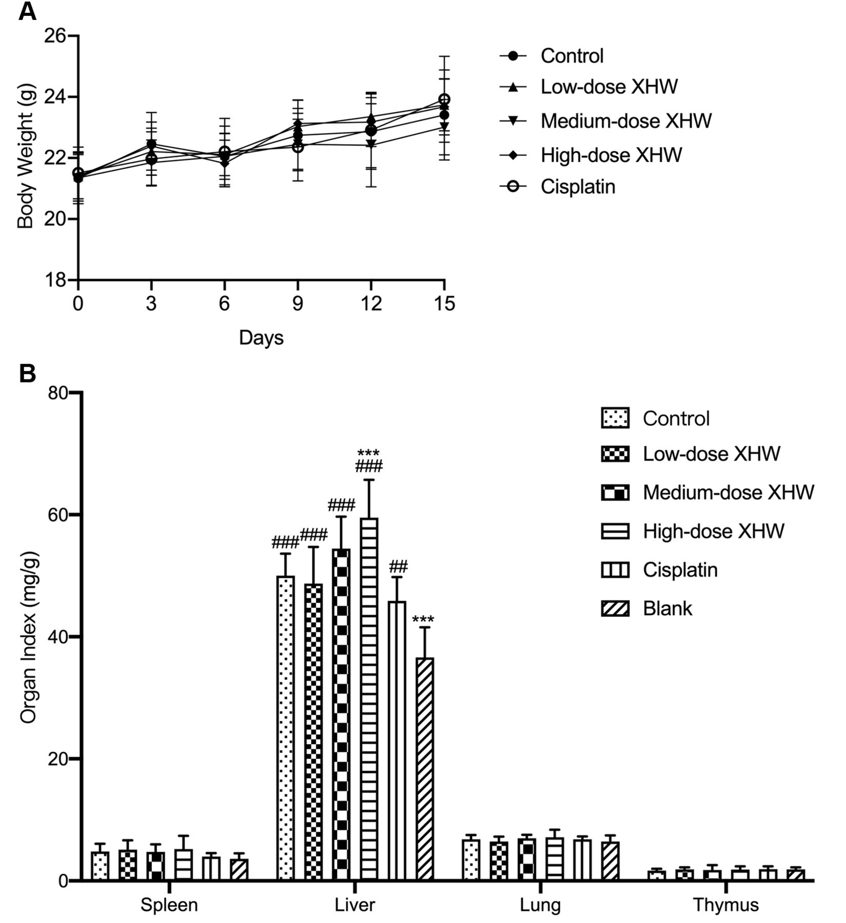
Effect of treatment on body weight and organ weight ratio. A: Body weight. B: Organ ratio (spleen, liver, lung and thymus). *p<0.05 vs. control; ##p<0.01 vs. blank; ###p<0.001 vs. blank.
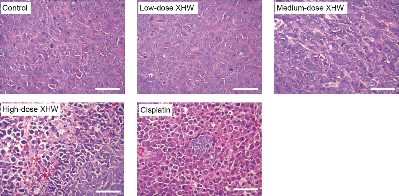
Effect of treatment on histology. Under light microscopy, LLC cells in the untreated control group were densely arranged with obvious anaplastic characteristics: various cell sizes and diverse morphologies; variable cell nuclei sizes and increased nucleus to cytoplasm ratio; granular chromatin and uneven distribution. Pathological mitoses such as multipolar and asymmetric nuclear divisions were observed. In the cisplatin group, more cells showed morphological changes associated with apoptosis including chromatin aggregation, nuclear consolidation, nuclear fragmentation and lysis. In the high-dose XHW and cisplatin group, necrotic areas were observed (Figure 3).

Effect of treatment on tumor histology. Hematoxylin and eosin staining was performed according to standard protocols. Under light microscopy, LLC cells in the untreated control group were densely arranged with obvious anaplastic characteristics: various cell sizes and diverse morphologies; variable cell nuclei sizes and increased nucleus to cytoplasm ratio; granular chromatin and uneven distribution. Pathological mitoses such as multipolar and asymmetric nuclear divisions were observed. In the cisplatin group, more cells showed morphological changes associated with apoptosis including chromatin aggregation, nuclear consolidation, nuclear fragmentation and lysis. In the high-dose XHW and cisplatin group, necrotic areas were observed. Scale bar: 50 μm.
Although the liver index was larger in each group compared to that in the blank group, there was no significant difference in liver histology among all groups. All groups had normal liver parenchyma with unremarkable hepatocytes having vacuolated cytoplasm, bland nuclei, and punctate nucleoli (Figure 4).

Effect of treatment on liver histology. Freshly-harvested livers were fixed in 10% formalin and embedded in paraffin and stained with hematoxylin and eosin. All groups had normal liver parenchyma with unremarkable hepatocytes having vacuolated cytoplasm, bland nuclei, and punctate nucleoli. There was no significant difference in liver histology among all groups. Scale bar: 50 μm.
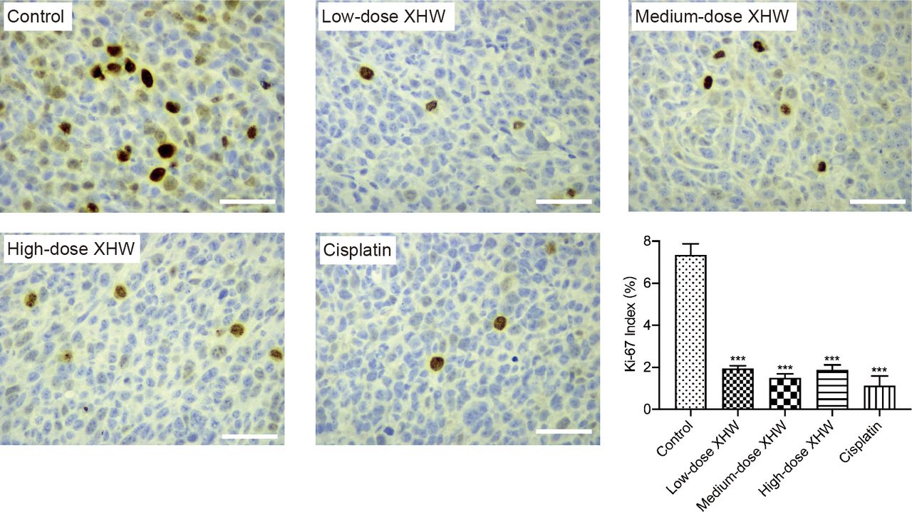
Effect of treatment on Ki-67 index. The Ki-67 index in the control group was 7.35%±0.90%, which was significantly greater than that in the cisplatin group (1.31%±1.04%, p<0.001), the low-dose XHW group (1.94%±0.25%, p<0.001), the medium-dose XHW group (1.50%±0.33%, p<0.001), and the high-dose XHW group (1.87%±0.43%, p<0.001) (Figure 5).

Effect of treatment on the Ki-67 index. Ki-67 staining was performed with a primary anti-Ki-67 antibody. The Ki-67 index was calculated as the percentage of positively stained cancer cells among all cancer cells. The Ki-67 index in the control group was significantly greater than that in the cisplatin, the low-dose XHW, the medium-dose XHW, and the high-dose XHW group.
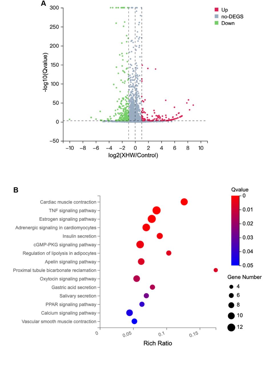
RNA-sequencing analysis. An average of 23.12 Mb clean reads, with an average genome mapping rate of 96.41%, was obtained. There were 406 differentially expressed genes between the control group and high-dose XHW group (|log2FC| ≥1, Q value ≤0.001), 130 genes were up-regulated, 276 genes were down-regulated (Figure 6A). The KEGG pathway cluster analysis showed that the 406 genes were closely associated with multiple signaling pathways, including tumor necrosis factor (TNF), estrogen, cyclic guanosine 3’, 5’-monophosphate-protein kinase G (cGMP-PKG), apelin and the peroxisome proliferator-activated receptor (PPAR) signaling pathways (Figure 6B).

Effect of high-dose XHW on gene expression. A) Volcano map of differentially expressed genes between the control group and the high-dose XHW group. There were 406 differentially expressed genes (|log2FC|≥1, Q value ≤0.01), 130 genes were up-regulated, 276 genes were down-regulated. B) KEGG pathway cluster analysis based on the 406 genes was significantly changed as described in A.
Discussion
The present study showed that XHW inhibited the growth of LLC in a syngeneic mouse model, and reduced the Ki-67 index, indicating that XHW alone could inhibit lung cancer. XHW alone was at least as effective as the first-line cytotoxic drug cisplatin both on tumor growth inhibition and decrease in the Ki-67 index. No toxicity was observed in histology among all groups. Thus, XHW is a candidate to replace cisplatin in lung cancer treatment. This may become very important clinically since many patients cannot tolerate platinum-based drugs.
A previous study on XHW treatment of LLC cells indicated that 11 metabolites were differentially expressed compared to the untreated control, including L-phenylalanine, L-tryptophan, farnesylcysteine, LysoPC(18:1(9Z)/0:0, L-adrenaline, 4-maleylacetoacetate, guanosine, aminoimidazole ribotide, corticosterone, sphingosine-1-phosphate and D-urobilinogen (48), suggesting that XHW plays a major role in altering aromatic amino-acid metabolism in the LLC mouse model.
Previous studies showed that XHW induced apoptosis of human glioblastoma U-87 MG cells via targeting the ROS-activated Akt/mTOR/FOXO1 pathway (49), and potentiated the efficacy of temozolomide on a glioblastoma xenograft through the Akt/mTOR pathway (50). XHW has also been shown to promote apoptosis of Treg cells in the tumor microenvironment in a 4T1 breast-cancer mouse model by upregulating the MEKK1/SEK1/JNK1/AP-1 pathway (51). A systems pharmacology and proteomics study of XHW on triple-negative breast cancer (TNBC) showed that XHW may be effective on non-small cell lung cancer and small cell lung cancer, as demonstrated by the KEGG signaling pathway enrichment analysis (52).
In the present study, to further explore the mechanism of XHW inhibition of LLC in a syngeneic mouse model, RNA-sequencing (RNA-seq) was performed. RNA-seq demonstrated that many signaling pathways were involved, including TNF, estrogen, cGMP-PKG, apelin and PPAR signaling pathways. A previous transcriptomics and chemical informatics study showed that the TNF and estrogen signaling pathways were also involved in XHW-mediated inhibition of TNBC (53). Estrogen and the estrogen receptor have the potential to become prognosticators and therapeutic targets in lung cancer (54). Another previous study examining the effect of XHW on multiple cancers by network pharmacology showed that one of the key targets of XHW is estrogen receptor 1 (ESR1) (55). The TNF signaling pathway is involved in many physiological and pathological processes, including cell proliferation, apoptosis, regulation of immune responses and inflammation (56), closely related to cancer, including lung cancer (57). Apelin, a transmembrane G protein-coupled receptor, is an endogenous ligand of the apelin receptor, and is upregulated in multiple cancer types including lung cancer, colon cancer, gastroesophageal cancer, hepatocellular carcinoma and brain cancer (58). Down-regulation of apelin has been shown to inhibit mammary and lung cancer growth as well as lung metastasis (59). PPARs are nuclear hormone receptors activated by fatty acids and their derivatives, PPARγ is expressed in various cancer types including lung cancer. Reduced PPARγ and elevated cyclooxygenase-2 (COX-2) expression are associated with poor prognosis in lung cancer patients. PPARγ is considered to be a therapeutic target for lung cancer (60-62). Cyclic GMP (cGMP) is an intracellular second messenger, mediating the action of nitric oxide (NO) and natriuretic peptides (NPs). cGMP has been shown to exert its physiological action through cGMP-dependent protein kinase (PKG), cGMP-regulated phosphodiesterases (PDE2, PDE3) and cGMP-gated cation channels. The cGMP-PKG pathway has been found to be closely related to lung cancer (63, 64).
The RNA-seq results and KEGG pathway enrichment analysis of the present study showed that many signaling pathways were altered following treatment of the syngeneic LLC mouse model with XHW, indicating that XHW inhibited the LLC carcinoma through many different pathways. These pathways need to be further studied, including their interaction, to develop an improved therapy for lung cancer.
Acknowledgements
This study was supported by Horizontal Scientific Research Project of Dongfang Hospital, Beijing University of Chinese Medicine (No. 040101003063).
Footnotes
This article is freely accessible online.
Authors’ Contributions
ZZ and ZT designed the study. ZZ, WJ, DH, LD, ZX, PH and SM performed the experiments. ZZ and LX analyzed the data. ZZ and ZX drafted the article. RMH revised the article and rewrote the Discussion. HK administrated and supervised the study.
Conflicts of Interest
All the Authors declare no conflicts of interest in relation to this study.
- Received May 16, 2021.
- Revision received May 27, 2021.
- Accepted May 31, 2021.
- Copyright © 2021 International Institute of Anticancer Research (Dr. George J. Delinasios), All rights reserved
References
In this issue


Jump to section
Related Articles
Cited By...
- No citing articles found.