
Main menu

  • Home
  • Current Issue
  • Archive
  • Info for
    • Authors
    • Editorial Policies
    • Advertisers
    • Editorial Board
    • Special Issues 2025
  • Journal Metrics
  • Other Publications
    • Anticancer Research
    • Cancer Genomics & Proteomics
    • Cancer Diagnosis & Prognosis
  • More
    • IIAR
    • Conferences
  • About Us
    • General Policy
    • Contact
  • Other Publications
    • In Vivo
    • Anticancer Research
    • Cancer Genomics & Proteomics

User menu

  • Register
  • Subscribe
  • My alerts
  • Log in
  • My Cart

Search

  • Advanced search
In Vivo
  • Other Publications
    • In Vivo
    • Anticancer Research
    • Cancer Genomics & Proteomics
  • Register
  • Subscribe
  • My alerts
  • Log in
  • My Cart
In Vivo

Advanced Search

  • Home
  • Current Issue
  • Archive
  • Info for
    • Authors
    • Editorial Policies
    • Advertisers
    • Editorial Board
    • Special Issues 2025
  • Journal Metrics
  • Other Publications
    • Anticancer Research
    • Cancer Genomics & Proteomics
    • Cancer Diagnosis & Prognosis
  • More
    • IIAR
    • Conferences
  • About Us
    • General Policy
    • Contact
  • Visit iiar on Facebook
  • Follow us on Linkedin
Research ArticleExperimental Studies

A Patient-Derived Orthotopic Xenograft Model of Gastroesophageal-Junction Adenocarcinoma Translated to the Clinic by Tumor-Targeting Fluorescent Antibodies to Carcinoembryonic-Antigen-Related Cell-Adhesion Molecules

MICHAEL A. TURNER, SIAMAK AMIRFAKHRI, HIROTO NISHINO, THINZAR M. LWIN, THOMAS J. SAVIDES, TONY R. REID, BERNHARD B. SINGER, ROBERT M. HOFFMAN and MICHAEL BOUVET
In Vivo July 2021, 35 (4) 1959-1963; DOI: https://doi.org/10.21873/invivo.12463
MICHAEL A. TURNER
1Department of Surgery, University of California San Diego, San Diego, CA, U.S.A.;
2VA San Diego Healthcare System, San Diego, CA, U.S.A.;
  • Find this author on Google Scholar
  • Find this author on PubMed
  • Search for this author on this site
SIAMAK AMIRFAKHRI
1Department of Surgery, University of California San Diego, San Diego, CA, U.S.A.;
2VA San Diego Healthcare System, San Diego, CA, U.S.A.;
  • Find this author on Google Scholar
  • Find this author on PubMed
  • Search for this author on this site
HIROTO NISHINO
1Department of Surgery, University of California San Diego, San Diego, CA, U.S.A.;
2VA San Diego Healthcare System, San Diego, CA, U.S.A.;
  • Find this author on Google Scholar
  • Find this author on PubMed
  • Search for this author on this site
THINZAR M. LWIN
1Department of Surgery, University of California San Diego, San Diego, CA, U.S.A.;
  • Find this author on Google Scholar
  • Find this author on PubMed
  • Search for this author on this site
THOMAS J. SAVIDES
3Department of Medicine, University of California San Diego, San Diego, CA, U.S.A.;
  • Find this author on Google Scholar
  • Find this author on PubMed
  • Search for this author on this site
TONY R. REID
3Department of Medicine, University of California San Diego, San Diego, CA, U.S.A.;
  • Find this author on Google Scholar
  • Find this author on PubMed
  • Search for this author on this site
BERNHARD B. SINGER
4Institute of Anatomy, Medical Faculty, University of Duisburg-Essen, Essen, Germany;
  • Find this author on Google Scholar
  • Find this author on PubMed
  • Search for this author on this site
ROBERT M. HOFFMAN
1Department of Surgery, University of California San Diego, San Diego, CA, U.S.A.;
2VA San Diego Healthcare System, San Diego, CA, U.S.A.;
5AntiCancer, Inc., San Diego, CA, U.S.A.
  • Find this author on Google Scholar
  • Find this author on PubMed
  • Search for this author on this site
MICHAEL BOUVET
1Department of Surgery, University of California San Diego, San Diego, CA, U.S.A.;
2VA San Diego Healthcare System, San Diego, CA, U.S.A.;
  • Find this author on Google Scholar
  • Find this author on PubMed
  • Search for this author on this site
  • For correspondence: mbouvet{at}ucsd.edu
  • Article
  • Figures & Data
  • Info & Metrics
  • PDF
Loading

Abstract

Background/Aim: During surgical resection of gastroesophageal-junction (GEJ) adenocarcinoma, the margin status is often difficult to visualize resulting in high recurrence rates. The aim of the present study was to develop a labelling technique that would allow improved visualization of GEJ tumor margins for surgeons to reduce recurrence rates in a patient-like model. Materials and Methods: A patient GEJ tumor was obtained from an endoscopic biopsy and implanted subcutaneously in a nude mouse. A patient-derived orthotopic xenograft (PDOX) model was established by implanting tumor fragments grown from a subcutaneous model to the cardia of the stomach of nude mice. CC1/3/5-SAB, an antibody to carcinoembryonic-antigen-related cell-adhesion molecules, was conjugated with infrared dye IRDye800 to create SAB-IR800. Forty-eight hours after i.v. injection of SAB-IR800, GEJ-PDOX mice were imaged. Results: Fluorescence imaging with SAB-IR800 brightly visualized the GEJ adenocarcinoma demonstrating specific targeting. In the PDOX model, injection of SAB-IR800 (50 μg) resulted in a tumor to background ratio of 1.78 at 48 hours and 1.86 at 72 hours. Conclusion: PDOX models of GEJ tumors can be established from patients by endoscopic biopsy without undergoing surgical resection. GEJ PDOX models should be useful for developing novel diagnostics and therapeutics for this recalcitrant disease.

  • PDOX
  • patient-derived orthotopic xenograft
  • gastro – esophageal junction
  • adenocarcinoma
  • carcinoembryonic-antigen-related cell adhesion
  • fluorescent antibodies

Gastroesophageal junction (GEJ) tumors are defined as tumors whose origins are within 5 cm of the anatomical GEJ (1). These tumors have been increasing in incidence in the Western World (1, 2). Currently, the only curative treatment for GEJ tumors is surgical resection (3). However, the majority of patients present with advanced disease, and less than 50% undergo surgery (3). There remains controversy regarding classification and treatment of this type of cancer with wide variations in operative management (2, 3). One challenge of surgical resection of GEJ tumors is achieving an R0 resection which is an independent predictor of survival (1). This present report describes an imaging PDOX model of adenocarcinoma of the GEJ.

Carcinoembryonic-antigen-related cell-adhesion molecules (CEACAMs) are a family of cell-surface glycoproteins. CEACAMs have many cellular functions including cell-cell recognition, cell adhesion, intra- and intercellular signaling as well as functions involved in inflammation, cancer progression and metastasis (4, 5). Several CEACAMs have been validated as biomarkers for melanoma, colorectal cancer, lung and pancreatic cancer (4). Specifically CEACAM1 and CEACAM5 are overexpressed in several cancers including gastric cancer (4). CC1/3/5-SAB is an antibody which selectively binds to human CEACAM1, CEACAM3 and CEACAM5.

In the present study, we used the CC1/3/5-SAB antibody conjugated to a near infrared 800 nm dye (SAB-IR800) to brightly label the PDOX model of GEJ adenocarcinoma.

Materials and Methods

Animal care. Athymic nude mice (Jackson Laboratory, Bar Harbor, ME) were maintained in a barrier facility with high-efficiency particulate air (HEPA) filtration and fed an autoclaved laboratory diet. All mice were anesthetized with intraperitoneal ketamine and xylazine reconstituted in phosphate-buffered saline (PBS) prior to surgical procedures. All mice post-laparotomy received buprenorphine subcutaneously the day of the procedure and when displaying signs of distress, for pain control. Mice were euthanized by CO2 inhalation and cervical dislocation at the conclusion of the study. All studies were approved by the San Diego Veterans Administration Medical Center Institutional Animal Care and Use Committee (IACUC, animal use protocol A17-020).

GEJ-PDOX establishment. The patient-derived GEJ-tumor specimen was obtained under standard sterile conditions via diagnostic esophagogastroduodenoscopy (Figure 1). Patient tumors and normal tissue were obtained with informed consent under the UCSD Institutional Review Board (IRB). To initially establish the patient tumor in nude mice, tumor fragments (1 mm3) were implanted over the bilateral upper and lower flanks and allowed to grow to approximately 5 mm in diameter. To establish a GEJ patient-derived orthotopic xenograft (PDOX) model, mice were anesthetized as described above and the stomach was delivered through a midline ventral incision. A 1 mm3 tumor fragment was sutured to the stomach’s lesser curve by the cardia using 8-0 nylon sutures (6). The peritoneum and skin were closed using 6-0 vicryl sutures. Tumor size was initially estimated via palpation.

Figure 1.
  • Download figure
  • Open in new tab
  • Download powerpoint
Figure 1.

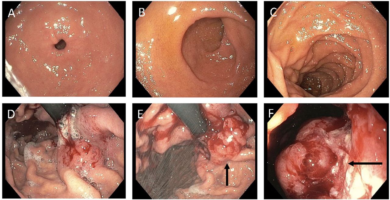
Endoscopic images from a GEJ patient. Panel A shows normal-appearing duodenum. Panels B and C show normal gastric rugae. Panels D, E and F show the endoscope retroflexed to demonstrate a near obstructing gastric cardia mass (black arrows).

Imaging. In vivo imaging was performed using the Pearl Trilogy Small Animal Imaging System (LI-COR, Lincoln, NE). The GEJ PDOX models were administered SAB-IR800 (50 μg) via tail vein injection. The PDOX models were anesthetized at 48 hours after dye injection and a small ventral incision was made to expose both the stomach and tumor which were imaged using the Pearl Trilogy Small Animal Imaging System. The incision was closed with a 6-0 vicryl suture. PDOX models were euthanized at 72 hours after dye injection and then a laparotomy was performed. Normal stomach was used as background to obtain a tumor to background ratio (TBR).

Statistical analysis. Statistical analysis was performed with R software (Free Software Foundation, Boston, MA, USA).

Results

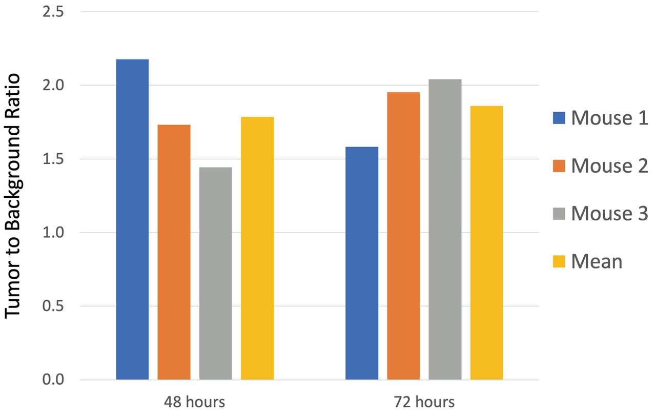
The GEJ PDOX model mimicked the clinical disease. A representative mouse treated with SAB-IR800 was imaged under brightfield and NIR fluorescence using the Pearl Imager (Figure 2). The bright fluorescence labelling of the tumor was evident. For the PDOX models (n=3), the mean tumor signal at 48 hours after dye injection was 0.244 (±0.036). The TBR at 48 hours ranged from 1.45 to 2.18 with a mean TBR of 1.78 (±0.37). At 72 hours, the mean tumor signal was 0.317 (±0.075). The mean TBR at 72 hours was 1.86 (±0.24) (Figure 3).

Figure 2.
  • Download figure
  • Open in new tab
  • Download powerpoint
Figure 2.

Laparotomy of a representative GEJ-PDOX mouse 72 hours after receiving SAB-IR800 (50 μg). Panel A is a brightfield image and Panel B is a fluorescence image. The yellow arrows indicate the patient-derived GEJ tumor growing over the mouse’s stomach. Fluorescence labeling with SAB-IR800 resulted in a tumor to background ratio (TBR) of 2.04.

Figure 3.
  • Download figure
  • Open in new tab
  • Download powerpoint
Figure 3.

Bar graphs represent the tumor to background ratios (TBRs) of the GEJ PDOX mouse models (n=3) as well as the mean TBR (yellow bar).

Discussion

A previous study described a PDOX model of a GEJ tumor (7). However, the present study is the first study to report a GEJ-PDOX mouse model that was successfully imaged with a tumor-specific antibody conjugated to a near-infrared (NIR) fluorophore. Importantly, endoscopically-obtained tissue was sufficient to establish the GEJ tumor in nude mice, thus avoiding a surgical procedure and all the risks inherent with it. The study demonstrated the ability of SAB-IR800 to image GEJ tumors with resultant TBRs that should be sufficient for fluorescence-guided surgery (FGS) in future experiments.

The patient in the present study had a Siewert Type III GEJ tumor originating >2 cm below the GEJ. de Graaf et al. reported a series of 416 patients thought to have potentially resectable GEJ tumors (8). After undergoing staging laparoscopy (SL), 84 (20.2%) patients were deemed not surgical candidates, thereby sparing them an unnecessary laparotomy. If some patients have metastatic deposits too small to detect on imaging or visual inspection with SL, FGS could aid the surgeon using the technology described in the present study.

It is difficult to determine the frequency of R0 resections as some series combine GEJ tumors with esophageal tumors or with gastric adenocarcinoma (2). Along with nodal status and tumor infiltration depth, an R0 resection is an independent predictor of survival (1). We expect SAB-IR800 to become clinically useful for both SL and FGS. Using CEACAMs as an appropriate tumor-targeting agent for FGS is consistent with our previous results with CEACAMs to image colorectal cancer (9).

There are potential indications to use fluorescently-labeled tumors to aid in screening. Iseki et al. demonstrated the effectiveness of using an NIR endoscope to accurately assess gastric-tumor depth to aid in endoscopic mucosal resection (10). Habibollahi et al. used a protease-activatable NIR fluorescent probe to improve detection of human esophageal adenocarcinoma in orthotopic mouse models (11). Tjalma et al. demonstrated that NIR with fluorescent tracers targeting VEGF-A and EGFR improved colorectal-polyp detection over bright light alone, continuing to demonstrate the validity of the principle of the present report in a different organ (12).

The SAB-IR800 has several benefits described in the present report, including high specificity for a GEJ tumor in the PDOX model and a relatively high TBR with potential as a GEJ diagnostic adjunct in SL and FGS. This may lead to improved rate of R0 resections and patient outcomes. A weakness is this antibody is not a genetic probe (e.g., telomerase-dependent adenovirus expressing GFP) which can genetically label tumors for FGS, as well as recurrence (13-16).

Future studies will be performed with a larger sample size to better characterize the binding and clearing of the SAB-IR800-dye conjugate to obtain higher TBRs.

Acknowledgements

This study was funded by VA Merit Review grant numbers 1 I01 BX003856-01A1 and 1 I01 BX004494-01 (MB) and NIH/NCI T32CA121938 (MAT and TML).

Footnotes

  • This article is freely accessible online.

  • Authors’ Contributions

    M.A.T., R.M.H. and M.B. designed the experiments and wrote the paper. M.A.T., S.A., H.N., T.M.L., T.J.S., T.R.R., and B.B.S., were involved in the acquisition of the data. All Authors were involved in final manuscript editing and approval.

  • Conflicts of Interest

    RMH is a non-salaried affiliate of AntiCancer, Inc which uses PDOX models for contract research.

  • Received April 5, 2021.
  • Revision received April 26, 2021.
  • Accepted May 10, 2021.
  • Copyright © 2021 The Author(s). Published by the International Institute of Anticancer Research.

References

  1. ↵
    1. Parry K,
    2. Haverkamp L,
    3. Bruijnen RC,
    4. Siersema PD,
    5. Ruurda JP and
    6. van Hillegersberg R
    : Surgical treatment of adenocarcinomas of the gastro-esophageal junction. Ann Surg Oncol 22(2): 597-603, 2015. PMID: 25190126. DOI: 10.1245/s10434-014-4047-1
    OpenUrlCrossRefPubMed
  2. ↵
    1. Rusch VW
    : Are cancers of the esophagus, gastroesophageal junction, and cardia one disease, two, or several? Semin Oncol 31(4): 444-449, 2004. PMID: 15297937. DOI: 10.1053/j.seminoncol.2004.04.023
    OpenUrlCrossRefPubMed
  3. ↵
    1. Barbour AP,
    2. Rizk NP,
    3. Gonen M,
    4. Tang L,
    5. Bains MS,
    6. Rusch VW,
    7. Coit DG and
    8. Brennan MF
    : Adenocarcinoma of the gastroesophageal junction: influence of esophageal resection margin and operative approach on outcome. Ann Surg 246(1): 1-8, 2007. PMID: 17592282. DOI: 10.1097/01.sla.0000255563.65157.d2
    OpenUrlCrossRefPubMed
  4. ↵
    1. Beauchemin N and
    2. Arabzadeh A
    : Carcinoembryonic antigen-related cell adhesion molecules (CEACAMs) in cancer progression and metastasis. Cancer Metastasis Rev 32(3-4): 643-671, 2013. PMID: 23903773. DOI: 10.1007/s10555-013-9444-6
    OpenUrlCrossRefPubMed
  5. ↵
    1. Kuespert K,
    2. Pils S and
    3. Hauck CR
    : CEACAMs: their role in physiology and pathophysiology. Curr Opin Cell Biol 18(5): 565-571, 2006. PMID: 16919437. DOI: 10.1016/j.ceb.2006.08.008
    OpenUrlCrossRefPubMed
  6. ↵
    1. Furukawa T,
    2. Fu X,
    3. Kubota T,
    4. Watanabe M,
    5. Kitajima M and
    6. Hoffman RM
    : Nude mouse metastatic models of human stomach cancer constructed using orthotopic implantation of histologically intact tissue. Cancer Res 53(5):1204-1208, 1993. PMID: 8439965.
    OpenUrlAbstract/FREE Full Text
  7. ↵
    1. Veeranki OL,
    2. Tong Z,
    3. Mejia A,
    4. Verma A,
    5. Katkhuda R,
    6. Bassett R,
    7. Kim TB,
    8. Wang J,
    9. Lang W,
    10. Mino B,
    11. Solis L,
    12. Kingsley C,
    13. Norton W,
    14. Tailor R,
    15. Wu JY,
    16. Krishnan S,
    17. Lin SH,
    18. Blum M,
    19. Hofstetter W,
    20. Ajani J,
    21. Kopetz S and
    22. Maru D
    : A novel patient-derived orthotopic xenograft model of esophageal adenocarcinoma provides a platform for translational discoveries. Dis Model Mech 12(12): dmm041004, 2019. PMID: 31732509. DOI: 10.1242/dmm.041004
    OpenUrlAbstract/FREE Full Text
  8. ↵
    1. de Graaf GW,
    2. Ayantunde AA,
    3. Parsons SL,
    4. Duffy JP and
    5. Welch NT
    : The role of staging laparoscopy in oesophagogastric cancers. Eur J Surg Oncol 33(8): 988-992, 2007. PMID: 17344017. DOI: 10.1016/j.ejso.2007.01.007
    OpenUrlCrossRefPubMed
  9. ↵
    1. Hollandsworth HM,
    2. Amirfakhri S,
    3. Filemoni F,
    4. Schmitt V,
    5. Wennemuth G,
    6. Schmidt A,
    7. Hoffman RM,
    8. Singer BB and
    9. Bouvet M
    : Anti-carcinoembryonic antigen-related cell adhesion molecule antibody for fluorescence visualization of primary colon cancer and metastases in patient-derived orthotopic xenograft mouse models. Oncotarget 11(4): 429-439, 2020. PMID: 32064046. DOI: 10.18632/oncotarget.27446
    OpenUrlCrossRefPubMed
  10. ↵
    1. Iseki K,
    2. Tatsuta M,
    3. Iishi H,
    4. Sakai N,
    5. Yano H and
    6. Ishiguro S
    : Effectiveness of the near-infrared electronic endoscope for diagnosis of the depth of involvement of gastric cancers. Gastrointest Endosc 52(6): 755-762, 2000. PMID: 11115912. DOI: 10.1067/mge.2000.110455
    OpenUrlCrossRefPubMed
  11. ↵
    1. Habibollahi P,
    2. Figueiredo JL,
    3. Heidari P,
    4. Dulak AM,
    5. Imamura Y,
    6. Bass AJ,
    7. Ogino S,
    8. Chan AT and
    9. Mahmood U
    : Optical imaging with a cathepsin B activated probe for the enhanced detection of esophageal adenocarcinoma by dual channel fluorescent upper GI endoscopy. Theranostics 2(2): 227-234, 2012. PMID: 22400064. DOI: 10.7150/thno.4088
    OpenUrlCrossRefPubMed
  12. ↵
    1. Tjalma JJ,
    2. Garcia-Allende PB,
    3. Hartmans E,
    4. Terwisscha van Scheltinga AG,
    5. Boersma-van Ek W,
    6. Glatz J,
    7. Koch M,
    8. van Herwaarden YJ,
    9. Bisseling TM,
    10. Nagtegaal ID,
    11. Timmer-Bosscha H,
    12. Koornstra JJ,
    13. Karrenbeld A,
    14. Kleibeuker JH,
    15. van Dam GM,
    16. Ntziachristos V and
    17. Nagengast WB
    : Molecular fluorescence endoscopy targeting vascular endothelial growth factor A for improved colorectal polyp detection. J Nucl Med 57(3): 480-485, 2016. PMID: 26678613. DOI: 10.2967/jnumed.115.166975
    OpenUrlAbstract/FREE Full Text
  13. ↵
    1. Yano S,
    2. Takehara K,
    3. Miwa S,
    4. Kishimoto H,
    5. Tazawa H,
    6. Urata Y,
    7. Kagawa S,
    8. Bouvet M,
    9. Fujiwara T and
    10. Hoffman RM
    : Fluorescence-guided surgery of a highly-metastatic variant of human triple-negative breast cancer targeted with a cancer-specific GFP adenovirus prevents recurrence. Oncotarget 7(46): 75635-75647, 2016. PMID: 27689331. DOI: 10.18632/oncotarget.12314
    OpenUrlCrossRefPubMed
    1. Yano S,
    2. Takehara K,
    3. Miwa S,
    4. Kishimoto H,
    5. Hiroshima Y,
    6. Murakami T,
    7. Urata Y,
    8. Kagawa S,
    9. Bouvet M,
    10. Fujiwara T and
    11. Hoffman RM
    : Improved resection and outcome of colon-cancer liver metastasis with fluorescence-guided surgery using in situ GFP labeling with a telomerase-dependent adenovirus in an orthotopic mouse model. PLoS One 11(2): e0148760, 2016. PMID: 26849435. DOI: 10.1371/journal.pone.0148760
    OpenUrlCrossRefPubMed
    1. Kishimoto H,
    2. Zhao M,
    3. Hayashi K,
    4. Urata Y,
    5. Tanaka N,
    6. Fujiwara T,
    7. Penman S and
    8. Hoffman RM
    . In vivo internal tumor illumination by telomerase-dependent adenoviral GFP for precise surgical navigation. Proc Natl Acad Sci U S A 106(34):14514-7, 2009. PMID: 19706537. DOI: 10.1073/pnas.0906388106
    OpenUrlAbstract/FREE Full Text
  14. ↵
    1. Kishimoto H,
    2. Aki R,
    3. Urata Y,
    4. Bouvet M,
    5. Momiyama M,
    6. Tanaka N,
    7. Fujiwara T and
    8. Hoffman RM
    . Tumor-selective, adenoviral-mediated GFP genetic labeling of human cancer in the live mouse reports future recurrence after resection. Cell Cycle 10(16):2737-2741, 2011. PMID: 21785265. DOI: 10.4161/cc.10.16.16756
    OpenUrlCrossRefPubMed
PreviousNext
Back to top

In this issue

In Vivo
Vol. 35, Issue 4
July-August 2021
  • Table of Contents
  • Table of Contents (PDF)
  • Index by author
  • Back Matter (PDF)
  • Ed Board (PDF)
  • Front Matter (PDF)
Print
Download PDF
Article Alerts
Sign In to Email Alerts with your Email Address
Email Article

Thank you for your interest in spreading the word on In Vivo.

NOTE: We only request your email address so that the person you are recommending the page to knows that you wanted them to see it, and that it is not junk mail. We do not capture any email address.

Enter multiple addresses on separate lines or separate them with commas.
A Patient-Derived Orthotopic Xenograft Model of Gastroesophageal-Junction Adenocarcinoma Translated to the Clinic by Tumor-Targeting Fluorescent Antibodies to Carcinoembryonic-Antigen-Related Cell-Adhesion Molecules
(Your Name) has sent you a message from In Vivo
(Your Name) thought you would like to see the In Vivo web site.
CAPTCHA
This question is for testing whether or not you are a human visitor and to prevent automated spam submissions.
2 + 6 =
Solve this simple math problem and enter the result. E.g. for 1+3, enter 4.
Citation Tools
A Patient-Derived Orthotopic Xenograft Model of Gastroesophageal-Junction Adenocarcinoma Translated to the Clinic by Tumor-Targeting Fluorescent Antibodies to Carcinoembryonic-Antigen-Related Cell-Adhesion Molecules
MICHAEL A. TURNER, SIAMAK AMIRFAKHRI, HIROTO NISHINO, THINZAR M. LWIN, THOMAS J. SAVIDES, TONY R. REID, BERNHARD B. SINGER, ROBERT M. HOFFMAN, MICHAEL BOUVET
In Vivo Jul 2021, 35 (4) 1959-1963; DOI: 10.21873/invivo.12463

Citation Manager Formats

  • BibTeX
  • Bookends
  • EasyBib
  • EndNote (tagged)
  • EndNote 8 (xml)
  • Medlars
  • Mendeley
  • Papers
  • RefWorks Tagged
  • Ref Manager
  • RIS
  • Zotero
Reprints and Permissions
Share
A Patient-Derived Orthotopic Xenograft Model of Gastroesophageal-Junction Adenocarcinoma Translated to the Clinic by Tumor-Targeting Fluorescent Antibodies to Carcinoembryonic-Antigen-Related Cell-Adhesion Molecules
MICHAEL A. TURNER, SIAMAK AMIRFAKHRI, HIROTO NISHINO, THINZAR M. LWIN, THOMAS J. SAVIDES, TONY R. REID, BERNHARD B. SINGER, ROBERT M. HOFFMAN, MICHAEL BOUVET
In Vivo Jul 2021, 35 (4) 1959-1963; DOI: 10.21873/invivo.12463
Twitter logo Facebook logo Mendeley logo
  • Tweet Widget
  • Facebook Like
  • Google Plus One

Jump to section

  • Article
    • Abstract
    • Materials and Methods
    • Results
    • Discussion
    • Acknowledgements
    • Footnotes
    • References
  • Figures & Data
  • Info & Metrics
  • PDF

Related Articles

  • No related articles found.
  • PubMed
  • Google Scholar

Cited By...

  • No citing articles found.
  • Google Scholar

More in this TOC Section

  • Effect of Decay-accelerating Factor on LPS-induced Acute Lung Injury in the Rat
  • Early and Late Effects of Difluorodeoxycytidine on Murine Cell Radiosensitivity In Vivo
  • Reduced Pain Responses With a Digital Automatic Syringe: Behavioral and Molecular Evidence in a Rodent Formalin Model
Show more Experimental Studies

Similar Articles

Keywords

  • PDOX
  • patient-derived orthotopic xenograft
  • gastro – esophageal junction
  • adenocarcinoma
  • carcinoembryonic-antigen-related cell adhesion
  • fluorescent antibodies
In Vivo

© 2025 In Vivo

Powered by HighWire