


Abstract
Background/Aim: The aim of this study was to detect circulating tumor cells (CTC) in the peripheral blood of gastrointestinal cancer patients using conditionally reprogrammed cell (CRC) culture. Materials and Methods: We confirmed the sensitivity of the CRC culture method. Five ml of blood were obtained from 81 cancer patients (56 colorectal and 25 gastric). The collected mononuclear cells were cultured for 4 weeks in the CRC condition. Finally, cultured cells were characterized by RT-PCR for the expression of hTERT and MAGE A1-6 mRNA. Results: The CRC method had a CTC detection limit of 6 cells for gastric cancer cells. After culture of 81 blood specimens, 38 formed visible cells, including 5 colonies. Among the 38 cells, 13 were hTERT positive and 4 were MAGE A1-6 positive. The final CTC detection rate was 16.0%. Conclusion: The CRC culture may potentially be used to evaluate the metastatic cancer cells in the circulation.
Detection of circulating tumor cells (CTCs) in the blood has been investigated using various methods based on antibodies, transcripts or proteins (1, 2). Recently, many researchers utilized cell-free DNA (cfDNA) instead of CTCs (3-5). Regarding some solid tumors, cfDNA has produced promising results (6). However, the approach is limited because cfDNA yields the same information regarding mutations and epigenetic modifications as the tumor tissue without providing any information about protein and RNA transcription profiles of the tumor phenotype (7).
The detection of CTCs has been refined by the progression from the conventional immunologic capture method using EpCAM and cytokeratin antibody to an in vitro cell culture method (8, 9). CTC culture consists of short-term culture, such as the epithelial immunospot approach, and long-term culture (1). Successful culture rates for patients with early lung cancer have been achieved using a three-dimensional co-culture system (10). However, CTC cultures for gastrointestinal cancers are rare (11-14).
The Georgetown University Medical Center (GUMC) has also developed a conditionally reprogrammed cell (CRC) culture method using 3T3 mouse fibroblasts (J2 cells) and a Rho-kinase inhibitor (Y27632) (15). The CRC culture method is very efficient for the culture of tumor cells in vitro.
In this study, we attempted to detect circulating colorectal and gastric cancer cells using the CRC culture. To characterize the cultured cells isolated from the peripheral blood, we used the Melanoma antigen-encoding gene (MAGE) and human telomerase reverse transcriptase (hTERT) mRNA as markers. MAGE is known to be a cancer-specific gene, but individual MAGE genes are poorly expressed in cancer cells (16, 17). Common MAGE primers that can detect MAGE A1 to A6 (MAGE A1-6) mRNA simultaneously were developed and have been used in various types of cancer (18, 19). The hTERT mRNA has also been studied in many types of cancer cells (20). Though the hTERT mRNA would be also expressed in activated lymphocytes (21), it has been utilized as a sensitive and specific tumor marker in the blood.
Materials and Methods
Sensitivity of CRC technique. To confirm the sensitivity of the CRC technique, we used SNU484 gastric cancer cells obtained from the Korean Cell Line Bank (Seoul, Republic of Korea), and colorectal cancer and gastric cancer cells isolated from cancer patients using CRC. J2 embryonic fibroblasts were kindly donated from the Georgetown University Medical Center (GUMC). Six, 12, 25, 50, 500 and 5,000 cells of the three cell types were incubated for 4 weeks in the CRC condition. The CRC condition was prepared using the same protocols of the GUMC (15).
Patient samples. Five ml of blood were obtained from each of the 81 cancer patients. The patients had been admitted at Daegu Catholic University Medical Center (Daegu, Korea) and diagnosed with gastric or colorectal cancer radiographically and pathologically. The 81 patients comprised 56 colorectal cancer and 25 gastric cancer patients. The stage of gastric and colorectal cancer was determined according to the American Joint Committee on Cancer 7th edition. Informed consent was obtained from each patient. The study protocol was approved by the Institutional Review Board (IRB) of Daegu Catholic University Medical Center. To collect CTCs from the blood specimens, red blood cells were lysed using RBC lysis buffer (Roche, Basel, Switzerland) and blood mononuclear cells were collected.
CRC culture and RNA extraction from the culture plates. The collected mononuclear cells were incubated for 4 weeks at 37°C in the CRC condition. Briefly, Swiss-3T3-J2 mouse fibroblasts were cultured to generate feeder cells and then, the isolated mononuclear cells were plated in F medium containing J2 feeder cells and Y-27632 (cat. no. 270-333M025, Enzo Life Sciences, Lausen, Switzerland). F medium was made by mixing Dulbecco’s modified Eagle’s medium (DMEM) (Invitrogen, Gaithersburg, MD, USA), 5% fetal bovine serum (Gibco; Thermo Fisher Scientific Inc., Waltham, MA, USA), L-glutamine (Gibco), penicillin/strepto mycin mix (Gibco), F12 nutrient mix (Gibco), 25 ng/ml hydrocortisone (Sigma-Aldrich, St. Louis, MO, USA), 0.125 ng/ml epidermal growth factor (EGF) (Invitrogen), 5 μg/ml insulin (Sigma-Aldrich), 250 ng/ml amphotericin B (Gibco), 10 μg/ml genta micin (Gibco) and 0.1 nM cholera toxin (Sigma-Aldrich). All cells were maintained at 37°C in a cell culture incubator with 95% humidity and 5% CO2. At the end of incubation, some colonies, cell aggregations, J2 cells or unknown cells persisted on the plate. All 81 samples were treated with 1 ml Trizol (Invitrogen), RNA was extracted, and RNA quality and quantity were measured using a spectrophotometer (NanoDrop Technologies, Wilmington, DE, USA).
Real time RT-PCR. The mRNAs of hTERT and MAGE A1-6 were amplified to determine the identity of the cultured cells. The mRNA of glyceraldehyde-3-phosphate dehydrogenase (GAPDH) was amplified to confirm the RNA integrity for all specimens. The hTERT and MAGE A1-6 mRNAs were amplified with nested PCR, whereas GAPDH mRNA was amplified with a single round of PCR amplification, using specific primers for each gene and the PCR conditions presented in Table I. Cells were determined as cancer cells if they expressed one cancer related mRNA. The amplification of all mRNAs was performed using the LightCycler 480 (Roche, Basel, Switzerland). We interpreted the PCR products as positive results using fluorescent signals and melting temperature analysis.
Gene-specific primers used in the polymerase chain reactions.
STR (short tandem repeat) analysis of culture cells and cancer tissue. To verify the cultured cell, we used STR analysis. In one colorectal cancer patient, some cells proliferated to form a colony (CR1520). We extracted DNA using the remnant of RNA extraction procedure and STR markers were analyzed using PowerPlex 16 system (Promega, Madison, WI, USA) with ABI 3500xL Genetic Analyzer (Applied Biosystems, Waltham, MA, USA). The matched cancer tissue of CR1520 obtained from the biopsy was also used for the STR analysis.
IRB approval and statistical analysis. This study was conducted in accordance with the standards of the Declaration of Helsinki and was approved by the IRB of Daegu Catholic University Medical Center (CR-16-002-L). All patients have provided written informed consent for their information to be stored and used in the hospital database.
For the statistical analysis, Chi-squared test was performed using MedCalc Version 14.12.0 (Ostend, Belgium).
Results
Sensitivity of CRC culture. Sensitivity data of spiked cancer cells cultured in the CRC condition are summarized in Table II. Gastric cancer cells were cultured from 6 to 5,000 cells, and colorectal cancer cells were cultured from 25 to 5,000 cells. Interestingly, even the lowest number of gastric cancer cells (6) grew in culture.
Sensitivity of cancer cell culture using conditionally reprogrammed cell method.
Clinical data, CRC culture and RT-PCR results. Demographic data of cancer patients are summarized in Table III. Most gastric cancer patients were stage I and II, while colorectal cancer patients were stage II and III.
Demographic data and circulating tumor cell culture results.
After 4 weeks of incubation, some cells were cultured in 38 of 81 blood samples. The cultured cells were observed as colonies, cell aggregations, J2 cells and unknown cells. Figure 1 shows the cultured colonies of gastric and colorectal cancer. Among the 38 cells, 13 cells showed positive results with MAGE A1-6 or hTERT RT-PCR analysis. The overall positive rates were 16.0% for hTERT and 4.9% for MAGE A1-6. The respective positive rates for each mRNA were 16.1% and 5.4% in colorectal cancer, and 16.0% and 4.0% in gastric cancer, respectively (Table III). All MAGE A1-6 positive cases were also positive forhTERT RT-PCR.
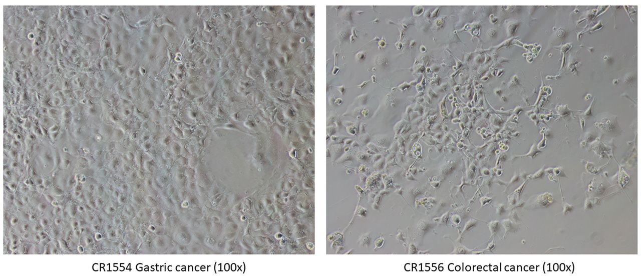
Light microscopic images of CR1554 and CR1556 cancer cells using conditionally reprogrammed cell culture.
STR analysis and cancer cell determination. In the STR analysis, 16 STR markers of CR1520 were completely consistent with those of cancer tissue (Figure 2), demonstrating that the cultured cells were derived from the cancer tissue. We determined the 13 cultured cells as cancer cells based on the results of hTERT or MAGE A1-6 RT-PCR and STR analysis.

STR marker analysis of CR1520 disclosed the same pattern between cultured cell (A) and cancer tissue (B).
The positive rates of hTERT and MAGE A1-6 RT-PCR according to cancer stages. The association of RT-PCR positive cases with cancer stages were also analyzed (Table IV). Though the positive rates of RT-PCR were not statistically significantly associated with cancer stage, the positive rates of hTERT and MAGE RT-PCR were associated with increased cancer stage. The hTERT RT-PCR was the most efficient in identifying cancer cells in the CRC culture.
Positive rates of RT-PCR results by cancer stage.
Discussion
CTCs are rare in the blood. Many methods have been used to increase CTC capture rates. These include immunomagnetic separation (22), microfluidic separation (23), microfiltration (24), electrophoresis (25), and acoustophoresis (26). Bobek et al. (27) and Zhang et al. (10) reported 66.7% and 73.7% detection rates from pancreatic and lung cancer patients, respectively. Zhang et al. (10) successfully isolated CTCs from 14 of 19 early-stage lung cancer patients using a microfluidic three-dimensional co-culture system.
In this study, we did not use any special CTC isolation kit and culture device. We collected mononuclear cells after red blood cells lysis without the depletion of CD45 positive leukocytes to avoid cancer cell loss. The collected cells were inoculated into the CRC culture plate using a fibroblast feeder layer. The CRC culture system effectively supports the growth of normal and tumor cells of epithelial origin (15, 28).
We evaluated the sensitivity of the CRC culture method. In a prior study (15), the minimum number of primary keratinocytes capable of forming a colony was four. Presently, the minimum number was similar (six) but the cell number depended on the cell type. The results indicate the potential of the CRC culture method for the study of CTCs.
To determine whether the cultured cells were cancer cells, we assayed for MAGE and hTERT mRNA. Though the hTERT mRNA might be expressed in the cells of the CRC culture (15), its expression could differentiate human cancer cells from mouse fibroblast J2 cells. The results regarding MAGE mRNA revealed that this gene is a cancer specific oncogene (17, 29).
The CRC method allowed the growth of some cells from the 38 blood samples. Among them, 13 samples showed the expression of MAGE or hTERT mRNA. Using RT-PCR, the hTERT mRNA was more frequently amplified than the MAGE A1-6 mRNA. The general expression rates of hTERT and MAGE A mRNA in cancer cells were 85% (30) and 50% (31), respectively. This could explain the difference in RT-PCR positive rates between hTERT and MAGE A1-6 mRNAs. The hTERT mRNA expressed in all cancer cells and its positive rates were dependent on cancer stage. Therefore, hTERT RT-PCR might be the best marker for the identification of cancer cells in the CRC culture.
After one-year follow-up of the 13 patients with a positive culture, two had multiple metastases (CR1554 and CR1560) and four had metastases to adjacent organs (CR 1600, CR1577, CR1546 and CR1632). Further studies are necessary to evaluate the clinical utility of CRC culture.
In the one case of positive culture, we isolated the tumor DNA from remnant of RNA extraction and analyzed the STR markers. In the CRC specimens, we could analyze DNA, RNA and protein levels. The CRC specimens can provide most of the information needed for cancer research. Therefore, the CRC culture might be the best method to study the metastatic event of cancer cells in the circulation.
The absence of a control group for comparison is a limitation of this study. The majority of circulating tumor cell studies using peripheral blood samples of cancer patients have not used a control group (27).
In summary, to isolate CTCs, we cultured blood cells of 81 cancer patients using the CRC culture method. The 38 cases displayed visible cells after 4 weeks. We characterized the culture cells using RT-PCR to detect hTERT and MAGE A1-6 mRNA. Of these, 13 cases showed cancer cell growth. Our study proved the potential of CRC culture to detect CTCs.
Acknowledgements
Supported by Basic Science Research Program through the National Research Foundation of Korea (NRF) funded by the Ministry of Education, Science and Technology (NRF-2013R1A1A2007189).
Footnotes
This article is freely accessible online.
Conflicts of Interest
All Authors have no conflicts of interest to declare regarding this study.
Authors’ Contributions
Chun-Seok Yang and Dae-Dong Kim collected blood samples and clinical information from the patients of colorectal cancer. In-Hwan Kim and Hyun-Dong Chae collected blood samples and clinical information from the patients of colorectal cancer. Chun-Seok Yang wrote the article. Chang-Ho Jeon performed the entire experiment, collected the experimental data, and revised the version to be submitted. All Authors designed the concept of this study and analyzed the experimental results.
- Received January 8, 2021.
- Revision received February 25, 2021.
- Accepted February 26, 2021.
- Copyright© 2021, International Institute of Anticancer Research (Dr. George J. Delinasios), All rights reserved
References
In this issue


Jump to section
Related Articles
Cited By...
- No citing articles found.