
Main menu

  • Home
  • Current Issue
  • Archive
  • Info for
    • Authors
    • Editorial Policies
    • Advertisers
    • Editorial Board
    • Special Issues
  • Journal Metrics
  • Other Publications
    • Anticancer Research
    • Cancer Genomics & Proteomics
    • Cancer Diagnosis & Prognosis
  • More
    • IIAR
    • Conferences
  • About Us
    • General Policy
    • Contact
  • Other Publications
    • In Vivo
    • Anticancer Research
    • Cancer Genomics & Proteomics

User menu

  • Register
  • Subscribe
  • My alerts
  • Log in
  • My Cart

Search

  • Advanced search
In Vivo
  • Other Publications
    • In Vivo
    • Anticancer Research
    • Cancer Genomics & Proteomics
  • Register
  • Subscribe
  • My alerts
  • Log in
  • My Cart
In Vivo

Advanced Search

  • Home
  • Current Issue
  • Archive
  • Info for
    • Authors
    • Editorial Policies
    • Advertisers
    • Editorial Board
    • Special Issues
  • Journal Metrics
  • Other Publications
    • Anticancer Research
    • Cancer Genomics & Proteomics
    • Cancer Diagnosis & Prognosis
  • More
    • IIAR
    • Conferences
  • About Us
    • General Policy
    • Contact
  • Visit iiar on Facebook
  • Follow us on Linkedin
Research ArticleExperimental Studies

Protective Effects of Baicalin on Arsenic Trioxide-induced Oxidative Damage and Apoptosis in Human Umbilical Vein Endothelial Cells

CHUNG-LIN TSAI, CHIA-WEN TSAI, WEN-SHIN CHANG, JIUNN-CHERNG LIN, TE-CHUN HSIA and DA-TIAN BAU
In Vivo January 2021, 35 (1) 155-162; DOI: https://doi.org/10.21873/invivo.12243
CHUNG-LIN TSAI
1Graduate Institute of Clinical Medical Science, China Medical University, Taichung, Taiwan, R.O.C.;
2Division of Cardiac and Vascular Surgery, Cardiovascular Center, Taichung Veterans General Hospital, Taichung, Taiwan, R.O.C.;
  • Find this author on Google Scholar
  • Find this author on PubMed
  • Search for this author on this site
CHIA-WEN TSAI
3Terry Fox Cancer Research Laboratory, Department of Medical Research, China Medical University Hospital, Taichung, Taiwan, R.O.C.;
  • Find this author on Google Scholar
  • Find this author on PubMed
  • Search for this author on this site
WEN-SHIN CHANG
3Terry Fox Cancer Research Laboratory, Department of Medical Research, China Medical University Hospital, Taichung, Taiwan, R.O.C.;
  • Find this author on Google Scholar
  • Find this author on PubMed
  • Search for this author on this site
JIUNN-CHERNG LIN
4Division of Cardiology, Department of Internal Medicine, Taichung Veterans General Hospital Chiayi Branch, Chiayi, Taiwan, R.O.C.;
  • Find this author on Google Scholar
  • Find this author on PubMed
  • Search for this author on this site
TE-CHUN HSIA
3Terry Fox Cancer Research Laboratory, Department of Medical Research, China Medical University Hospital, Taichung, Taiwan, R.O.C.;
  • Find this author on Google Scholar
  • Find this author on PubMed
  • Search for this author on this site
DA-TIAN BAU
1Graduate Institute of Clinical Medical Science, China Medical University, Taichung, Taiwan, R.O.C.;
3Terry Fox Cancer Research Laboratory, Department of Medical Research, China Medical University Hospital, Taichung, Taiwan, R.O.C.;
5Department of Bioinformatics and Medical Engineering, Asia University, Taichung, Taiwan, R.O.C.
  • Find this author on Google Scholar
  • Find this author on PubMed
  • Search for this author on this site
  • For correspondence: datian{at}mail.cmuh.org.tw artbau2{at}gmail.com
  • Article
  • Figures & Data
  • Info & Metrics
  • PDF
Loading

Abstract

Background/Aim: Arsenic trioxide (As2O3) is an environmental pollutant. However, the detailed mechanisms about As2O3-induced loss of endothelial integrity are unknown. This study aimed at investigating how As2O3 causes endothelial dysfunction and whether baicalin can reverse such dysfunction. Materials and Methods: Human umbilical vein endothelial cells (HUVECs) were used to examine As2O3-induced oxidative stress, and apoptosis. The influence of baicalin on As2O3-induced endothelial dysfunction were investigated. Results: The viability of HUVECs was inhibited by As2O3 and cells underwent apoptosis. As2O3 treatment increased NADPH oxidase activity, and elevated the level of reactive oxygen species (ROS). Formamidopyrimidine DNA-glycosylase- and endonuclease III-digestible adducts were accumulated. Baicalin reversed As2O3-induced apoptosis and As2O3-suppressed cell viability. Baicalin caused a decrease in NADPH oxidase activity, and re-balanced the ROS level. As2O3-induced formamidopyrimidine DNA-glycosylase- and endonuclease III-digestible adducts were down-regulated. Conclusion: Baicalin was found to have the potential capacity to protect endothelial cells from As2O3-induced cytotoxicity.

  • Apoptosis
  • arsenic trioxide
  • baicalin
  • human umbilical vein endothelial cells
  • oxidative damage

Arsenic has been recognized as a major inorganic pollutant, and drinking contaminated groundwater is a major source of arsenic toxicity for humans. The level of inorganic arsenic in contaminated groundwater has been found to be as high as 5300 μg/l in Latin America and Asian countries (1-5). From the viewpoint of epidemiology, mounting evidence has shown that chronic exposure to inorganic arsenic from drinking water is closely related to many human disorders, including atherosclerosis (6), diabetes mellitus (7), hypertension (8), ischemic heart disease (9, 10) peripheral vascular disease (11), and cancer (12, 13). However, extremely little information is available in the literature on the effects of inorganic arsenic exposure on the vascular system. Translational scientists are also interested in potential anti-toxic traditional Chinese medicine for protection against these disorders caused by chronic exposure to arsenic. Endothelial cells in blood vessels are the key targets of vasculopathy caused by inorganic arsenic exposure. Arsenic has been reported to increase vascular oxidative stress, which has been reported to promote cell apoptosis and dysfunction of endothelial cells (14). Oxidative stress is thought to be a major part of the intracellular mechanism of arsenic toxicity (15, 16), while its effects on DNA has, as far as we are aware, never been studied.

Scutellaria baicalensis Georgi has been used as traditional Chinese medicine for more than 2,000 years, and has been reported to have antioxidant, anti-inflammatory, antibacterial, and immune-enhancing properties (17-21). Baicalin is a flavonoid representing the major bioactive component from the roots of Scutellaria baicalensis Georgi and is reported to have a series of pharmacological activities, including antioxidant, anti-inflammatory, antiviral, anti-human immunodeficiency virus and antitumor properties (17, 22-27). However, to our knowledge, it has never been studied whether it may protect from arsenic toxicity. The aim of the present study was to evaluate the effects of As2O3 exposure on endothelial apoptosis and oxidative stress, and to investigate whether baicalin can effectively prevent such oxidative endothelial damage.

Materials and Methods

Cell culture and chemicals. Human umbilical vein endothelial cells (HUVECs) (CRL-1730; American Type Culture Collection, Manassas, VA, USA) were maintained in RPMI-1640 Medium (Hyclone, South Logan, UT, USA), supplemented with 10% fetal calf serum. Baicalin, As2O3, dimethyl sulfoxide (DMSO), 3-(4,5-dimethylthiazol-2-yl)-2,5-diphenyl-tetrazolium bromide (MTT) and propidium iodide (PI) were obtained from Sigma Chemical Co. (St. Louis, MO, USA) and Aldrich Chemical Co. (Milwaukee, WI, USA). Baicalin was freshly dissolved in DMSO and the final concentration of DMSO in any experimental treatment was less than 0.05%.

Cell viability assay. Cell viability were performed by tetrazolium 3-(4,5-dimethylthiazole-2-yl)-2,5-diphenyltetrazolium bromide (MTT) colorimetric assay as previously published (28, 29). Briefly, HUVECs were cultured on 96-well plates at a density of 3×10⁴ cells/well, and then exposed to 0-20 μM As2O3, with or without pre-treatment of 0-200 μg/ml baicalin for 1 h prior to As2O3 exposure. After As2O3 exposure, the cells were re-incubated with 0.5 mg/ml MTT. The plates were further incubated in the dark for 4 h at 37°C. The color intensity was then measured at 570 nm using a Multiskan MS ELISA reader (Labsystems, Helsinki, Finland). Each experiment was repeated at least in triplicate.

Cell apoptosis measurement. HUVECs were seeded in 10-cm dishes at 2×10⁶/ml and treated with 0-20 μM As2O3, with or without 0-200 μg/ml baicalin pre-treatment, and cells were incubated for 24 h. Then the cells were fixed with 70% ethanol and incubated with PI buffer [4 μg/ml PI, 0.5 μg/ml RNase, and 1% Triton X-100 in phosphate-buffered saline (PBS)] for 30 min in the dark. After the fixing and staining processes, the HUVECs were filtered through a 40-μm nylon filter and 10,000 HUVECs were analyzed for appearance of sub-G1 phase cells using a FACS Calibur instrument (BD Biosciences, San Jose, CA, USA) equipped with Cell Quest software as described elsewhere (30, 31). The sub-G1 population was deemed to be representative of the percentage of cells in apoptosis. Each experiment was repeated at least in triplicate.

Measurement of ROS production. Cells were plated at a density of 2×10⁵ cells/well into 12-well plates and incubated with 0-20 μM As2O3 alone, 0-200 μg/ml baicalin alone, or pre-treatment 0-200 μg/ml of baicalin followed by co-treatment for 12 h. The cells were harvested, re-suspended in 10 μM 2’,7’-dichlorodihydrofluorescein diacetate, incubated at 37°C for 30 min and then analyzed by flow cytometry (FACS Calibur; BD Bioscience). Results are expressed as a fold of the value of the control and each experiment was repeated at least in triplicate.

Detection of NADPH oxidase activity. HUVECs were plated at a density of 2×10⁵ cells/well into 12-well plates and incubated with 0-20 μM As2O3 alone, 0-200 μg/ml baicalin alone, or pre-treatment 0-200 μg/ml of baicalin followed by co-treatment for 24 h, and were washed with PBS. Then cells were scraped from the plate in 1 ml of PBS and centrifuged for 10 min at 4°C at 9,100 × g. The supernatant was discarded and the pellet was resuspended to measure the activity of NADPH oxidase. The reaction buffer contained 100 mM of Tris-HCl, 1 mM of EDTA and 0.2 mM of NADPH. NADPH oxidase activity was calculated by monitoring the alteration in absorbance at 340 nm. Each experiment was repeated at least in triplicate.

Formamidopyrimidine DNA-glycosylase (FPG) and endonuclease III-incorporated comet assay. A glass microscope slide was placed on a hot plate at 60°C, and 100 μl of 1% normal-melting point agarose at 65°C was firstly applied. A coverslip was applied immediately and the slide was put on ice. Then the coverslips were removed. A second layer of agarose containing HUVECs in a volume of 80 μl was applied. This second layer was prepared by mixing 400 μl of 1.5% low-melting point agarose with 200 μl of PBS containing HUVECs. A coverslip was applied to each slide with HUVECs. After the gel had solidified, the coverslips were removed. In a similar manner as the first layer, 100 μl of 1.5% low-melting-point agarose was then applied as a third layer.

For enzyme digestion, as performed previously (32, 33), slides were washed after lysis treatment with distilled water then incubated at 37°C for 30 min in enzyme reaction buffer. Then one unit of FPG or one unit of endonuclease III in 10 μl of enzyme buffers supplied by Trevigen (Gaithersburg, MD, USA) [FDG: 10 mM HEPES-KOH (pH 7.4), 100 mM KCl; endonuclease III: 10 mM EDTA, 0.1 mg/ml bovine serum albumin; and 10 mM HEPES-KOH (pH 7.4), 100 mM KCl, and 10 mM EDTA] was added. A coverslip was applied and the slides were further incubated at 37°C for 2 h.

After removing coverslips, slides were immersed in cell lysis solution (2.5 M NaCl, 100 mM EDTA and 10 mM Tris, pH adjusted to 10.0 with NaOH, and 1% N-lauroylsarcosine, 1% Triton X-100, and 10% DMSO added immediately before use) at 4°C for 18 h. DNA was totally denatured by incubating in 0.3 M NaOH, 1 mM EDTA pH 13.4 for 20 min. Electrophoresis was performed at 25 V, 300 mA for 25 min using a tank. The slides were stained by adding 12.5 μl of 200x SYBR Green I. A coverslip was applied and the comets were observed under a fluorescence microscope (wavelength 450-490 nm, farb teiler 510 nm, long-pass filter 520 nm). The comet moment was calculated using the formula ∑0→n [(amount of DNA at distance X)×(distance X)]/total DNA.

Statistical analysis. The results were expressed as the mean±standard error of the mean of at least three independent experiments in which triplicate samples were performed. Statistical significance was assessed using Student’s t-test and one-way ANOVA followed by post hoc test using SPSS (version 15.0) software (SPSS Inc., Chicago, IL, USA). A value of p<0.05 was considered statistically significant.

Results

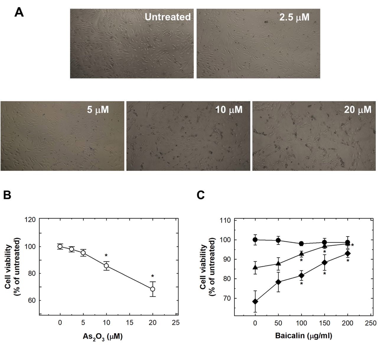
As2O3-induced cytotoxicity and apoptosis were suppressed by baicalin in HUVECs. Originally, we found that As2O3 induced cell cytotoxicity, with dose-dependent reduction of cell viability (Figure 1A). Treatment of HUVECs with 10 μM and 20 μM reduced viability by 14.3% and 31.7%, respectively (Figure 1B). Pre-treatment with baicalin restored cell viability to almost normal levels (Figure 1C).

Figure 1.
  • Download figure
  • Open in new tab
  • Download powerpoint
Figure 1.

The effects of As2O3 and baicalin on viability of human umbilical vein endothelial cells (HUVECs). A: The effects of 24-h treatment of HUVECs with 0-20 μM As2O3. HUVECs were photographed under a microscope at ×40, representative images are shown. B: The quantification of the effect of 0-20 μM As2O3 treatment for 24 h in HUVECs. C: The effect on HUVEC viability after pretreatment with baicalin for 1 h, and co-treatment of baicalin with 20 μM (◆), 10 μM (▲) or 0 μM (●) As2O3 for 24 h. Data are the mean±SD for three independent experiments. *Significantly different at p<0.05 compared with untreated control cells.

Treatment with As2O3 at 5 μM or more for 24 h was capable of inducing a significant sub G1 population of cells, i.e. significant apoptosis (Figure 2A). Pre-treatment with baicalin abrogated the activation of apoptosis dose-dependently (Figure 2B).

Figure 2.
  • Download figure
  • Open in new tab
  • Download powerpoint
Figure 2.

The effects of As2O3 and baicalin onapoptosis of human umbilical vein endothelial cells (HUVECs). A: The effects of 24-h treatment with 0-20 μM As2O3 on HUVECs. Apoptotic cells were detected by flow cytometry by the appearance of a sub-G1 cell population. B: The effect on apoptosis of HUVECs after pretreatment with baicalin for 1 h, and co-treatment of baicalin with 20 μM (◆), 10 μM (▲) or 0 μM (●) As2O3 for 24 h. Data are the mean±SD for three independent experiments. *Significantly different at p<0.05 compared with untreated cells.

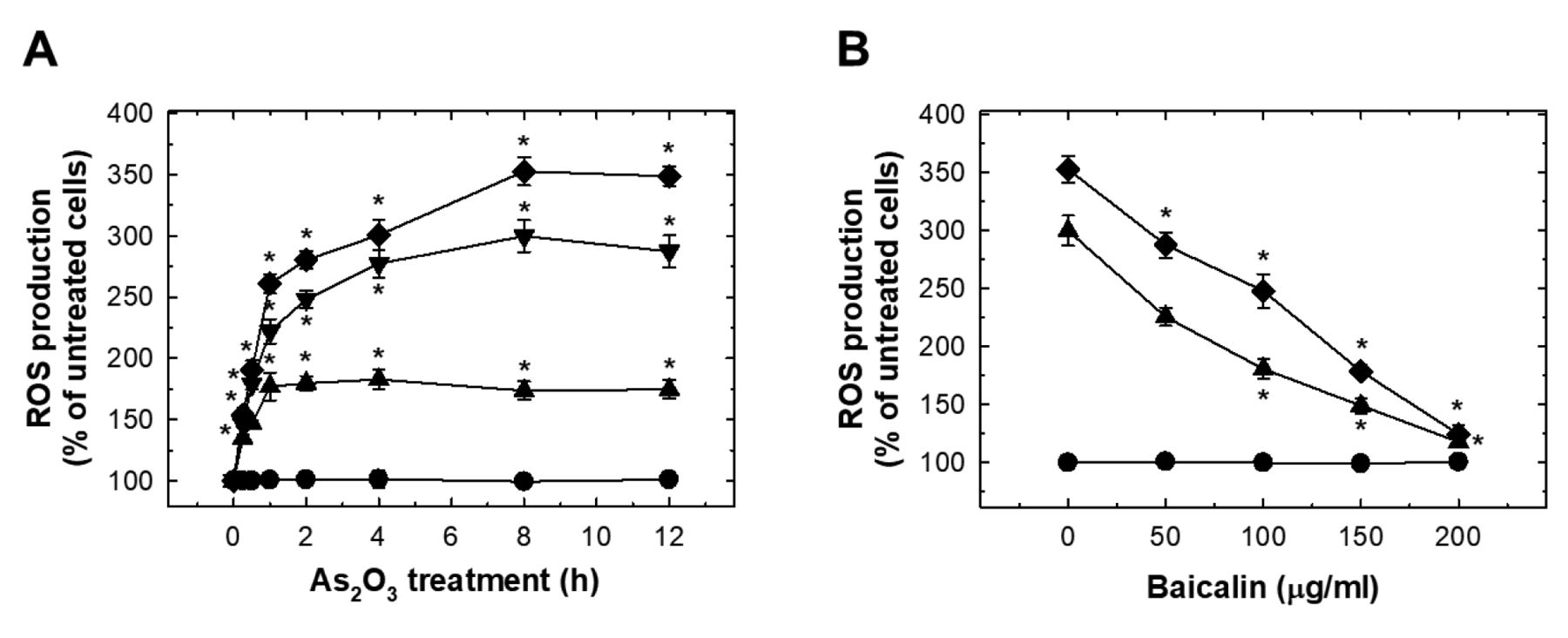
As2O3 induced oxidative stress in HUVECs which was suppressed by baicalin. As2O3 treatment dose-dependently induced significant ROS production at 15 min (Figure 3A). The peak ROS level was reached at 8 h, and plateau at 12 h (Figure 3A). The effect of As2O3 treatment at 10 and 20 μM on ROS was suppressed by pre-treatment with baicalin (Figure 3B). Exposure to As2O3 increased NADPH oxidase activity dose-dependently (Figure 4A). Pre-treatment with baicalin abrogated As2O3-induced activation of NADPH oxidase (Figure 4B).

Figure 3.
  • Download figure
  • Open in new tab
  • Download powerpoint
Figure 3.

The effects of As2O3 and baicalin on reactive oxygen species (ROS) production in human umbilical vein endothelial cells (HUVECs). A: HUVECs were treated with 0, 5, 10, and 20 μM As2O3 for the indicated time, and ROS were detected with 2’,7’-dichlorodihydrofluorescein diacetate. B: The effect on ROS production in HUVECs after pretreatment with baicalin for 1 h, and co-treatment of baicalin with 20 μM (◆), 10 μM (▼), 5 μM (▲) or 0 μM (●) As2O3 for 12 h. Data are the mean±SD for three independent experiments. *Significantly different at p<0.05 compared with untreated cells.

Figure 4.
  • Download figure
  • Open in new tab
  • Download powerpoint
Figure 4.

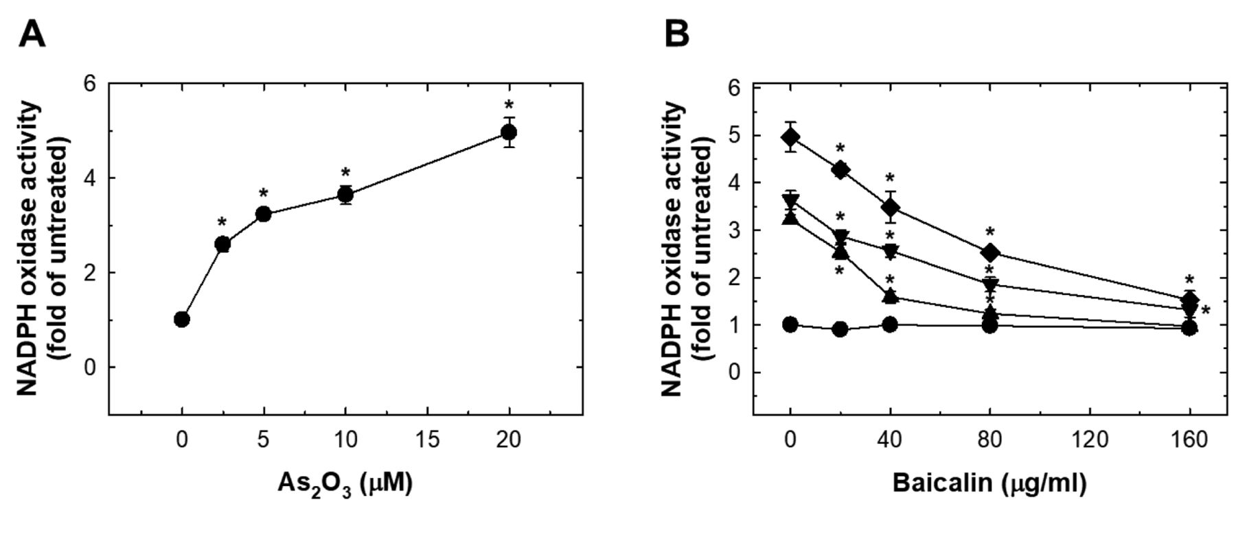
The effects of As2O3 and baicalin on NADPH oxidase activity in HUVECs. A: HUVECs were treated with 0-20 μM As2O3 for 24 h and NADPH oxidase activity was evaluated as fold that of untreated cells. B: The effect on NADPH oxidase activity in HUVECs after pretreatment of baicalin for 1 h, and co-treatment of baicalin with 20 μM (◆), 10 μM (▼), 5 μM (▲) or 0 μM (●) As2O3 for 24 h. Data are the mean±SD for three independent experiments. *Significantly different at p<0.05 compared with untreated cells.

As2O3 induced oxidative FPG- and endonuclease III-digestible DNA adducts in HUVECs which was suppressed by baicalin. As2O3 treatment induced a significant level of FPG- and endonuclease III-digestible adducts (Figures 5A and 6A). The levels of these oxidative adducts were effectively reduced by pre-treatment with baicalin but did not reach basal levels (Figures 5B and 6B).

Figure 5.
  • Download figure
  • Open in new tab
  • Download powerpoint
Figure 5.

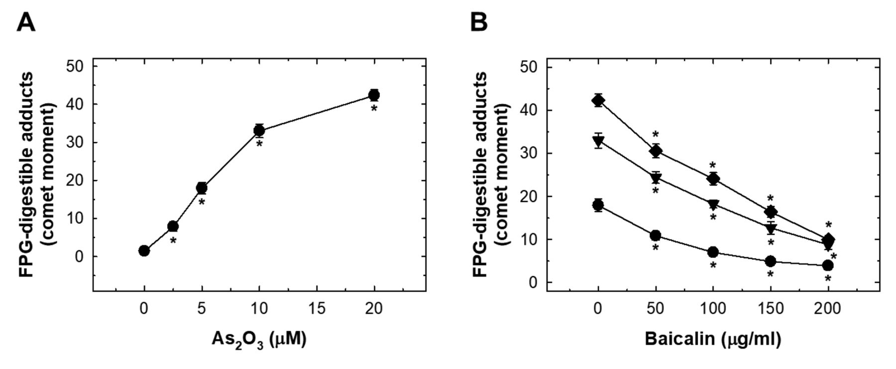
The effects of As2O3 and baicalin on formamidopyrimidine-DNA glycosylase (FPG)-digestible adducts in human umbilical vein endothelial cells (HUVECs). A: HUVECs were treated with 0-20 μM As2O3 for 24 h and FPG-digestible adducts produced were detected with comet assay. The data are presented as comet moments. B: The effect on FPG-digestible adducts in HUVECs after pretreatment with baicalin for 1 h, and co-treatment of baicalin with 20 μM (◆), 10 μM (▼) or 0 μM (●) As2O3 for 24 h. Data are the mean±SD for three independent experiments. *Significantly different at p<0.05 compared with untreated cells.

Figure 6.
  • Download figure
  • Open in new tab
  • Download powerpoint
Figure 6.

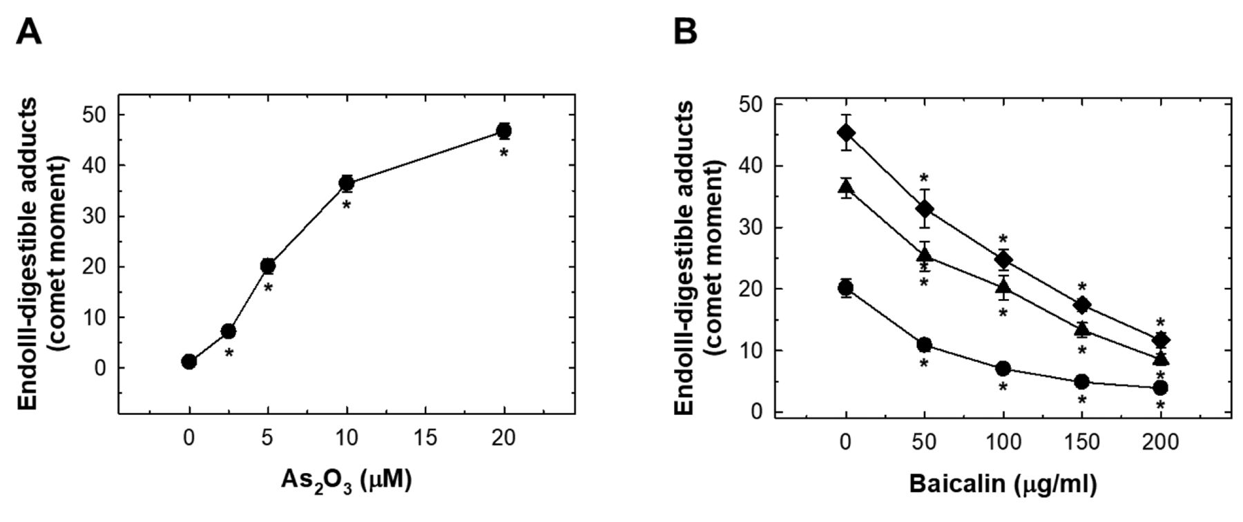
The effects of As2O3 and baicalin on endonuclease III-digestible adducts in human umbilical vein endothelial cells (HUVECs). A: HUVECs were treated with 0-20 μM As2O3 for 24 h and FPG-digestible adducts produced were detected with comet assay. The data are presented as comet moments. B: The effect on endonuclease III-digestible adducts in HUVECs after pretreatment with baicalin for 1 h, and co-treatment of baicalin with 20 μM (◆), 10 μM (▼) or 0 μM (●) As2O3 for 24 h. Data are the mean±SD for three independent experiments. *Significantly different at p<0.05 compared with untreated cells.

Discussion

Vascular endothelium not only serves as a physical monolayer between blood and tissue but plays an important role in detection and reaction to blood-borne stresses. Impaired endothelial function maybe an irreversible process in the initiation event of various cardiovascular diseases and it is worth determining the practical cardio-protective effects of Chinese traditional medicine, such as baicalin. Arsenic exposure induced HUVEC dysfunction through proapoptotic and oxidative stress, which might be responsible for its adverse effects on blood vessels. The protective role of Chinese traditional medicine may be of great help in prevention of cardiovascular disorders. In this study, the effects of baicalin on As2O3-induced endothelial cell apoptosis, oxidative stress and oxidative DNA adduct production were revealed for the first time.

Increased endothelial cell apoptosis may contribute to the loss of endothelial integrity and lead to vascular disorders (34, 35). Our study confirmed a significant increase in HUVEC apoptosis when cells were treated with As2O3, while pre-treatment with baicalin reduced As2O3-induced HUVEC apoptosis (Figure 2B). The degree of As2O3-induced apoptosis was not as strong as in Yu et al.’s finding, reporting that 5 μM As2O3 was capable suppressed cell viability by about 40% of (36). Our results are consistent with Ma et al.’s finding that 5 μM As2O3 induced apoptosis in less than 20% of HUVECs (37). The As2O3 induced generation of ROS was rapidly detected, and sustained for as long as 12 h (Figure 3A). Pre-treatment with baicalin did not alter the level of ROS itself but did suppress ROS production induced by As2O3. A number of risk factors for cardiovascular disorders were reported to be associated with increased vascular ROS (38, 39). Our studies also showed that As2O3 activated NADPH oxidase, which was reversed by pre-treatment with baicalin. Activation of NADPH oxidase may be the major mechanism related to As2O3-mediated intracellular generation of ROS.

One of the highlights of this was the detection of FPG-digestible adducts and endonuclease III-digestible adducts. FPG specifically cleaves oxidized purines, including 8-oxoguanine, 5-hydroxycytosine, 5-hydroxyuracil, 2,6-diamino-4-hydroxy-5-N-methylformamidopyrimidine, and 4,6-diamino-5-formamidopyrimidine (40), while endonuclease III specifically cleaves oxidized pyrimidines, including thymine glycol, 5,6-dihydrothymine, 5-hydroxydihydrothymine, 5-hydroxycytosine, 5-hydroxyuracil, and uracil glycol (41). We also demonstrated that baicalin pre-treatment reduced the formation of As2O3-induced oxidative adducts, although the detailed mechanisms of this need further investigation.

In conclusion, at a non-toxic dose, baicalin is of reducing the As2O3-induced apoptosis, oxidative stress and DNA adducts. The underlying mechanisms may provide novel insights into potential clinical cardioprotective effects.

Acknowledgements

Yun-Chi Wang and Yu-Chen Hsiau helped in the experiments and analysis. This study was supported mainly by grants from Taichung Veterans General Hospital (TCVGH-VHCY1098604).

Footnotes

  • Authors’ Contributions

    C.L.T. and C.W.T. conceived and designed the experiments. W.S.C. and C.W.T. performed the experiments. J.C.L. and T.C.H. analyzed the data. C.L.T. and J.C.L. contributed reagents/materials/analysis tools. C.W.T. and D.T.B. wrote the article.

  • This article is freely accessible online.

  • Conflicts of Interest

    The Authors declare no conflicts of interest in regard to this study.

  • Received November 9, 2020.
  • Revision received November 23, 2020.
  • Accepted November 24, 2020.
  • Copyright © 2021 The Author(s). Published by the International Institute of Anticancer Research.

References

  1. ↵
    1. Jadhav SV,
    2. Bringas E,
    3. Yadav GD,
    4. Rathod VK,
    5. Ortiz I and
    6. Marathe KV
    : Arsenic and fluoride contaminated groundwaters: A review of current technologies for contaminants removal. J Environ Manage 162: 306-325, 2015. PMID: 26265600. DOI: 10.1016/j.jenvman.2015.07.020
    OpenUrlCrossRef
    1. Guzman A,
    2. Nava JL,
    3. Coreno O,
    4. Rodriguez I and
    5. Gutierrez S
    : Arsenic and fluoride removal from groundwater by electro-coagulation using a continuous filter-press reactor. Chemosphere 144: 2113-2120, 2016. PMID: 26583293. DOI: 10.1016/j.chemosphere.2015.10.108
    OpenUrlCrossRef
    1. Pi K,
    2. Wang Y,
    3. Xie X,
    4. Su C,
    5. Ma T,
    6. Li J and
    7. Liu Y
    : Hydrogeochemistry of co-occurring geogenic arsenic, fluoride and iodine in groundwater at Datong Basin, northern China. J Hazard Mater 300: 652-661, 2015. PMID: 26282220. DOI: 10.1016/j.jhazmat.2015.07.080
    OpenUrlCrossRef
    1. Rasool A,
    2. Xiao T,
    3. Baig ZT,
    4. Masood S,
    5. Mostofa KM and
    6. Iqbal M
    : Co-occurrence of arsenic and fluoride in the groundwater of Punjab, Pakistan: Source discrimination and health risk assessment. Environ Sci Pollut Res Int 22(24): 19729-19746, 2015. PMID: 26278901. DOI: 10.1007/s11356-015-5159-2
    OpenUrlCrossRef
  2. ↵
    1. Gonzalez-Horta C,
    2. Ballinas-Casarrubias L,
    3. Sanchez-Ramirez B,
    4. Ishida MC,
    5. Barrera-Hernandez A,
    6. Gutierrez-Torres D,
    7. Zacarias OL,
    8. Saunders RJ,
    9. Drobna Z,
    10. Mendez MA,
    11. Garcia-Vargas G,
    12. Loomis D,
    13. Styblo M and
    14. Del Razo LM
    : A concurrent exposure to arsenic and fluoride from drinking water in Chihuahua, Mexico. Int J Environ Res Public Health 12(5): 4587-4601, 2015. PMID: 25918912. DOI: 10.3390/ijerph120504587
    OpenUrlCrossRef
  3. ↵
    1. Hsieh YC,
    2. Lien LM,
    3. Chung WT,
    4. Hsieh FI,
    5. Hsieh PF,
    6. Wu MM,
    7. Tseng HP,
    8. Chiou HY and
    9. Chen CJ
    : Significantly increased risk of carotid atherosclerosis with arsenic exposure and polymorphisms in arsenic metabolism genes. Environ Res 111(6): 804-810, 2011. PMID: 21605854. DOI: 10.1016/j.envres.2011.05.003
    OpenUrlCrossRefPubMed
  4. ↵
    1. Brauner EV,
    2. Nordsborg RB,
    3. Andersen ZJ,
    4. Tjonneland A,
    5. Loft S and
    6. Raaschou-Nielsen O
    : Long-term exposure to low-level arsenic in drinking water and diabetes incidence: A prospective study of the diet, cancer and health cohort. Environ Health Perspect 122(10): 1059-1065, 2014. PMID: 24927198. DOI: 10.1289/ehp.1408198
    OpenUrlCrossRefPubMed
  5. ↵
    1. Abhyankar LN,
    2. Jones MR,
    3. Guallar E and
    4. Navas-Acien A
    : Arsenic exposure and hypertension: A systematic review. Environ Health Perspect 120(4): 494-500, 2012. PMID: 22138666. DOI: 10.1289/ehp.1103988
    OpenUrlCrossRefPubMed
  6. ↵
    1. Chen Y,
    2. Wu F,
    3. Graziano JH,
    4. Parvez F,
    5. Liu M,
    6. Paul RR,
    7. Shaheen I,
    8. Sarwar G,
    9. Ahmed A,
    10. Islam T,
    11. Slavkovich V,
    12. Rundek T,
    13. Demmer RT,
    14. Desvarieux M and
    15. Ahsan H
    : Arsenic exposure from drinking water, arsenic methylation capacity, and carotid intima-media thickness in Bangladesh. Am J Epidemiol 178(3): 372-381, 2013. PMID: 23788675. DOI: 10.1093/aje/kwt001
    OpenUrlCrossRefPubMed
  7. ↵
    1. Moon KA,
    2. Guallar E,
    3. Umans JG,
    4. Devereux RB,
    5. Best LG,
    6. Francesconi KA,
    7. Goessler W,
    8. Pollak J,
    9. Silbergeld EK,
    10. Howard BV and
    11. Navas-Acien A
    : Association between exposure to low to moderate arsenic levels and incident cardiovascular disease. A prospective cohort study. Ann Intern Med 159(10): 649-659, 2013. PMID: 24061511. DOI: 10.7326/0003-4819-159-10-201311190-00719
    OpenUrlCrossRefPubMed
  8. ↵
    1. Tseng CH
    : Cardiovascular disease in arsenic-exposed subjects living in the arseniasis-hyperendemic areas in Taiwan. Atherosclerosis 199(1): 12-18, 2008. PMID: 18367191. DOI: 10.1016/j.atherosclerosis.2008.02.013
    OpenUrlCrossRefPubMed
  9. ↵
    1. Zimta AA,
    2. Schitcu V,
    3. Gurzau E,
    4. Stavaru C,
    5. Manda G,
    6. Szedlacsek S and
    7. Berindan-Neagoe I
    : Biological and molecular modifications induced by cadmium and arsenic during breast and prostate cancer development. Environ Res 178: 108700, 2019. PMID: 31520827. DOI: 10.1016/j.envres.2019.108700
    OpenUrlCrossRefPubMed
  10. ↵
    1. Wei S,
    2. Zhang H and
    3. Tao S
    : A review of arsenic exposure and lung cancer. Toxicol Res 8(3): 319-327, 2019. PMID: 31160966. DOI: 10.1039/c8tx00298c
    OpenUrlCrossRefPubMed
  11. ↵
    1. Li H,
    2. Horke S and
    3. Forstermann U
    : Vascular oxidative stress, nitric oxide and atherosclerosis. Atherosclerosis 237(1): 208-219, 2014. PMID: 25244505. DOI: 10.1016/j.atherosclerosis.2014.09.001
    OpenUrlCrossRefPubMed
  12. ↵
    1. Dwivedi N,
    2. Flora G,
    3. Kushwaha P and
    4. Flora SJ
    : Alpha-lipoic acid protects oxidative stress, changes in cholinergic system and tissue histopathology during co-exposure to arsenic-dichlorvos in rats. Environ Toxicol Pharmacol 37(1): 7-23, 2014. PMID: 24291368. DOI: 10.1016/j.etap.2013.10.010
    OpenUrlCrossRefPubMed
  13. ↵
    1. Kesavan M,
    2. Sarath TS,
    3. Kannan K,
    4. Suresh S,
    5. Gupta P,
    6. Vijayakaran K,
    7. Sankar P,
    8. Kurade NP,
    9. Mishra SK and
    10. Sarkar SN
    : Atorvastatin restores arsenic-induced vascular dysfunction in rats: Modulation of nitric oxide signaling and inflammatory mediators. Toxicol Appl Pharmacol 280(1): 107-116, 2014. PMID: 25058445. DOI: 10.1016/j.taap.2014.07.008
    OpenUrlCrossRefPubMed
  14. ↵
    1. Huang WH,
    2. Lee AR and
    3. Yang CH
    : Antioxidative and anti-inflammatory activities of polyhydroxyflavonoids of Scutellaria baicalensis GEORGI. Biosci Biotechnol Biochem 70(10): 2371-2380, 2006. PMID: 17031041. DOI: 10.1271/bbb.50698
    OpenUrlCrossRefPubMed
    1. Jeong K,
    2. Shin YC,
    3. Park S,
    4. Park JS,
    5. Kim N,
    6. Um JY,
    7. Go H,
    8. Sun S,
    9. Lee S,
    10. Park W,
    11. Choi Y,
    12. Song Y,
    13. Kim G,
    14. Jeon C,
    15. Park J,
    16. Lee K,
    17. Bang O and
    18. Ko SG
    : Ethanol extract of Scutellaria baicalensis Georgi prevents oxidative damage and neuroinflammation and memorial impairments in artificial senescence mice. J Biomed Sci 18(1): 14, 2011. PMID: 21299906. DOI: 10.1186/1423-0127-18-14
    OpenUrlCrossRefPubMed
    1. Xu C and
    2. Ji GE
    : Bioconversion of flavones during fermentation in milk containing Scutellaria baicalensis extract by Lactobacillus brevis. J Microbiol Biotechnol 23(10): 1422-1427, 2013. PMID: 23851266. DOI: 10.4014/jmb.1305.05001
    OpenUrlCrossRef
    1. Yoon JJ,
    2. Jeong JW,
    3. Choi EO,
    4. Kim MJ,
    5. Hwang-Bo H,
    6. Kim HJ,
    7. Hong SH,
    8. Park C,
    9. Lee DH and
    10. Choi YH
    : Protective effects of Scutellaria baicalensis Georgi against hydrogen peroxide-induced DNA damage and apoptosis in HaCaT human skin keratinocytes. EXCLI J 16: 426-438, 2017. PMID: 28694748. DOI: 10.17179/excli2016-817
    OpenUrlCrossRef
  15. ↵
    1. Zhou Y,
    2. Yang ZY and
    3. Tang RC
    : Bioactive and UV protective silk materials containing baicalin – The multifunctional plant extract from Scutellaria baicalensis Georgi. Mater Sci Eng C Mater Biol Appl 67: 336-344, 2016. PMID: 27287129. DOI: 10.1016/j.msec.2016.05.063
    OpenUrlCrossRef
  16. ↵
    1. Lee YC,
    2. Chuah AM,
    3. Yamaguchi T,
    4. Takamura H and
    5. Matoba T
    : Antioxidant activity of traditional Chinese medicinal herbal materials. Food Sci Technol Res 14(2): 205-210, 2008. DOI: 10.3136/fstr.14.205
    OpenUrlCrossRef
    1. Li BQ,
    2. Fu T,
    3. Gong WH,
    4. Dunlop N,
    5. Kung H,
    6. Yan Y,
    7. Kang J and
    8. Wang JM
    : The flavonoid baicalin exhibits anti-inflammatory activity by binding to chemokines. Immunopharmacology 49(3): 295-306, 2000. PMID: 10996027. DOI: 10.1016/s0162-3109(00)00244-7
    OpenUrlCrossRefPubMed
    1. Li BQ,
    2. Fu T,
    3. Yan YD,
    4. Baylor NW,
    5. Ruscetti FW and
    6. Kung HF
    : Inhibition of HIV infection by baicalin–a flavonoid compound purified from Chinese herbal medicine. Cell Mol Biol Res 39(2): 119-124, 1993. PMID: 7693133.
    OpenUrlPubMed
    1. Pan TL,
    2. Wang PW,
    3. Leu YL,
    4. Wu TH and
    5. Wu TS
    : Inhibitory effects of Scutellaria baicalensis extract on hepatic stellate cells through inducing G2/M cell-cycle arrest and activating ERK-dependent apoptosis via BAX and caspase pathway. J Ethnopharmacol 139(3): 829-837, 2012. PMID: 22210104. DOI: 10.1016/j.jep.2011.12.028
    OpenUrlCrossRefPubMed
    1. Park JR,
    2. Lee MC,
    3. Moon SC,
    4. Kim J,
    5. Ha KT,
    6. Park EJ,
    7. Hong C,
    8. Seo BD and
    9. Kim BJ
    : Scutellaria baicalensis Georgi induces caspase-dependent apoptosis via mitogen-activated protein kinase activation and the generation of reactive oxygen species signaling pathways in MCF-7 breast cancer cells. Mol Med Rep 16(2): 2302-2308, 2017. PMID: 28627691. DOI: 10.3892/mmr.2017.6798
    OpenUrlCrossRef
  17. ↵
    1. Wei L,
    2. Lin J,
    3. Xu W,
    4. Cai Q,
    5. Shen A,
    6. Hong Z and
    7. Peng J
    : Scutellaria barbata D. Don inhibits tumor angiogenesis via suppression of Hedgehog pathway in a mouse model of colorectal cancer. Int J Mol Sci 13(8): 9419-9430, 2012. PMID: 22949805. DOI: 10.3390/ijms13089419
    OpenUrlCrossRefPubMed
  18. ↵
    1. Hsu SW,
    2. Hsu PC,
    3. Chang WS,
    4. Yu CC,
    5. Wang YC,
    6. Yang JS,
    7. Tsai FJ,
    8. Chen KY,
    9. Tsai CW and
    10. Bau DT
    : Protective effects of valproic acid on 6-hydroxydopamine-induced neuroinjury. Environ Toxicol 35(8): 840-848, 2020. PMID: 32167238. DOI: 10.1002/tox.22920
    OpenUrlCrossRefPubMed
  19. ↵
    1. Chang WS,
    2. Lin EY,
    3. Hsu SW,
    4. Hu PS,
    5. Chuang CL,
    6. Liao CH,
    7. Fu CK,
    8. Su CH,
    9. Gong CL,
    10. Hsiao CL,
    11. Bau DT and
    12. Tsai CW
    : Baicalin scavenged reactive oxygen species and protected human keratinocytes against UVB-induced cytotoxicity. In Vivo 30(5): 605-610, 2016. PMID: 27566079.
    OpenUrlAbstract/FREE Full Text
  20. ↵
    1. Lin CC,
    2. Chen KB,
    3. Tsai CH,
    4. Tsai FJ,
    5. Huang CY,
    6. Tang CH,
    7. Yang JS,
    8. Hsu YM,
    9. Peng SF and
    10. Chung JG
    : Casticin inhibits human prostate cancer DU 145 cell migration and invasion via RAS/AKT/ NF-kappaB signaling pathways. J Food Biochem 43(7): e12902, 2019. PMID: 31353708. DOI: 10.1111/jfbc.12902
    OpenUrlCrossRefPubMed
  21. ↵
    1. Wu KM,
    2. Hsu YM,
    3. Ying MC,
    4. Tsai FJ,
    5. Tsai CH,
    6. Chung JG,
    7. Yang JS,
    8. Tang CH,
    9. Cheng LY,
    10. Su PH,
    11. Viswanadha VP,
    12. Kuo WW and
    13. Huang CY
    : High-density lipoprotein ameliorates palmitic acid-induced lipotoxicity and oxidative dysfunction in H9c2 cardiomyoblast cells via ROS suppression. Nutr Metab 16: 36, 2019. PMID: 31149020. DOI: 10.1186/s12986-019-0356-5
    OpenUrlCrossRefPubMed
  22. ↵
    1. Wang SC,
    2. Chen SF,
    3. Lee YM,
    4. Chuang CL,
    5. Bau DT and
    6. Lin SS
    : Baicalin scavenges reactive oxygen species and protects human keratinocytes against UVC-induced cytotoxicity. In Vivo 27(6): 707-714, 2013. PMID: 24292572.
    OpenUrlAbstract/FREE Full Text
  23. ↵
    1. Tsai CW,
    2. Yang MD,
    3. Hsia TC,
    4. Chang WS,
    5. Hsu CM,
    6. Hsieh YH,
    7. Chung JG and
    8. Bau DT
    : Dithiothreitol enhanced arsenic-trioxide-induced cell apoptosis in cultured oral cancer cells via mitochondrial dysfunction and endoplasmic reticulum stress. Environ Toxicol 32(1): 17-27, 2017. PMID: 26494474. DOI: 10.1002/tox.22208
    OpenUrlCrossRef
  24. ↵
    1. Liu S,
    2. Shen H,
    3. Xu M,
    4. Liu O,
    5. Zhao L,
    6. Liu S,
    7. Guo Z and
    8. Du J
    : FRP inhibits ox-LDL-induced endothelial cell apoptosis through an AKT-NF-{kappa}B-BCL-2 pathway and inhibits endothelial cell apoptosis in an apoE-knockout mouse model. Am J Physiol Endocrinol Metab 299(3): E351-363, 2010. PMID: 20530739. DOI: 10.1152/ajpendo.00005.2010
    OpenUrlCrossRefPubMed
  25. ↵
    1. Liu M,
    2. Xiang G,
    3. Lu J,
    4. Xiang L,
    5. Dong J and
    6. Mei W
    : TRAIL protects against endothelium injury in diabetes via AKT-eNOS signaling. Atherosclerosis 237(2): 718-724, 2014. PMID: 25463111. DOI: 10.1016/j.atherosclerosis.2014.10.013
    OpenUrlCrossRefPubMed
  26. ↵
    1. Yu CX,
    2. Zhang YY,
    3. Wu XY,
    4. Tang HX,
    5. Liang XQ,
    6. Xue ZM,
    7. Xue YD,
    8. Li J,
    9. Zhu H,
    10. Huo R and
    11. Ban T
    : Transient receptor potential melastatin 4 contributes to early-stage endothelial injury induced by arsenic trioxide. Toxicol Lett 312: 98-108, 2019. PMID: 31054354. DOI: 10.1016/j.toxlet.2019.04.035
    OpenUrlCrossRefPubMed
  27. ↵
    1. Ma Y,
    2. Ma Z,
    3. Yin S,
    4. Yan X and
    5. Wang J
    : Arsenic and fluoride induce apoptosis, inflammation and oxidative stress in cultured human umbilical vein endothelial cells. Chemosphere 167: 454-461, 2017. PMID: 27750169. DOI: 10.1016/j.chemosphere.2016.10.025
    OpenUrlCrossRefPubMed
  28. ↵
    1. States JC,
    2. Srivastava S,
    3. Chen Y and
    4. Barchowsky A
    : Arsenic and cardiovascular disease. Toxicol Sci 107(2): 312-323, 2009. PMID: 19015167. DOI: 10.1093/toxsci/kfn236
    OpenUrlCrossRefPubMed
  29. ↵
    1. Tousoulis D,
    2. Briasoulis A,
    3. Papageorgiou N,
    4. Tsioufis C,
    5. Tsiamis E,
    6. Toutouzas K and
    7. Stefanadis C
    : Oxidative stress and endothelial function: therapeutic interventions. Recent Pat Cardiovasc Drug Discov 6(2): 103-114, 2011. PMID: 21513492. DOI: 10.2174/157489011795933819
    OpenUrlCrossRefPubMed
  30. ↵
    1. Nickoloff JA,
    2. Spirio LN and
    3. Reynolds RJ
    : A comparison of calcium phosphate coprecipitation and electroporation. Implications for studies on the genetic effects of DNA damage. Mol Biotechnol 10(2): 93-101, 1998. PMID: 9819809. DOI: 10.1007/BF02760857
    OpenUrlCrossRefPubMed
  31. ↵
    1. Sarker AH,
    2. Ikeda S,
    3. Nakano H,
    4. Terato H,
    5. Ide H,
    6. Imai K,
    7. Akiyama K,
    8. Tsutsui K,
    9. Bo Z,
    10. Kubo K,
    11. Yamamoto K,
    12. Yasui A,
    13. Yoshida MC and
    14. Seki S
    : Cloning and characterization of a mouse homologue (mNthl1) of Escherichia coli endonuclease III. J Mol Biol 282(4): 761-774, 1998. PMID: 9743625. DOI: 10.1006/jmbi.1998.2042
    OpenUrlCrossRefPubMed
PreviousNext
Back to top

In this issue

In Vivo
Vol. 35, Issue 1
January-February 2021
  • Table of Contents
  • Table of Contents (PDF)
  • Index by author
  • Back Matter (PDF)
  • Ed Board (PDF)
  • Front Matter (PDF)
Print
Download PDF
Article Alerts
Sign In to Email Alerts with your Email Address
Email Article

Thank you for your interest in spreading the word on In Vivo.

NOTE: We only request your email address so that the person you are recommending the page to knows that you wanted them to see it, and that it is not junk mail. We do not capture any email address.

Enter multiple addresses on separate lines or separate them with commas.
Protective Effects of Baicalin on Arsenic Trioxide-induced Oxidative Damage and Apoptosis in Human Umbilical Vein Endothelial Cells
(Your Name) has sent you a message from In Vivo
(Your Name) thought you would like to see the In Vivo web site.
CAPTCHA
This question is for testing whether or not you are a human visitor and to prevent automated spam submissions.
5 + 2 =
Solve this simple math problem and enter the result. E.g. for 1+3, enter 4.
Citation Tools
Protective Effects of Baicalin on Arsenic Trioxide-induced Oxidative Damage and Apoptosis in Human Umbilical Vein Endothelial Cells
CHUNG-LIN TSAI, CHIA-WEN TSAI, WEN-SHIN CHANG, JIUNN-CHERNG LIN, TE-CHUN HSIA, DA-TIAN BAU
In Vivo Jan 2021, 35 (1) 155-162; DOI: 10.21873/invivo.12243

Citation Manager Formats

  • BibTeX
  • Bookends
  • EasyBib
  • EndNote (tagged)
  • EndNote 8 (xml)
  • Medlars
  • Mendeley
  • Papers
  • RefWorks Tagged
  • Ref Manager
  • RIS
  • Zotero
Reprints and Permissions
Share
Protective Effects of Baicalin on Arsenic Trioxide-induced Oxidative Damage and Apoptosis in Human Umbilical Vein Endothelial Cells
CHUNG-LIN TSAI, CHIA-WEN TSAI, WEN-SHIN CHANG, JIUNN-CHERNG LIN, TE-CHUN HSIA, DA-TIAN BAU
In Vivo Jan 2021, 35 (1) 155-162; DOI: 10.21873/invivo.12243
Twitter logo Facebook logo Mendeley logo
  • Tweet Widget
  • Facebook Like
  • Google Plus One

Jump to section

  • Article
    • Abstract
    • Materials and Methods
    • Results
    • Discussion
    • Acknowledgements
    • Footnotes
    • References
  • Figures & Data
  • Info & Metrics
  • PDF

Related Articles

Cited By...

  • Protective Effects of Crocetin on Arsenic Trioxide-induced Oxidative Stress in Human Umbilical Vein Endothelial Cells
  • Google Scholar

More in this TOC Section

  • Antagonistic Effect of Erythropoietin Receptor Signaling in Macrophages and Type II Alveolar Epithelial Cells on Pulmonary Fibrosis in Mice
  • Association of TIMP2 Gene Variants With Development of Diabetic Retinopathy
  • PLLA and PDLLA Fillers: Linking Crystallinity and Mechanical Stability to Aggregation, Redispersion, and Collagen Formation
Show more Experimental Studies

Keywords

  • apoptosis
  • arsenic trioxide
  • baicalin
  • human umbilical vein endothelial cells
  • oxidative damage
In Vivo

© 2026 In Vivo

Powered by HighWire