


Abstract
Background/Aim: Radiation (RT) induced ERK/NF-κB in hepatocellular carcinoma (HCC) has been reported in our previous works; it weakens the toxicity of RT or triggers a radioresistance effect. Thus, combining RT with a suitable NF-κB inhibitor may sensitize HCC to RT. Magnolol, a bioactive compound, was known to have anti-inflammatory and anti-tumor functions. Here, we aimed to investigate whether magnolol may enhance anti-HCC efficacy of RT in vivo. Materials and Methods: We established a Hep3B bearing mouse to evaluate the efficacy of the combination treatment of magnolol and RT. Results: Most significantly, tumor volume and tumor weight inhibition was found in the combination group. Tumor immunohistochemistry staining also illustrated the suppression of RT-induced ERK/NF-κB-related proteins expression by magnolol. In addition, intrinsic apoptosis-related proteins, such as caspase-3 and -9, were markedly increased in the combination group. Conclusion: Magnolol may effectively enhance anti-HCC ability of RT by downregulating the expression of ERK/NF-κB-related proteins and increasing the expression of apoptosis-related proteins.
Hepatocellular carcinoma (HCC), the most common type of liver cancer in adults, ranks as the fourth leading cause of cancer-related mortality worldwide (1). Patients with advanced HCC are unsuitable candidates for curative treatment options including surgery, liver transplantation, and percutaneous ablation (2). The low tolerance of whole liver to radiation has restricted application of radiotherapy (RT) for HCC in the past. Recent technology advancements in RT such as intensity-modulated RT (IMRT), image-guided RT (IGRT), hypofractionated radiotherapy (HFRT), and stereotactic body RT (SBRT) can precisely deliver high-dose radiation to HCC while reducing noncancerous liver tissue irradiation (3-5). RT has been shown to provide excellent local control and palliative efficacy in unresectable or metastatic HCC (6).

Tumor growth is suppressed by the combination of magnolol with RT in a Hep3B tumor bearing model. (A) The experimental flow chart of the in vivo study. (B) Tumor growth trend was recorded by calliper every three days. (C) The extracted tumors on day 18 are photographed and are displayed. (D) Whole-body CT image from each treatment group on day 18. (E) The weight of the extracted tumors is measured and quantified. (a1p<0.05 and a2 p<0.01 vs. CTRL; b1 p<0.05 and b2 p<0.01 vs. single treatment)
In addition to RT technology, radiosensitizers that sensitize HCC to radiation have been developing. Natural products such as ellagic acid, curcumin, and mesima have been indicated as potential radiosensitizers to boost anti-HCC efficacy of radiation. Induction of mitochondrial apoptosis, arrest of cell cycle G2/M phase, and inhibition of NF-κB activation have been associated with the radiosensitizing effect of natural products in HCC (7-9). Magnolol, one of major components of Magnolia officinalis, has been shown to trigger apoptosis through death receptor (extrinsic)/mitochondrial (intrinsic) pathways and to down-regulate extracellular signal-regulated kinase (ERK)/NF-κB signaling in HCC in vitro and in vivo (10, 11). Magnolol was also recognized to have an anti-inflammation and anti-tumor potential (12). However, whether magnolol functions as radiosensitizer and enhances anti-HCC efficacy of radiation has not yet been elucidated. Therefore, the main purpose of the present study was to investigate the therapeutic efficacy and possible underlying mechanism of magnolol combined with radiation in HCC in vivo.

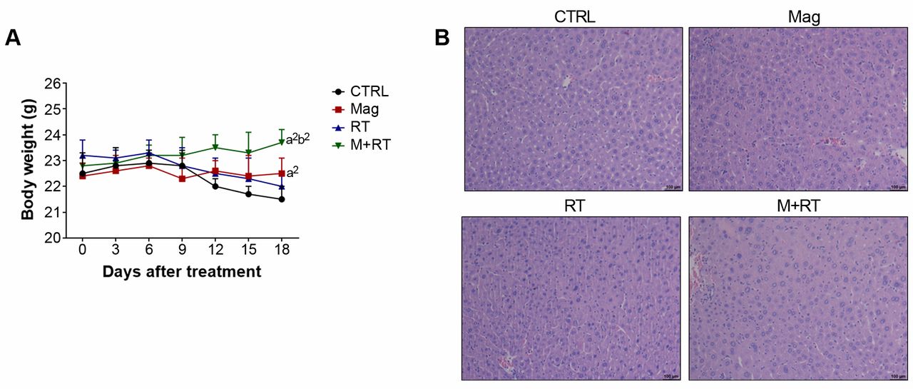
No general toxicity is observed following treatment with the combination of magnolol with RT in Hep3B tumor bearing model. (A) Body weight of each mouse was recorded every three days. (B) Representative liver H&E staining results from each group. (a2 p<0.01 vs. CTRL; b2 p<0.01 vs. single treatment)
Materials and Methods
Cell lines. Hep3B cells were purchased from Bioresource Collection and Research Center (BCRC, Hsinchu, Taiwan). Cells were cultured in DMEM (Dulbecco's Modified Eagle Medium), containing with 10% fetal bovine serum, 1% penicillin and streptomycin and 2 mM L-glutamine (Thermo Fisher Scientific, Fremont, CA, USA) at 37°C in 5% CO2 humidified atmosphere.
Animal experiment. All animal experiments were performed in accordance with the protocols approved by the Animal Care and Use Committee at China Medical University (approval number: CMU IACUC-2019-206). Six-week-old male BALB/cAnN.Cg-Foxn1nu/CrlNarl nude mice were purchased from the National Laboratory Animal Centre (Taipei, Taiwan) and housed in a pathogen-free animal facility. The animals were fed sterilised mouse chow and water. Mice (20-25 g) were subcutaneously injected with Hep3B cells (5×10⁶) on the right leg. When tumor volume reached 80 mm3, mice were randomly separated into 4 groups (n=16), including CTRL group (0.1% DMSO daily gavage treatment), Mag group (75 mg/kg magnolol daily gavage treatment), RT group (once dosage of 6 Gy radiotherapy), and combination therapy. All treatments were initiated at day 0, which is indicated in Figure 1A. Tumor volume was measured every three days and calculated using the following formula: Volume=Height×Weight2×0.523. Computer tomography for tumor size assessment was performed on day 18. Mice were sacrificed on day 18 for further liver pathology and tumor protein expression evaluation. All animal experiments were repeated twice.
Radiation exposure. After anesthesia, mice were irradiated by using clinical linear accelerator (6 MV photons, Elekta Synergy linear accelerator, Crawley, UK). Prescribed dose to gross tumor volume was 6 Gy and 1.0 cm bolus was used to cover tumor for dose build up.
Computer tomography (CT) scanning. After eighteen days of treatment, mice were assessed with CT scan (Mediso Ltd., Budapest, Hungary) to evaluate their tumor size. Hep3B bearing animals were sedated by 1-3% isoflurane during whole-body CT examination. The scanning parameters were as follow: tube energy=55 kVp×145 μA; direction=360°; Voxel size=145×145×145 μM (13).
Immunohistochemistry (IHC) staining. After eighteen days of treatment, mice were sacrificed and tumor tissues were extracted for IHC stain. Formalin-fixed (4%), paraffin-embedded tissues were cut into 5 μm sections and subjected to IHC staining, as described previously (14). The sections were immunohistochemically stained using antibodies against cleaved-caspase-3 (E-AB-30004, Elabscience, Houston, TX, USA), cleaved-caspase-8 (E-AB-22107, Elabscience), cleaved-caspase-9 (10380-1-AP, Proteintech, Rosemont, IL, USA), Phospho-p44/42 MAPK (Erk1/2) (Thr202/Tyr204) (E-AB-21303, Elabscience), Phospho-NF-κB p65 (Ser276) (#3037, Cell Signaling Technology, Danvers, MA, USA), Ki-67 (E-AB-63523, Elabscience), C-FLIP (E-AB-16437, Elabscience), and MCL-1 (E-AB-33430, Elabscience). IHC stained sections were then imaged by microscope (Nikon ECLIPSE Ti-U, Tokyo, Japan) at 100´ magnification. Finally, the positive staining result of each protein was quantified by ImageJ software version 1.50 (National Institutes of Health, Bethesda, MD, USA).

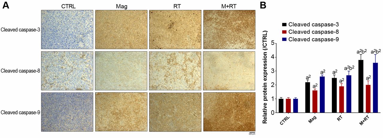
Intrinsic apoptosis related proteins are enhanced by the combination treatment with magnolol and RT in Hep3B tumor bearing model. (A) Representative tumor IHC staining results of cleaved-caspase-3, -8 and -9 from each group is displayed. (B) Quantification of the results of each group is presented as a bar chart. (a2 p<0.01 vs. CTRL; b2 p<0.01 vs. single treatment) Scale bars: 100 μm.
Hematoxylin and eosin (H&E) staining. Liver tissue extracted from mice on day 18 was prepared for H&E staining. The fixation, paraffin-embedding and slicing procedure was the same as for IHC staining. Hematoxylin and eosin (H&E) staining was executed by Bio-Check Laboratories Ltd (New Taipei, Taiwan) as previously described (15). H&E stained sections were also photographed by Nikon microscope at 100´ magnification.
Statistical analysis. Excel 2017 was used to analyse data. All data are presented as mean±SD. Statistical significance was evaluated through the conventional Student's t-test and one-way ANOVA. p<0.05 was regarded as statistically significant.
Results
Magnolol markedly enhanced the anti-HCC growth effect of RT. To identify whether magnolol may increase the therapeutic efficacy of RT, we established a Hep3B tumor bearing animal model. The experimental flow chart is displayed in Figure 1A. Mice were randomly separated into four groups, including CTRL, MAG (magnolol 75 mg/kg), RT (6 Gy) and magnolol combined with RT (M+RT), when mice tumor reached 80 mm3. Tumor volume and tumor size were recorded every three days. As shown in Figure 1B, the slowest tumor growth trend was shown in the M+RT group. Tumors extracted from the mice on the final day of therapy were also photographed and were displayed in Figure 1C. The smallest tumor size was found in the M+RT group, which corresponded to the tumor volume results. Furthermore, mice whole-body CT images were taken before sacrifice. Notable tumor growth inhibition was observed in the M+RT group compared to single treatment (Figure 1D). Finally, we also measured the weight of the extracted tumors. The lightest tumor was presented in the M+RT group compared to all single therapies (Figure 1E). Taken together, the above results indicated that magnolol effectively sensitized HCC to RT.
Mice body weight and liver pathology were not affected by magnolol combined with RT in HCC bearing mice. To investigate whether the combination of magnolol with RT may induce general toxicity in mice, we recorded mouse body weight and extracted liver for H&E staining. As shown in Figure 2A, both the M+RT and the magnolol single treatment groups of mice maintained their body weight during the whole treatment process. The non-treated control and the RT group showed marked body weight loss compared to M+RT. In addition, we performed H&E staining on mice liver to investigate liver pathology after treatment. There was no obvious change on liver pathology following the different therapies (Figure 2B). In conclusion, magnolol and RT combination therapy may not induce general toxicity in Hep3B bearing mice.
Magnolol markedly enhanced RT-induced intrinsic apoptosis signalling in HCC bearing mice. Next, we investigated whether apoptosis related mechanisms were affected by the combination of magnolol with RT using IHC staining of the mouse tumor tissue. As indicated in Figure 3A and B, protein expression of the apoptosis marker cleaved caspase-3 was markedly increased 1 to 1.5 times by M+RT compared to single treatments. Furthermore, protein expression of cleaved caspase-9, a recognized intrinsic apoptosis marker, was also augmented by M+RT compared to the single treatment. Similar induction of cleaved caspase-8 protein, an extrinsic apoptosis marker, was found in all treatment groups. Taken together, magnolol may effectively enhance the induction of intrinsic apoptosis by RT.

RT induced phosphorylation of ERK/NF-κB and expression of their downstream proteins was blocked by magnolol in Hep3B tumor bearing model. (A, C, E) Representative tumor IHC staining results of P-ERK, P-NF-κB, Ki-67, C-FLIP, MCL-1, XIAP, IL-1β and TNF-α from each group. (B, D, F) Quantification of the results of each group is shown as bar chart. (a1 p<0.05 and a2 p<0.01 vs. CTRL; b2 p<0.01 vs. single treatment) Scale bars: 100 μm.
Magnolol diminished RT-activated ERK/NF-κB and its downstream signalling in HCC bearing mice. A previous study has suggested that RT may activate ERK/NF-κB and expression of their downstream proteins in HCC (16). Here, we aimed to evaluate whether magnolol may suppress RT-induced ERK/NF-κB mediated signal transduction. As illustrated in Figure 4A and B, RT-induced expression of phospho-ERK and phospho-NF-κB were diminished by combination of RT with magnolol. The proliferation marker, Ki-67 was effectively reduced in the M+RT group compared to single treatment (Figure 4C and D). Furthermore, NF-κB mediated expression of anti-apoptosis proteins including C-FLIP, MCL-1 and XIAP was markedly reduced by M+RT treatment. NF-κB-mediated expression of inflammation proteins such as IL-1β and TNF-α were also markedly suppressed by the combination of magnolol with RT (Figure 4E and F). These results indicated that magnolol may effectively suppress RT-induced ERK/NF-κB pathway and the expression of the related anti-apoptosis and inflammation proteins.

The proposed mechanism of the effect of the combination of magnolol with RT. Arrow indicates induction. Magnolol may trigger the apoptosis pathway and suppress ERK/NF-κB related signal transduction.
Magnolol enhanced RT efficacy via the induction of apoptosis signalling and inhibition of ERK/NF-κB mediated pathways in HCC bearing mice. As showed in Figure 5, both RT and magnolol may induce the expression of the extrinsic and intrinsic apoptosis-related proteins, included cleaved-caspase-3, -8 and -9. In addition, the ERK/NF-κB signalling transduction that induced by RT can be mitigated by magnolol. The NF-κB downstream signalling that is involved in anti-apoptosis and inflammation was decreased by magnolol. In summary, magnolol may facilitate RT's anti-HCC potential through triggering apoptosis and inhibiting ERK/NF-κB pathway.
Discussion
In our study, the therapeutic efficacy of the combination of magnolol with radiation (high-energy x-rays) was evaluated by using HCC bearing mice. We found that magnolol was a radiosensitizer that significantly promoted radiation-mediated inhibition of tumor growth in HCC in vivo. Cleaved-caspase-3, -8, and -9 are required for radiation-induced apoptosis through the extrinsic and intrinsic pathways (17). Our findings showed that the combination of magnolol and radiation significantly increased expression of both cleaved-caspase-3 and caspase-9 compared to magnolol or radiation alone treatment (Figure 3). Magnolol may enhance radiation-induced apoptosis through the intrinsic apoptotic pathway in HCC.
Nuclear factor kappa-light-chain-enhancer of activated B cells (NF-κB) is the key transcription factor which mediates cancer cell radioresistance by regulating the expression of anti-apoptotic genes. Anti-apoptotic proteins C-FLIP, MCL-1, and XIAP limit the anti-cancer efficacy of radiation by inhibiting the extrinsic and intrinsic apoptosis signaling transduction (18, 19). In a previous study, inhibitor of κBα mutant vector(p-IκBαM), a super repressor of NF-κB, not only augmented radiation-induced cytotoxicity but also inhibited expression of radiation-triggered NF-κB-related proteins in HCC (16). The increased expression of proinflammatory cytokines TNF-α and IL-1β as unfavorable prognostic markers was associated with recurrence of HCC (20). QNZ, an inhibitor of NF-κB signaling, has been shown to decrease protein levels of C-FLIP, MCL-1, XIAP, TNF-α, and IL-1 β in HCC in vitro (21, 22). Our data demonstrated that magnolol promoted radiation--decreased phosphorylation of NF-κB, protein levels of C-FLIP, MCL-1, XIAP, TNF-α, and IL-1 β in HCC ex vivo (Figure 4).
ERK, a member of activated protein kinase (MAPK) family, is the downstream component of RAF/mitogen-activated protein/ERK kinase (MEK)/ERK signaling pathway involved in tumor progression (23). ERK has been demonstrated to regulate expression of proliferation, anti-apoptotic, and invasion-related proteins through activation of the NF-κB signaling in HCC (10, 20). Furthermore, radioresistance of cancer cells has been linked to the phosphorylation of ERK (24). Sorafenib, an inhibitor of RAF/MEK/ERK signaling, has been shown to reverse the radiation-induced NF-κB activity and expression of NF-κB-related proteins in HCC (16). Magnolol has been shown to reduce NF-κB activation and expression of NF-κB-related proteins through upregulating dephosphorylation of ERK in HCC in vitro and in vivo (10, 11). In this study, magnolol inhibited radiation-induced ERK phosphorylation in HCC in vivo (Figure 4A).
In conclusion, magnolol is a radiosensitizer that enhances the therapeutic effect of radiation in HCC in vivo. The radiosensitizing effect of magnolol on tumor growth of hepatocellular carcinoma in vivo is mediated through the induction of the intrinsic apoptosis pathway and suppression of NF-κB signaling.
Acknowledgements
This study was funded by Taipei Medical University, Taipei, Taiwan (Grant ID: 107TMU-TMUH-18). We also want to thanks to “TMU Research Center of Cancer Translational Medicine” from The Featured Areas Research Center Program within the framework of the Higher Education Sprout Project by the Ministry of Education (MOE). This study was also supported by Show Chwan Memorial Hospital, Cathay General Hospital and China Medical University Hospital, respectively. (Grant ID: RD108023, CGH-MR-A10823 and DMR-109-172)
Footnotes
Authors' Contributions
YSC, RS, WLC and YCY performed the experiments. FTH, JGC and CJT prepared the initial version of the paper. CLH, YMC and JHC designed the study, performed the literature review, and prepared the final versions of the paper.
This article is freely accessible online.
Conflicts of Interest
The Authors declare no competing financial interests regarding this study.
- Received April 9, 2020.
- Revision received April 15, 2020.
- Accepted April 17, 2020.
- Copyright© 2020, International Institute of Anticancer Research (Dr. George J. Delinasios), All rights reserved


