


Abstract
Background/Aim: Results of Guided Bone Regeneration (GBR) primarily depend on the membrane used. The aim of this study was to compare biocompatibility of different absorbable and non-absorbable membranes by using unrestricted somatic stem cells (USSCs) as an indicator for biocompatibility. Materials and Methods: Five absorbable membranes (Bio-Gide®, RESODONT®, GENTA-FOIL resorb®, BioMend® and BioMend® Extend™) and one non-absorbable alternative (GORE-TEX®) were colonized with USSCs. After 24 h, 3 days and 7 days, cell proliferation, cell viability, and cytotoxicity were assessed. Moreover, cell morphology was evaluated by electron microscopy. Results: Significantly higher cell proliferation and cell viability rates were observed in Bio-Gide® and RESODONT® membranes. Cell toxicity was highest on GENTA-FOIL resorb® membranes. The electron microscopical assessment showed a better cell attachment on porous surfaced membranes. Conclusion: This study shows that USSCs can be used for assessments of biocompatibility, and that absorbable membranes with collagenous composition and porous structure tend to positively impact biocompatibility and enhance cell proliferation.
Bony defects involving the head and face often result not only in limited functionality, but also present esthetic deficiencies reducing patients' quality of life (1). Depending on defect size, different covering techniques are applied. In cases of greater bony defects, augmentative procedures become necessary (2). Current gold standards for bone augmentation are autologous bone grafts mainly retrieved from the scapula, the fibular bone, and the iliac crest (3). Disadvantages are donor site morbidity and limited availability (4). Alternatives to autologous bone grafts present various methods of tissue engineering and guided bone regeneration (GBR) (5, 6). GBR has been described in research over the last 20 years (7-10). In GBR, occlusive membranes are used to prevent soft tissue ingrowth into the bone, allowing osteogenic cells originating from the adjacent bone to immigrate the restoration site (5). Results of GBR primarily depend on the membrane used (11). Ideally the membrane material should be biocompatible, provide a way for revascularization of the defect area, and allow nutrition of target cells (12). Nowadays, mainly absorbable membranes are used (13), and their indications have been expanded to periodontal flap surgery (14) and third molar extraction (15), providing appealing results. Nevertheless, their clinical application is still restricted due to high costs, difficult clinical handling, infection, and collapse of membranes (16).
The aim of this study was to compare biocompatibility of different absorbable and non-absorbable membranes. As indicator for biocompatibility unrestricted somatic stem cells (USSCs) were used. USSCs retrieved from the umbilical cord can be cultivated without losing their potential of pluripotent differentiation (17). They seem to hold an epigenetic state in between a terminally differentiated cell type and that of embryonic stem cells (18). Under certain conditions USSCs can be differentiated into osteoblasts, chondroblasts, adipocytes, hematopoietic, and neural cells (17). Dexamethasone, ascorbic acid, and β-glycerol phosphate (DAG) are used for differentiation into osteoblasts in vitro (19) and in vivo, as well (20).
Characteristics of the different membranes (Bio-Gide®, RESODONT®, GENTA-FOIL resorb®, BioMend®, BioMend® Extend™ und GORE-TEX®).
Materials and Methods
This study was approved by the Ethics-Committee of the Heinrich-Heine University Dusseldorf, Germany (No.: 3376).
All membranes used have already been tested in vivo and are approved medical products. For absorbable membranes Bio-Gide® (Geistlich Pharma AG, Wolhusen, Switzerland), RESODONT® (RESORBA Wundversorgung GmbH & Co. KG, Nuernberg, Germany), GENTA-FOIL resorb® (RESORBA Wundversorgung GmbH & Co. KG), BioMend® and BioMend® Extend™ (Zimmer Dental GmbH, Freiburg, Germany) were used. As a non-absorbable alternative a (GORE-TEX® W.L. Gore & Associates, Inc., Flagstaff, USA) membrane was tested (detailed membrane characteristics are shown in Table I).
First, the membranes were sterilized, then cut into pieces (n=27) containing an area of 0.785 cm2 in order to fit the holes of a well plate. A total of 9 pieces were used per cell line per day of testing. Those 9 pieces were used for the measurement of cell proliferation (n=5), the measurement of cytotoxicity and cell activity (n=3), and for measurements with the raster electron microscope (n=1). Because of their buoyancy in the culture medium, pieces of the RESODONT®- and the GENTA-FOIL resorb® membranes had to be fixed on the well plate surface. As a control a standard cell culture surface was used. All wells (either containing membranes or the control group) were filled with 610 μl culture medium and placed into the incubator at 37°C, 21% O2 and 5% CO2 saturation.
USSCs were provided by the José Carreras stem cell bank of the University of Duesseldorf. The following cell lines were used: USSC-18 (female, Passage 8), USSC-8 (female Passage 9), USSC-8/17 (male, Passage 8). To prepare the nutrient medium, 350 ml DMEM (Dulbecco's modified eagle medium, Lonza Cologne GmbH, Cologne, Germany), 150 ml fetal bovine serum (FBS, PAN-Biotech GmbH, Aidenbach, Germany), 5 ml penicillin/streptomycin (10,000 U/10,000 μg/ml, Biochrom GmbH, Berlin, Germany), and 5 ml L-glutamine (200 mM, Biochrom GmbH) were used. The differentiation was induced using DAG (50 μM Dexamethasone in DMEM (Sigma-Aldrich Chemie GmbH, Steinheim, Germany), 50 mM Ascorbic acid (Sigma-Aldrich Chemie GmbH) in phosphate buffered saline (PBS) and 1 M β-Gycerolphasphate in PBS) using a standardized protocol (20). Cells were cultivated in an incubator according to the standard protocol (37°C, 21% O2 and 5% CO2 saturation).
Every membrane was cultivated with a total of 19×103 cells for specific time intervals. After 24 h, 3 days, and 7 days representative membranes were selected, frozen and fixated for examination. The culture medium and the embedded well plates were renewed after 24 h and after 4 days. Cell viability was measured directly in the culture using CellTiter-Blue® Cell Viability Assay (Promega GmbH, Mannheim, Germany). Cytotoxicity was measured indirectly through the cell culture supernatant using CytoTox-ONE™ Homogeneous Membrane Integrity Assays (Promega GmbH). For cell count and membrane fixation (for REM-evaluation), membranes were washed with attenuated PBS solution and frozen (−80°C). Cell count was performed with the help of CyQuant® Cell Proliferation Assay Kit (Life Technologies GmbH, Darmstadt, Germany). For REM-evaluation the membranes were placed into 5% Glutardialdehydle, dried by an ascending acetone series (50%-70%-90-100%) and Critical Point Dryer CPD 030 (BAL-TEC GmbH, Schalksmuhle, Germany). For electrification, the membranes were sputtered using Sputter Coater 108 auto (Ted Pella, Inc., Redding, CA, USA). For evaluation a Scanning-Electron-Microscope S-3000N was used.

Attachment and proliferation of the osteogenic USSC-18, -8 and -8/77 cell lines growing on different membranes (Bio-Gide®, RESODONT®, GENTA-FOIL resorb®, BioMend®, BioMend® Extend™ und GORE-TEX®) for 1, 3, and 7 days. ***p<0.0005.
Statistical analysis. In total 27 pieces of each membrane were examined (n=189). Nine pieces were used for each cell line (n=9). Five of those were used for the measurement of cytotoxicity (n=5). Three were used for cell viability (n=3), and one piece was used for measurements with the raster electron microscope (n=1). A Shapiro-Wilk test was used for evaluation of normal distribution. In order to detect statistically significant differences a one-way ANOVA and the Bonferroni correction was performed as post-hoc test. Furthermore, t-tests were performed to find significant differences in comparison to the control group (specific membrane in comparison to the control group only). Calculations were made by the use of SPSS 21 for Mac (SPSS Inc., Chicago, IL, USA). Differences with p<0.05 were considered statistically significant. Data is presented as mean±standard deviation.
Results
The aim of this study was to evaluate biocompatibility of 6 different membrane types. Twenty-four hours after cell seeding, 7,617±1,040 USSC-18 cells, 5,824±425 USSC-8 cells, and 6,000±600 USSC-8/77 cells could be found on the control standard surface. In the control wells, significantly increased proliferation was observed in all cell lines, between days 1 and 3 (p>0.0005) and days 3 and 7 (p<0.05).
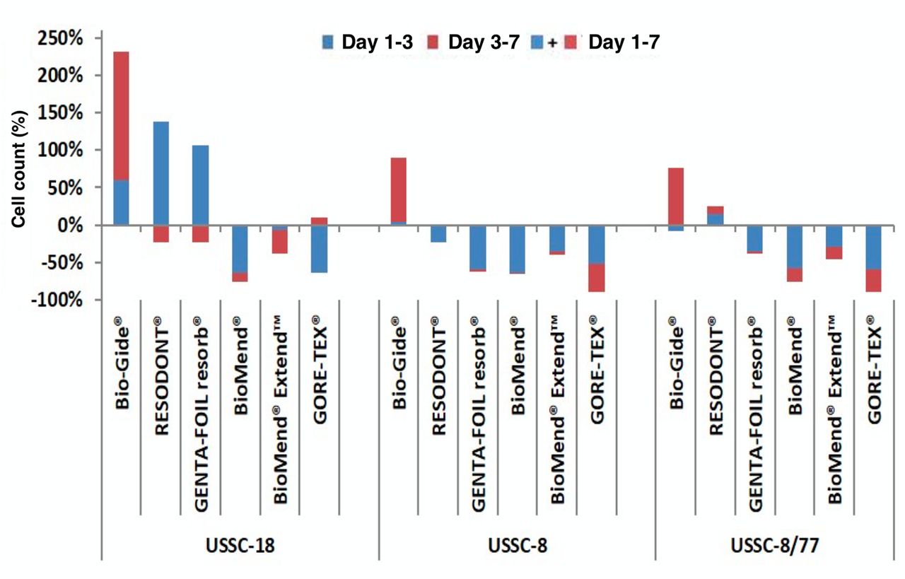
After 24 h, in cell line USSC-18, the RESODONT® membrane surface showed significantly more cell attachment (3,756±336; p>0.0005) followed by the Bio-Gide® membrane (2,715±361; p>0.0005), compared to the other membranes. Highest cell counts were found on day 7 on Bio-Gide® membranes (USSC-18: 9,015±1,491 cells, USSC-8: 4,358±542 cells, and USSC-8/77: 3,875±457 cells). In comparison to the numbers of cells on day 1, they showed significant differences (p>0.0005) in cell lines USSC-8 and USSC-8/77 on day 7. Relative differences (%) in cell count on the distinct membrane surfaces after 3 and 7 days in comparison to day 1 and day 3 are shown in Figure 1.
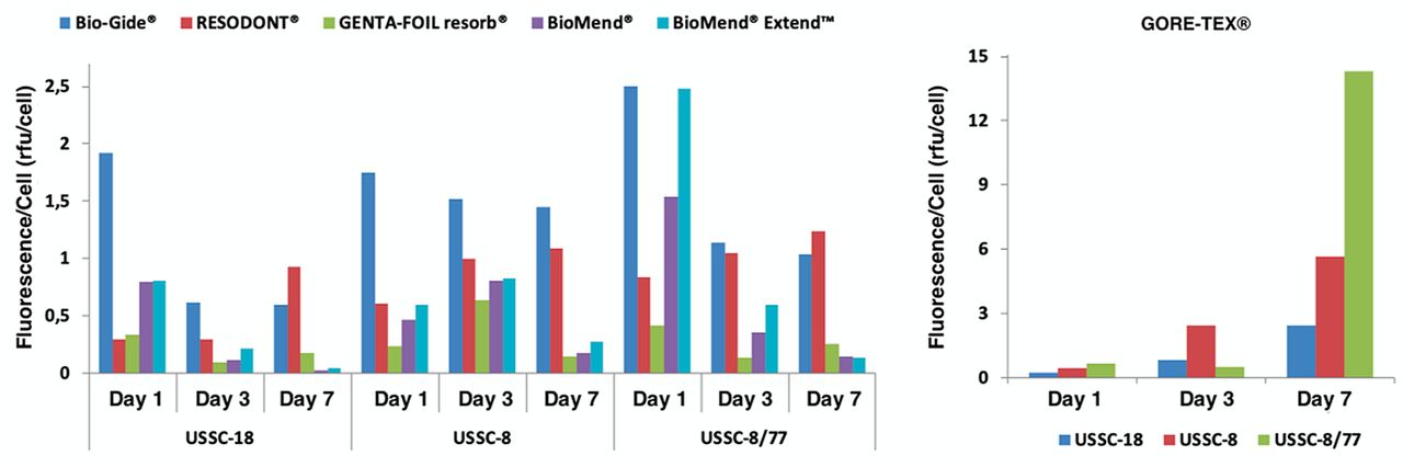
Cell viability was measured fluoroscopically (560Ex/590Em) and assessed after 1, 3 and 7 days. The control group showed significant rises in cell viability in every measurement, USSC-18 performed significantly better than the other two cell lines (p<0.0005; Day 1: USSC-18: 9,324±442 RFU, USSC-8: 7,942±447 RFU, and USSC-8/77 5,795±660 RFU; Day 7: USSC-18 17,222±395 RFU, USSC-8 14,019±457 RFU, and USSC-8/77 13,926±910 RFU). After 24 h, a significantly higher overall viability (in every cell line) could be observed on the Bio-Gide® membrane, in comparison to the other membranes tested (p<0.0005). This viability declined on day 3, but recovered significantly on day 7. The membranes, on which least cell viability was observed, were the GENTA-FOIL resorb® and the GORE-TEX® (Figure 2). In terms of viability per cell after 24 h, the greatest viability could be observed on the Bio-Gide® membrane (USSC-18: 1.92±0.51 RFU/cell, USSC-8: 1.75±0.38 RFU/cell, USSC-8/77: 2.50±0.64 RFU/cell). On day 3 and 7, viability per cell was greatest on GORE-TEX® membranes (Day 3: USSC-18: +2.19±0.52 RFU/cell, USSC-8: +5.24±1.25 RFU/cell and USSC-8/77: +13.67±3.32 RFU/cell and day 7: USSC-18: 0.60±0.30RFU/cell, USSC-8: 1.45±0.53 RFU/cell, and USSC-8/77: 1.04±0.47 RFU/cell) (Figure 3).

Viability of the osteogenic USSC-18, -8 and -8/77-cell lines growing on different membranes (Bio-Gide®, RESODONT®, GENTA-FOIL resorb®, BioMend®, BioMend® Extend™ and GORE-TEX®) for 1, 3, and 7 days. ***p<0.0005, Fluorescence (560Ex/590Em).
In terms of cytotoxicity, only the 4 membranes with the highest proliferation/viability rates (Bio-Gide®, RESODONT®, GENTA-FOIL resorb® and GORE-TEX®) were compared to each other and to the control group. Cytotoxicity on day 3 was significantly higher in cells grown on membranes, compared to the control wells. On day 3, the highest cytotoxicity was measured on GENTA-FOIL resorb® membranes (USSC-18: 21,613±2,364 RFU, USSC-8: 33,242±2,181 RFU, and USSC-8/77: 23,623±1,879 RFU, p<0.0005), while the lowest cytotoxicity was measured on GORE-TEX® membranes (USSC-18: 15,998±1,212 RFU, USSC-8: 30,662±928 RFU and USSC-8/77: 19,769±3,187 RFU). After 7 days, in cell lines USSC-18 (9,334±1.082 RFU) and USSC-8/77 (11,192±1.352 RFU) lowest cytotoxicity rates were measured (p<0.005) in the control group, whereas in cell line USSC-8 (12,959±633 RFU) highest cytotoxicity rates were measured in the control group (p<0.005). In USSC-8 they were significantly higher than those on RESODONT® (10,991±781 RFU), GENTA-FOIL resorb® (11,451±364 RFU) and GORE-TEX® (10,526±841 RFU) (p<0.0005). In the USSC-18 cell line highest cytotoxicity was found on RESODONT® membranes (21,230±2,622 RFU) and in USSC-8/77 on GORE-TEX® (17,210±1,470 RFU) (Figure 4).
Additionally, raster electron microscope images of the cultivated membranes were compared to each other and to non-colonized membranes (Figure 5). The Bio-Gide® membrane comes with a porous side facing the defect. After 24 h, the USSCs showed long cell extensions attaching to the collagenous fibers of the membrane. After 7 days, the cells aligned with the collagenous fibers. In comparison to the Bio-Gide®, the other membranes presented a smooth surface. After 24 h, the RESODONT® membrane surface changed and appeared spongey and porous. The USSCs showed long cell extensions attaching to the RESODONT® membrane, similarly to Bio-Gide®, while a continuous cell layer had developed after 7 days. The GENTA-FOIL resorb® membrane changed into a less porous surface after 24 h. A few elongated USSCs with small extensions could be observed. Up to day 3, they enlarged and become radial. After 7 days, USSC appeared elongated and smaller again. BioMend® and BioMend® Extend™ showed almost no change in membrane surface and only weakly adhering USSCs with few, short cell extensions showed after 7 days. Finally, the GORE-TEX® membrane was the only one showing collagenous polytetrafluoroethylene fibers (PTFE-Fibers). After 7 days, there was only little cell attachment and a few cell extensions could be observed.
Relative proliferation rates of the osteogenic USSC-18, -8 and -8/77-cell lines on different membranes (Bio-Gide®, RESODONT®, GENTA-FOIL resorb®, BioMend®, BioMend® Extend™ and GORE-TEX®) between days 1 and 3, days 3 and 7 and between days 1 and 7 in comparison to the cell count on day 1 (p<0.005).

Cytotoxicity on a standard cell surface and on the membranes (Bio-Gide®, RESODONT®, GENTA-FOIL resorb® and GORE-TEX®) on the osteogenic differentiated USSC-18, -8 and -8/77-cell lines after days 1, 3 and 7. ***p<0.0005, **p<0.005.

Raster electron microscope images of the studied membranes, native and colonized with USSC-18, for days 1, 3 and 7 (magnification 500×). For day 7, a representative cell has been marked in red. Cell structure, form, and size differed depending on the membrane surface. White arrow: cell, black arrow: cell extension.
Discussion
A recent review by Carballé-Serrano et al. (21) stated that biocompatibility is the crucial factor taken into account when choosing a membrane for guided bone regeneration. In this study the attachment, proliferation, and cell activity of osteoblast-differentiated USSCs growing on five resorbable membranes and one non-resorbable membrane were evaluated, as well as the cytotoxic effects on these cells. For cell attachment a membrane has to be biocompatible (22). In vivo cell attachment depends not only on the membrane material, but also on the location and timing of placement, together with condition and age of a patient (11). A rough membrane surface supports attachment and proliferation of osteoblasts, whereas fibroblasts tend to attach to smooth surfaces more easily (23, 24). The rough surfaces of the RESODONT® and Bio-Gide® membranes might be a reason for their high rates of cell attachment. Furthermore, cell proliferation was highest after 24 h on Bio-Gide® membranes, after 3 days on RESODONT®, and after 7 days on Bio-Gide® membranes again. Proliferation and cell numbers were significantly higher than on the other membranes. This could also be due to their composition. Bio-Gide® membranes consist of a mixture of collagen type I and III, whereas the others only contain collagen type III. Therefore, collagen type I or the mixture of collagens might have a positive impact on cell proliferation. In a similar study by Rothamel et al. (25) using osteobleasts, it was demonstrated that Bio-Gide® membranes had the highest proliferation rates. In accordance with the findings of this study, sparsely cell proliferation rates have been reported for BioMend® membranes (25, 26). This might be due to the glutaraldehyde used for cross-linking in these membranes (27, 28). Cross-linking treatments involving glutaraldehyde or formaldehyde are included to overcome rapid membrane degradation (12, 29). Glutaraldahyde may also be a reason for reduced cell viability, as cell metabolic activity was significantly highest on Bio-Gide® membranes at all sample points. UV-irradiation for cross-linking might present a more compatible solution (30). On RESODONT®, cell viability was significantly higher than on the other membranes. Gentamycin is added to GENTA-FOIL resorb® membranes, which might reduce cell viability in those membranes. GENTA-FOIL resorb® also showed significantly high cytotoxicity rates. In contrast, high cytotoxicity rates were found on Bio-Gide®, RESODONT® and GORE-TEX® membranes as well. Osteoblasts are very sensitive to surface structures (31, 32). Altered surface structures might, therefore, not only result in enhanced cell proliferation and attachment, but also in higher cytotoxicity rates. Furthermore, in collagenous membranes high costs and weak tensile strength in wet conditions show limitations for wide clinical application, resulting in testing of other materials such as silk (16). Lately, coated membranes including magnesium and chitosan or gelatin-chitosan nano-composed membranes have proven biocompatibility, osteogenic-conductive potential, and low biodegradation rates (30, 33). These new membranes might present excellent alternatives for GBR.
In conclusion, our findings suggested that USSCs can be used for biocompatibility assessment, and moreover, membranes with collagenous composition and porous structure tend to positively impact biocompatibility and enhance cell proliferation. Clinicians should be aware of membrane characteristics and choose a patient-specific membrane when performing GBR.
Acknowledgements
This research did not receive any specific grant from funding agencies in the public, commercial, or not-for-profit sectors.
Footnotes
Authors' Contributions
Jörg Handschel made substantial contributions to conception and study design; Felix Paulssen von Beck performed the laboratory work. Henrik Holtmann, Julian Lommen and Lara Schorn and analyzed and interpreted the data; Lara Schorn wrote the manuscript. Rita Depprich and Norbert Kübler were involved in revising the manuscript critically. All authors read and approved the final manuscript.
This article is freely accessible online.
Conflicts of Interest
The Authors have no conflicts of interest to declare.
- Received June 11, 2019.
- Revision received July 10, 2019.
- Accepted July 19, 2019.
- Copyright© 2019, International Institute of Anticancer Research (Dr. George J. Delinasios), All rights reserved
References
In this issue


Jump to section
Related Articles
Cited By...
- No citing articles found.