


Abstract
Background/Aim: Paradontopathy and neoplasms of the oral cavity represent one of the greatest challenges in human and animal dentistry. EGF plays a key role in maintaining the integrity and proper rate of cell proliferation in normal oral epithelium. The aim of the present study was to study serum levels of EGF in dogs diagnosed with periodontal diseases and oral cavity tumours. Materials and Methods: The samples comprised of cancerous tissue sections and serum obtained from dogs of various breeds, aged between 5-13 years. Serum EGF concentrations were measured by an immunoenzymatic method. Results: The median for EGF concentration in serum of dogs suffered from severe periodontal diseases was greater when compared to the control group. EGF concentration in dogs with malignant tumours was significantly higher than in those with non-malignant growths. A positive correlation between EGF concentration and tumour size was also observed. EGF level in dogs diagnosed with benign tumours was comparable to the control group. Conclusion: The blood serum level of EGF increases significantly in patients with malignant oral tumours and advanced periodontal disease. In malignant tumours, the high level of EGF correlates with the size and invasiveness of the neoplasm.
Pathological lesions in the oral cavity require comprehensive dental treatment, both in human and animal patients. Inflammatory processes in this region are usually caused by bacterial, less frequently viral or mycotic infections. The oral cavity is more susceptible than other regions of the body to mechanical damage of the natural protective barrier – oral mucosa due to e.g.: invasive dental procedures, presence of deep gingival pockets or periapical abscesses (1). Due to the constant temperature, humidity and presence of recesses that hinder self-cleaning, the oral cavity provides a nearly ideal environment for the growth of microorganisms and development of soft tissue inflammations. One of the key causes of periodontal diseases is the presence of dental plaque which undergoes mineralisation and saturation with calcium phosphate and turns into tartar (2). The rough surface of dental tartar provides ideal conditions for infiltration and multiplication of pathogenic microorganisms which are among the main causes of lesions in the periodontium and gingival connective tissue (3). Bacterial gingivitis is the initial and reversible form of periodontal disease which, unless properly treated, may lead to the development of periodontitis, and in consequence to the destruction of the alveolar process bone and development of bacteraemia.
Apart from periodontal diseases, another key problem faced by dentistry is cancer. Despite the constant development of diagnostic methods and targeted treatments, malignant neoplasms continue to rank as one of the main causes of human and animal death. The formation and development of cancer cells is conditioned by the incidence of proliferation, apoptosis, and differentiation disorders. The processes are regulated primarily by the activity of oncogenes and faulty regulation of the production of growth factor proteins and their receptors on the surface of a tumour cell (4). The neoplastic transformation is conditioned by disorders in the expression of factors directly influencing the process of neoangiogenesis including VEGF, FGF, IGF, TGF and many others. In turn, the excessive proliferation of pathological cells and the impairment of its apoptosis is caused by the epidermal growth factor – EGF.
EGF was first isolated in 1961 from mouse submandibular gland extract as a factor inducing premature eyelid opening and eruption of incisors (5). EGF is present in many tissues and shows mitogenic activity towards a number of cell lines (6). It induces epidermal keratosis and proliferation, multiplication and differentiation of pulmonary alveolar epithelium, facilitates corneal epithelium regeneration, causes hypertrophy and hyperplasia of liver cells, inhibits the secretion of gastric juice, retards ovary development and hair growth (7).
The biological activity of EGF depends on its bonding with a suitable receptor. The EGFR receptor partakes in the organogenesis of numerous organs formed from mesoderm and ectoderm such as the brain, heart and lungs (8). EGF activates routes that increase proliferation, migration and differentiation in epithelial cells, fibroblasts, smooth muscles, hepatocytes, and endothelial cells (6, 9). EGF receptors are present in the oral epithelium but no evidence of the same in dental pulp or periodontal tissues has been reported (6).
A significant contribution of the epidermal growth factor and its receptor in neoplastic angiogenesis has been confirmed. The mechanism of the pro-angiogenic activity of EGF entails its contribution, together with TGFα, to the increased expression of VEGF and consequently to the formation of the growing tumour's blood vessel network (10).
The Epidermal Growth Factor (EGF) not only plays a key role in the maintenance of epithelial cell homeostasis but it is also an important element of the immunological response to inflammatory states developing in the oral cavity (9). In humans, EGF is primarily synthesised in submandibular and parotid salivary glands and kidneys. Saliva is a natural potential source of the factor although the exact mechanism through which its secretion is controlled has yet to be determined and is currently under investigation by numerous researchers (9, 11). Together with mucins, bicarbonates and eicosanoids, EGF plays an important role in the maintenance of anatomic continuity of oral mucosa and its functional regularity. It acts cytoprotectively against mucosa lesions and protects it against mechanical and chemical damage during mastication (chewing) (9). In experimental animals, when EGF sources are limited by removing the submandibular salivary gland, pathological lesions to gingival epithelium occur accompanied by overall slower healing of oral mucosa injuries (12). Furthermore, a massive increase in EGF content in saliva was observed directly after periodontal procedures and extraction of molar teeth (6).
EGF also participates in numerous biological processes occurring outside the oral cavity. Under in vivo conditions it contributes to the healing of skin and gastric mucosa, protects the mucous membrane against harmful agents such as bile, pepsin and trypsin acids (6).
High concentrations of EGF were also observed in the exudate from burn wounds where it was produced by the neutrophils and macrophages flooding the wounded area. The factor's concentration increases significantly directly after an injury. Due to the fact that EGF is a strong keratinocyte and fibroblast mitogen, it plays a vital role in the processes of epidermal reconstruction and development of granulation tissue in slow-healing wounds (13).
The epidermal growth factor participates in physiological processes and inflammatory conditions. Its elevated expression has also been reported in relation to numerous types of human and animal tumours. Elevated EGF concentrations have been observed to correlate with a more aggressive course of the disease and worse prognosis in cases of head and neck cancer, ovary cancer, breast cancer, oesophageal cancer, bladder cancer, and cervical cancer (6, 8, 10, 14).
The aim of the presented study was to determine the EGF level in the blood serum of animals with oral lesions and to verify whether EGF levels may be used in the diagnostics and monitoring of cancer patients.
Materials and Methods
The study material was provided by 30 sick dogs of various breeds, aged between 5 and 13 years, selected from the patients of the dental clinic at the Department and Clinic of Animal Surgery at the University of Life Sciences in Lublin. In most of the sick animals the dominant clinical symptoms included: lack of appetite, unpleasant odour in the oral cavity, and presence of pathological lesions in the mandibular and maxillary mucosa.
Dogs with symptoms of periodontitis without a pathological growth were selected as a sub-group of the studied population (10 animals). In order to diagnose and evaluate the stage of the periodontal disease in those animals, detailed periodontological and radiological examinations were performed. Dental examinations were performed under general anaesthesia and entailed probing of gingival pocket depth (PPD), measurement of the degree of clinical attachment loss (CAL), as well as determination of the extent of bleeding during probing and tooth mobility. The depth of gingival pockets (PPD) and clinical attachment loss (CAL) were evaluated using a calibrated periodontological Williams probe introduced parallel to the tooth's long axis. Examinations were performed in four measurement points (mesially and distally on the buccal/labial side and mesially and distally on the palatal/glossal side at teeth 104, 106, 108, 404, 406, 409). The total of the obtained values was divided by four to obtain the index value for the given tooth. Values obtained for respective teeth were added up and divided by the number of teeth examined to obtain the PPD value.
Tooth mobility was measured using a probe and classified on a four-tier mobility scale:
0 – no mobility;
1 – lateral mobility under 1 mm;
2 – lateral mobility over 1 mm;
3 – lateral and vertical mobility.
Animals with at least 5 teeth showing both lateral and vertical mobility were included in the study.
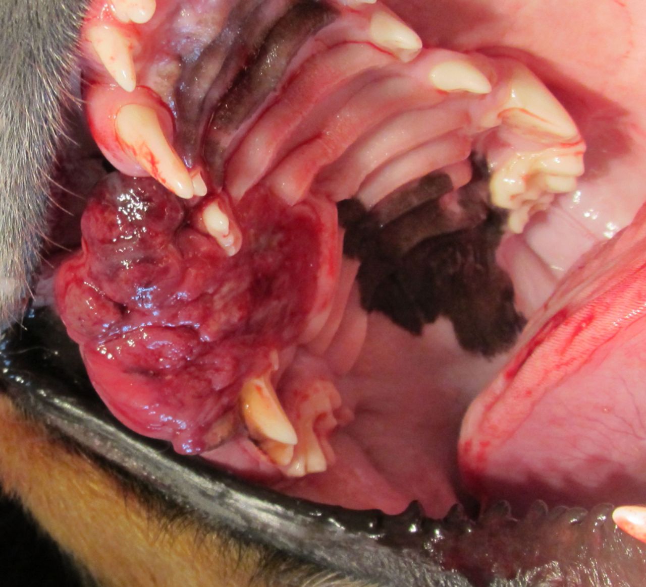
The aforementioned parameters allowed the assessment of the degree of soft tissue damage due to periodontal disease. The subsequent radiological examination provided imaging to illustrate the extent and type (lateral/vertical) of alveolar process bone loss. Dogs diagnosed in the course of the periodontological examination with advanced periodontal disease, i.e. PPD >7 mm, >50% clinical attachment loss around the examined teeth (>1 mm minimum at one measurement point), and lateral and vertical mobility >1 mm of at least 5 teeth – Figure 1.

Dog -Dachshund ♂ 4 l – advanced of the periodontal disease.

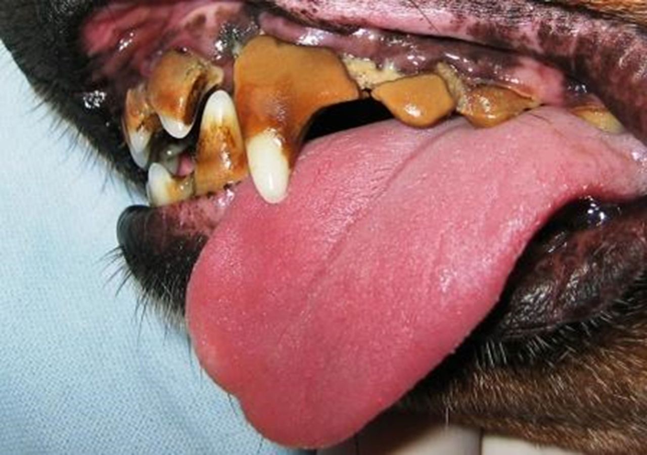
Dog-Mixed breed ♂ 10 l – epulides.

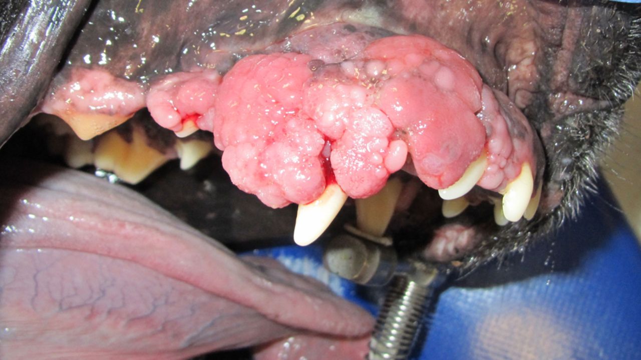
Dog-German Shepherd ♀ 10 l – squamous cell carcinoma.
Dog-Doberman ♂ 12 l – fibrosarcoma.
Results of the immunoenzimatic examination of EGF levels in group I and the control.
Results of histopathological examinations, immunoenzimatic EGF levels, and radiological examinations in Group II and Group III.
The other 20 dogs qualified for the study group were animals with pathological growths which provided material for histopathological examinations. In 7 patients from this group the pathological tissue (with an image similar to epulis) was located around the teeth and was removed surgically. In the remaining 13 cases, the size and infiltrative character of the growth suggested a malignant tumour – in 4 dogs the growth was between 1 and 2 cm and could be removed surgically with a due margin of healthy tissue, in 3 cases due to the size of 2-4 cm and location of the lesion in the vicinity of mandibular incisors, a partial mandibulectomy was performed. In the remaining 6 cases the tumours were larger than 4 cm and due to their advancement, size and infiltrative character they were deemed inoperable. In these patients, the specimen collected for analysis and final diagnosis consisted only of a fragment of the tumour.
The tissue material obtained during the surgical procedure was delivered to the Department of Pathological Anatomy at the Faculty of Veterinary Medicine, University of Life Sciences in Lublin. Tumour specimens were solidified in 10% buffered formalin for 24 hours. Based on an analysis of specimens stained with hematoxylin and eosin the diagnosis was confirmed and the histopathological type of the tumour was determined in accordance with the applicable World Health Organisation (WHO) classification (15).
The control group was composed of 10 healthy animals aged between 1 and 5 years brought in for sterilisation. Apart from specimens of the affected tissue, the research material also included blood samples drawn from all the study group and control group animals, from which serum was obtained for immunoenzimatic analysis to determine the concentration of the epidermal growth factor (EGF). Blood serum was frozen at −70°C and stored until shortly before analysis.
In all patients diagnosed with pathological growths in the oral cavity a lung x-ray was performed to exclude or confirm metastasis.
The concentrations of the epidermal growth factor in the blood serum of studied animals was determined with the ELISA method using readymade analytical kits (Canine Total Epidermal Growth Factor ELISA Kit -MyBioSource, Inc., USA). All stages of the EGF assay were performed in accordance with the vendor's recommendations.
All conducted procedures in the study group (blood drawing and tissue sampling, collection of tumour sections) and in the control group (blood drawing) were approved by the Local Ethics Committee No. II in Lublin (permit no. 26/2015, 28.04.2015 to conduct experiments on animals; experiments on tissues and animal organs). Furthermore, the owners of the animals were informed about the goal of the study and gave their consent to the experimental procedures.
A comparison of EGF levels: Groups I, II and the Control.
A comparison of EGF levels in Group III – patients with malignant tumours – with results from other study groups.
The results obtained from clinical tests and the final histopathological diagnoses translated into the following final division of the study population:
Group I – patients with advanced periodontal disease (10 animals);
Group II – patients with non-malignant tumours (7 animals);
Group III – patients with malignant tumours (13 animals); Control (10 animals).
A statistical analysis was conducted to determine whether significant correlations could be identified between the blood serum EGF concentrations observed in dogs with developing oral diseases and the control group animals. The comparison of the respective groups was conducted with the use of Statistica 10 software and the non-parametric Mann-Whitney test. The correlation between the EGF level and the incidence of metastasis and tumour size was verified with the use of the Kruskal-Wallis test.
Results
The results of histopathological, immunoenzymatic and radiological examinations are presented in Tables I and II.
In 7 cases, histopathological examinations confirmed the non-malignant character of the growths (Table I), which were subsequently diagnosed as epulides – 5 cases (Figure 2) and fibromas -2 cases. Malignant tumours were identified in 13 cases of which 7 were squamous carcinomas (Figure 3), 4 were fibrosarcomas – 4 (Figure 4), and 2 were amelanotic melanomas. Detailed results are presented in Table II.
In Group I composed of dogs with advanced periodontal disease the blood serum EGF levels fluctuated between 3.81 and 12.93 pg/ml (Me=7.93). Compared to the control group the result was statistically significant [Z=3.44 (***) p<0.001] - Table III.
The median EGF concentration in the blood serum of dogs suffering from oral tumours was 2.15 (range=1.08-4.14 pg/ml) for malignant tumours and 20.08 (range=7.19-82.09 pg/ml) for benign tumours. The EGF level in Group II was comparable to the Control (Table III) and statistically insignificant [Z=1.66 (−) p>0.05]. The EGF value was also statistically lower [Z=3.32 (***) p<0.001] in Group II composed of dogs with non-malignant tumours when compared to the respective results obtained in Group I – animals suffering from periodontitis – Table III.
Of all the analysed groups, the highest EGF level was observed in Group III, in animals suffering from squamous carcinoma, and ranged between 13.40 and 82.09 pg/ml with Me=20.08. A comparison between Group III, Group II, Group I and the Control revealed a statistically significant elevation in the epidermal growth factor levels in malignant tumour cases (Table IV).
EGF levels in metastatic and non-metastatic patients.
Correlation between tumour size and blood serum EGF level in Group III.
In 7 Group III animals, metastasis was observed. Kruskal-Wallis test results revealed that the EGF level in those animals was four times higher than in the group without metastasis. The difference was statistically significant at the level of p<0.01(Table V).
The correlation between EGF levels in malignant tumour cases (Group III) and tumour size was also analysed. A positive correlation between EGF concentration and tumour size was observed (Table VI).
Discussion
Periodontal diseases and head and neck tumours constitute a significant epidemiological and clinical problem. Untreated, severe periodontitis can often lead to serious local complications (tooth loss) or even potentially life-threatening systemic bacteraemia. Chronic inflammation in the area of teeth and periodontium as well as chronic irritation of oral mucosa can also facilitate the development of neoplastic lesions in this region (16-18).
From a pathomorphological perspective oral tumours constitute a relatively homogenous group with most cases diagnosed as epithelial tumours, including mostly squamous cell carcinoma. Lesions are most often located in the larynx, oral section of the pharynx, oral cavity, lips, sinuses or nasal cavity. Less common diagnoses include adenocarcinomas originating from the salivary gland epithelium or microcellular carcinomas located in the sinus region. Non-epithelial tumours include soft tissue and bone sarcomas, melanomas and lymphomas (17).
The incidence of malignant oral tumours is on the increase also in animals, and correspond to approximately 6% of all tumours diagnosed in dogs (18).
The clinical symptoms accompanying oral tumours occur relatively quickly and the increasing mass of the tumour tends to hinder feeding and have a destructive effect on tooth roots and tooth mobility in their sockets. Other characteristic symptoms include unpleasant odour and excessive salivation (17).
Neoplastic growths in the area of mandibular and maxillary teeth are characterised by fast development and high invasiveness. Metastasis to splanchnocranial bones, regional lymph nodes or lungs are common and fast (16, 18). The risk of metastasis depends on the location of the original tumour. Tumours located at the front of the oral cavity usually entail a better prognosis and are less metastatic than those located near the base of the tongue and tonsils (16).
The treatment of choice in oral tumour cases is radical resection along with a margin of healthy tissue. The extent of surgical intervention depends on the size and shape of the tumour. In cases where no metastasis to lymph nodes or lungs is observed, a partial mandibulectomy or maxillectomy is performed. The incidence of local relapse after mandibulectomy is 10% (16). Although surgery is rarely a guarantee of full recovery, it can improve the animal's quality of life for several months. The alternative treatment, particularly in cases of squamous carcinoma, entails radiotherapy or photodynamic therapy.
The growth and differentiation of tumours is particularly affected by cytokines, interleukins, and growth factors such as VEGF and EGF. VEGF is responsible for neoangiogenesis while EGF affects the proliferation and migration of tumour cells.
The epidermal growth factor can be isolated in humans and animals from a number of tissues and bodily fluids. Its highest concentrations are observed in saliva (12 μg/ml), milk (80 μg/ml), urine (100 μg/ml), plasma (2 μg/ml) and amniotic fluid (1 μg/ml). Its presence has also been reported in glandular cell secretions in the respiratory and digestive systems (6, 12, 19).
The physiological role of EGF is multidirectional and related to a number of systems: it conditions the development and maturation of intestines and the absorption of nutrients, promotes the growth of epithelial tissue, facilitates the development of the lactiferous gland and regulates lactation (6). A number of researchers also described it as a strong stimulant for the gastrin secretion and absorption of glucose in the small intestine (20).
A number of publications report the presence of the EGF receptor on neoplastic cells. Overexpression of the EGF receptor (EGFR) has been observed in the cells of bladder, breast, colon, ovary, prostate and kidney tumours and 80% of head and neck squamous carcinoma (14, 21, 22, 23). Elevated EGFR expression in the tumours was often correlated with a more invasive character and smaller patient survival rate.
EGFR expression in tumour cells was compared to that in healthy tissue and hyperplastic tissue surrounding the tumour, as well as its correlation to pathological characteristics and patient survival. The conducted research confirmed EGFR overexpression in cases of milk gland cancer in dogs – it oscillated between 16 and 29% but was not correlated with the mitotic activity of the tumour or patient survival (24). The results are contrary to those reported by Dutr, who observed that EGF receptor expression was related to milk gland carcinogenesis and correlated with the indicators of histological malignancy and negative prognoses (25).
Elevated EGF levels were also observed in homogenized cellular material collected from milk gland tumours. A significantly higher level of the factor was observed in malignant tumours, where it could reach up to even 3,081 ng/g. In non-malignant tumours EGF concentration (317.5 ng/g) was comparable to the levels observed in healthy milk gland tissue (26).
In humans, the presence and elevated expression of the EGF receptor was reported in neoplastically altered cells of tumours of the head and neck, larynx, and maxillary sinuses (21, 27). The conducted study confirmed the presence of EGFR in over 50% of squamous cancers and its overexpression constituted a strong, independent prognostic factor for remission (p=0.0016) and overall survival (p=0.0006) (2). Low expression was observed for leucoplakia and very low in healthy animals (27).
As confirmed in other studies, EGFR evaluation may be used for the purposes of differentiating between cancer and epithelium lesions related to non-malignant conditions. The conducted study confirmed its value in the diagnostics of lesions difficult to classify as inflammatory or neoplastic with the EGFR expression clearly reaching higher levels when compared to healthy tissue and non-malignant oral lesions (27, 28).
In the veterinary context, EGF studies focus on confirming its influence on laboratory animals and identifying its receptors on neoplastic cells. Expression of its receptor was observed in most studied cases of squamous skin cancers in cats – it correlated with shorter life expectancy but not with the tumour's mitotic activity (29). Incorrect activation of the EGFR receptor was also identified in epithelial tumours with high metastatic potential located in the lungs, nasal cavity and mamma in dogs (30-32) and oral cavity in cats (33).
Contradictory data with regard to EGF concentration in blood serum has also been reported. In a group of patients with a suspicion of ovarian cancer, significantly higher EGF levels in the blood serum of women with cancer were observed, compared to the respective values recorded in the control group. However, no correlation was observed between overexpression of the growth factor and the clinical advancement or histopathological differentiation of the tumour. Consequently, a test for EGF in the blood serum of patients with a suspicion of ovary cancer was deemed as usable only for the purposes of differential diagnoses, as a method supplementary to other tests (10).
Measurement of blood serum EGF levels was employed in tumour diagnostics in rodents. The study conformed presence of the factor in rats and mice at concentrations around 1 ng/ml (20).
Although data confirmed by Dutr confirmed the correlation between EGF levels and neoplasia (26), research data published by Campos suggested that the blood serum level of EGF could not be used effectively used as a neoplastic marker as its concentration was not statistically significantly different in dogs with tumours when compared to the control group (34).
The results obtained in our own research are parallel to those reported by Dutr. The EGF level in the blood serum of dogs suffering from malignant tumours was found to be statistically significantly higher than the levels observed in the control and other study groups. Furthermore, its correlation with the size of the tumour and its invasiveness may prove to be a valuable prognostic factor. Determining the blood serum level of EGF is considerably less invasive than collecting a tissue specimen. Elisa test is relatively easy to use as it can be performed at any time, without the need for tumour biopsy, i.e. also in situations where a specimen of the original tumour is difficult or indeed impossible to collect.
EGF serves a very specific function in the oral cavity, as evidenced by its presence in the saliva of both humans and animals. Under correct conditions, the natural flow of saliva facilitates mechanical rinsing of the oral cavity, throat and oesophagus of various carcinogenic substances while the presence of EGF further improves protection of the mucosa against injury, facilitates proliferation and differentiation of epithelial cells, and stimulated self-repair processes in the mucosa.
EGF is considered as a high-risk factor in cases of oral cancer. It was confirmed that the concentration of EGF in the saliva was statistically significantly higher in patients with squamous cancer (oral SCC) when compared to the control group, while at the same time the blood serum EGF level was lower compared to the control and did not yield a statistically significant result (9).
In our study, elevated EGF levels in patients showing symptoms of severe periodontitis and damage to the mucosa accompanying the development of a malignant tumour may constitute indirect evidence for EGF's participation in the processes of oral epithelium regeneration.
Other researchers suggested that the lower concentration of EGF in the saliva of patients with oral cancer prior to surgery and its tendency to grow after the surgery may suggest an important role of this growth factor in neoplasia. They also observed that low EGF levels in the saliva may be correlated with impaired self-healing ability of the mucosa (35).
Numerous studies conducted in the context of human oncology confirmed that overexpression of EGF and its receptors was related to shorter remission times, increased risk of metastasis, and immunity with a number of therapeutic methods. It was confirmed that the use of antibodies against EGFR receptors may inhibit or even entirely arrest tumour growth (24).
The determination of EGF levels in oral cavity disease in dogs may constitute a first step in further research concerned with the role of the epidermal growth factor as a potential marker of neoplasia.
Conclusion
The blood serum level of EGF increases significantly in patients with malignant oral tumours. It can be used as a marker for differentiation with non-malignant tumours. Also, EGF levels observed in non-malignant tumours are comparable to those recorded in healthy animals. Elevated EGF levels in cases of advanced periodontal disease confirm its participation in the processes of oral mucosa repair. Finally, in malignant tumours, the high level of EGF correlates with the size and invasiveness of the neoplasm.
Footnotes
This article is freely accessible online.
Conflicts of Interest
The Authors have no conflicts of interest to declare.
- Received January 18, 2018.
- Revision received February 15, 2018.
- Accepted February 16, 2018.
- Copyright© 2018, International Institute of Anticancer Research (Dr. George J. Delinasios), All rights reserved
References
In this issue


Jump to section
Related Articles
Cited By...
- No citing articles found.