
Main menu

  • Home
  • Current Issue
  • Archive
  • Info for
    • Authors
    • Advertisers
    • Editorial Board
  • Other Publications
    • Anticancer Research
    • Cancer Genomics & Proteomics
    • Cancer Diagnosis & Prognosis
  • More
    • IIAR
    • Conferences
  • About Us
    • General Policy
    • Contact
  • Other Publications
    • In Vivo
    • Anticancer Research
    • Cancer Genomics & Proteomics

User menu

  • Register
  • Subscribe
  • My alerts
  • Log in
  • My Cart

Search

  • Advanced search
In Vivo
  • Other Publications
    • In Vivo
    • Anticancer Research
    • Cancer Genomics & Proteomics
  • Register
  • Subscribe
  • My alerts
  • Log in
  • My Cart
In Vivo

Advanced Search

  • Home
  • Current Issue
  • Archive
  • Info for
    • Authors
    • Advertisers
    • Editorial Board
  • Other Publications
    • Anticancer Research
    • Cancer Genomics & Proteomics
    • Cancer Diagnosis & Prognosis
  • More
    • IIAR
    • Conferences
  • About Us
    • General Policy
    • Contact
  • Visit iiar on Facebook
  • Follow us on Linkedin
Research ArticleExperimental Studies

Corpus luteum Regression Induced by Prostaglandin F2α in Microminipigs During the Normal Estrous Cycle

MICHIKO NOGUCHI, MASAYA HIRATA, HIROAKI KAWAGUCHI and AKIHIDE TANIMOTO
In Vivo November 2017, 31 (6) 1097-1101;
MICHIKO NOGUCHI
1Laboratory of Theriogenology, Department of Veterinary Medicine, Azabu University, Sagamihara, Japan
2Laboratory of Domestic Animal Internal Medicine, Joint Faculty of Veterinary Medicine, Kagoshima University, Korimoto, Japan
  • Find this author on Google Scholar
  • Find this author on PubMed
  • Search for this author on this site
MASAYA HIRATA
2Laboratory of Domestic Animal Internal Medicine, Joint Faculty of Veterinary Medicine, Kagoshima University, Korimoto, Japan
  • Find this author on Google Scholar
  • Find this author on PubMed
  • Search for this author on this site
HIROAKI KAWAGUCHI
3Department of Hygiene and Health Promotion Medicine, Kagoshima University Graduate School of Medical and Dental Sciences, Sakuragaoka, Japan
  • Find this author on Google Scholar
  • Find this author on PubMed
  • Search for this author on this site
  • For correspondence: k3038952@kadai.jp akit09@m3.kufm.kagoshima-u.ac.jp
AKIHIDE TANIMOTO
4Department of Pathology, Kagoshima University Graduate School of Medical and Dental Sciences, Sakuragaoka, Japan
  • Find this author on Google Scholar
  • Find this author on PubMed
  • Search for this author on this site
  • For correspondence: k3038952@kadai.jp akit09@m3.kufm.kagoshima-u.ac.jp
  • Article
  • Figures & Data
  • Info & Metrics
  • PDF
Loading

Abstract

Induction of corpus luteum regression and subsequent estrus using prostaglandin F2α (PGF2α) in microminipigs was investigated. Microminipigs with normal estrous cycle were treated with PGF2α as 0.75 mg (0.75 PG group, n=3) or 1.5 mg (1.5 PG group, n=4) dinoprost injected into the vulva at 24-h intervals at 10 days after the onset of estrus (D0), D1 and D2. Three microminipigs were not treated (control group). The estrous interval in the 1.5 PG group was significantly shortened compared to the control and 0.75 PG groups. Plasma progesterone levels started to decline and reached the base line in the 1.5 PG group significantly faster than in the control group. In conclusion, we demonstrate that multiple PGF2α treatments can induce corpus luteum regression and estrous synchronization in female microminipigs.

  • Miniature pig
  • estrous synchronization
  • prostaglandin F2α

In 2011, the smallest miniature pig in the world, the microminipig was developed (1) and this strain has similar general biochemical and hematological (2-5) profiles to humans. The utility of microminipigs as experimental animals has been demonstrated in research on atherosclerosis (6-9), cardiovascular pharmacology (10-12), sleep conditions (13), coagulatory activity (2), toxicology (14, 15), genomic sequencing (16) and developmental engineering (17). Because microminipigs are accordingly expected to be an important animal model for future human health research, effective and stable production of microminipigs is needed. Unlike popular mouse models, the microminipig represents a potentially more appropriate experimental model, since their lipoprotein metabolism (9, 18), as well as their anatomy, physiology and feeding and sleep habits (13), are much more similar to those of humans.

The development of estrous synchronization in female animals is needed to improve the efficiency of animal production using novel reproductive techniques such as artificial insemination and embryo transfer. In the swine industry, the benefits of new reproductive technologies are well known in transferring germ cells (sperm and embryo) with a low risk of disease transmission and low costs compared with live animal transfer (19). In particular, embryo transfer is essential for the application of other reproductive biotechnologies, including transgenesis and cloning, not only in domestic pigs (20) but also miniature pigs (17, 21).

It is well known that prostaglandin F2α (PGF2α) is an effective regulator of the corpus luteum (CL) function in domestic animals (22). In domestic pigs, a single PGF2α treatment cannot induce luteolysis prior to the first 12 days of the estrous cycle, thus multiple PGF2α treatment is needed to induce regression of functional CL before 12 days of the estrous cycle (23-25). This estrous synchronization using multiple PGF2α treatment is an effective method for the preparation of recipients for artificial insemination (26) and embryo transfer (27, 28) in domestic pigs. However, multiple treatments for luteolysis induction and estrous exhibition in cyclic domestic pigs are an important issue in animal welfare. It was recently demonstrated that the effective dose for inducing regression of CL and subsequent farrowing in pregnant domestic pigs through intra-vulval injection of PGF2α is 25-50% of that injected intramuscularly (29, 30). PGF2α is secreted in pulses from the endometrium in many animal species, including cattle, pig and horses (22). The passage of PGF2α from the uterus to the CL involves close apposition of efferent uterine vessels (venous and lymphatic) with ovarian arterial vessels. Because prostaglandins are rapidly metabolized mainly in the lungs but also in other organs such as the liver and kidney, intra-vulval PGF2α injection is more effective in increasing the local concentration of PGF2α in the female reproductive tract without first-pass effects in the lung than intramuscular treatment. These reports (29, 30) suggest it may be possible to control luteal function through reduction of PGF2α doses and treatment times not only in domestic pigs but also experimental minipigs, such as microminipigs, as both have similar hormonal profiles during normal estrous cycles (31). In the present study, we examined the effect of multiple PGF2α treatment into the vulva on the luteal lifespan and subsequent estrous exhibition associated with hormonal changes in female microminipigs.

View this table:
  • View inline
  • View popup
  • Download powerpoint
Table I.

Effect of prostaglandin F2α (PG) treatment on regression of corpora lutea and length of estrus.

Materials and Methods

Animals and estrous detection. Eleven mature microminipig gilts were purchased from Fuji Micra Inc. (Shizuoka, Japan) and maintained at Kagoshima University in a dedicated, climate-controlled room with a temperature of 18-25°C and humidity of 30-65%, with a 12-h light/dark cycle at the breeder's facility. They were kept in individual cages with water available ad libitum and were fed a commercial diet for domestic pigs (Kodakara 73; Marubeni Nissin Feed Co., Tokyo, Japan) in quantities of 1.8-2.2% of body weight once daily. Estrous detection was performed twice daily using a mature male microminipig. At least one estrous cycle of normal length (18-22 days) (31) was observed in each microminipig before the start of each experiment. The ages and weights (mean±SEM) of the study animals at treatment were 15.8±0.8 months and 14.5±0.3 kg, respectively. All protocols were approved by the Kagoshima University Ethics Committee of Animal Care and Experimentation (VM13037) and the research was performed according to the Institutional Guidelines for Animal Experiments and in compliance with the Japanese Law Concerning the Protection and Control of Animals (Law No. 105 and Notification No. 6).

Study design. We investigated the effect of multiple intra-vulval administrations of PGF2α on estrus exhibition and ovarian steroid profiles in microminipigs. They were given PGF2α (5 mg dinoprost/ml, Panaceran Hi; Meiji Seika Falma Co., Ltd., Kyobashi, Tokyo, Japan) as subcutaneous injection of 0.75 mg (0.75 PG group, n=3) or 1.5 mg (1.5 PG group, n=4) dinoprostin into the vulva with a 22-G needle at 24-h interval at 10 days after the onset of estrus (D0), D1 and D2. The effective dose of PGF2α for regression of CL was derived from previous research (32). Three animals were no treated with PGF2α (control group). Each animal was fitted with a heparin-coated polyurethane catheter (CBAS-C70; Solomon Scientific, Plymouth Meeting, PA, USA) in the jugular vein at least 7 days from the onset of estrus. Blood samples were collected daily beginning at D −2 continuing until 7 days after the onset of the subsequent estrus. Additionally, blood samples were collected at 12-h intervals from D0 until the end of the subsequent estrus. Estrous detection in all female microminipigs was carried out every 12 h starting from D3 to the end of the subsequent estrus. Plasma was recovered after centrifugation of the blood samples and stored at −30°C until analysis.

Hormone assay. Time-resolved fluoroimmunoassay was used to measure plasma progesterone and estradiol-17β with DELFIA estradiol and progesterone kits (PerkinElmer Japan, Yokohama, Japan) as previously reported (33). The intra-assay and inter-assay CVs were 8.1% and 8.1% for estradiol-17β and 10.0% and 9.1% for progesterone, respectively.

Statistical analyses. Statistical analyses were performed using the general linear model procedure in SAS version 9.4 (SAS Institute Inc. Cary, NC., USA). Data pertaining to the hormonal profiles were subjected to repeated measures analysis of variance (ANOVA) (34). When a significant effect was detected with ANOVA, the significance of the difference between the means was determined using Tukey's test. The differences in intervals from D0 to the day of progesterone concentration >1 ng/ml among groups were also tested for significance using the Tukey's test. Because we were unable to detect estrous exhibition or increased estradiol-17β level in one microminipig in the 0.75 PG group after treatment, the differences in the estrous duration, the estrous length and the interval from D0 to the day of peak estradiol-17β concentrations were compared between the control and 1.5 PG groups and tested for significance using the Student's t-test. Differences with a p-value of less than 0.05 were considered statistically significant.

Results

The interval from D0 to the day of luteolysis in the 1.5 PG group was shorter (p<0.05) than that in the control and 0.75 PG groups (Table I). Although there was no difference in the estrous duration after treatment between the control and 1.5 PG groups, the estrous length after treatment in the 1.5 PG group was significantly shorter compared with the control group (p<0.05).

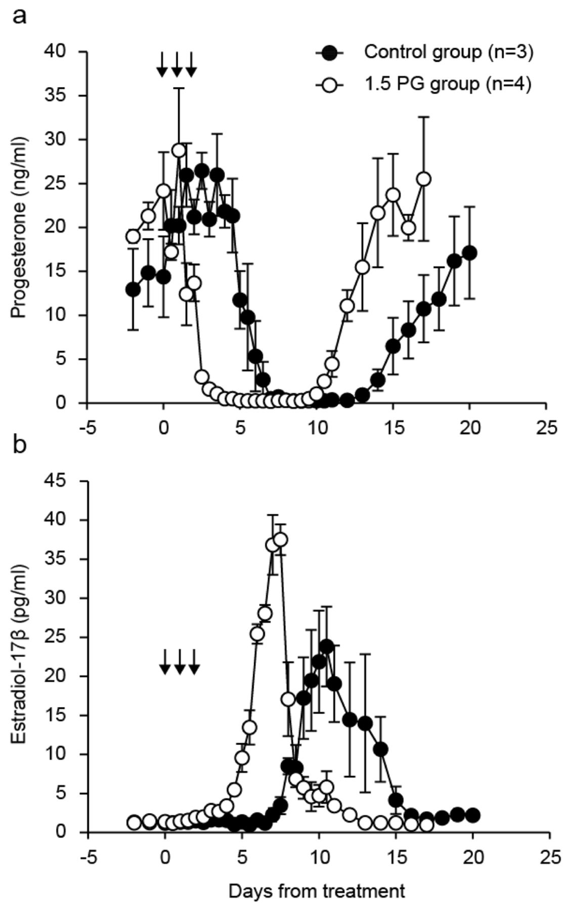
Ovarian steroid hormone profiles in the microminipigs after treatment are shown in Figure 1. Peripheral progesterone concentrations started to decrease (p<0.05) on day 6 in the control group and day 2.5 in the 1.5 PG group compared with concentrations at D0. Progesterone concentrations between D2 and D6.5 in the 1.5 PG group were significantly lower than those in the control group during the same period (p<0.05). There was a significant difference in progesterone levels on days 10, 10.5, 12, 15, 16 and 17 between the control and 1.5 PG groups (p<0.05). Estradiol-17β concentrations were significantly greater (p<0.05) on D9.5 in the control group and D5.5 in the 1.5 PG group than between D−2 and 0, respectively. The maximum estradiol-17β levels reached 30.7±1.0 pg/ml (day 11.3±0.9) in the control group and 38.8±2.8 pg/ml (day 7.3±0.1) in the 1.5 PG group. Concentrations of estradiol-17β from day 3 to 7.5 and from day 10 to 10.5 in the 1.5 PG group were higher and lower (p<0.05), respectively, than those in the control group.

Discussion

We clearly showed that triple administration of 1.5 mg PGF2α into the vulva during the functional luteal phase can shorten the inter-estrous interval in microminipigs. When female pigs were given 12.5 mg PGF2α 12 times (25) or 15 mg PGF2α six times intramuscularly (35, 36) during the luteal phase of the estrous cycle, inter-estrous intervals in the treatment groups (13.3-15.7 days) were significantly shortened compared to the control groups (19.8-21.2 days). Our results are in accordance with these inter-estrous intervals (25, 35, 36), thereby demonstrating that the estrous length in cyclic microminipigs can indeed be controlled with the fewer injections than that needed in domestic pigs.

Intramuscular treatment of a total of 90 mg dinoprost (6×15 mg dinoprost treatments) between 8 and 10 days from the last day of the estrous cycle induced luteolysis within 3 days after the first injection in all cyclic domestic pigs (average body weight of 176 kg) (36). In contrast the efficiency of shortening the estrous length in domestic pigs (average body weight of 167-199 kg) with treatment of 60 or 30 mg total dinoprost (6×5 or 10 mg dinoprost treatments) for the same period was 70% (36). From previous reports in domestic pigs, at least 75 mg dinoprost (approximately 0.6 mg/kg/experimental period) during the experimental period may be needed to induce luteolysis during the early luteal phase of the estrous cycle with high efficiency. Plasma progesterone concentrations, a general index of CL condition (37), in microminipigs treated with 1.5 mg intra-vulva dinoprost injection on D0 to D2 reached baseline 3.7 days from the first treatment day, which is similar to previous reports (25, 35, 36). In our research, total treatment dose (4.5 mg dinoprost) per experimental period (approximately 0.3 mg/kg/experimental period) in microminipigs treated with intra-vulval administration of PGF2α induced the decline of progesterone levels in the 1.5 PG group but not in the 0.75 PG group. In domestic pigs, it has been demonstrated that the effective dose for regressing CL function using intra-vulval PGF2α injection can be reduced to 25-50% of the effective intramuscular dose (29, 30). Our results and previous reports suggest that reaction of CL following exogenous PGF2α treatment is similar in microminipigs and domestic pigs.

Figure 1.
  • Download figure
  • Open in new tab
  • Download powerpoint
Figure 1.

Plasma concentrations of progesterone (a) and estradiol-17β (b) in the control group (n=3) and the group treated with 1.5 mg prostaglandin F2α (PG) (n=4) groups. Each arrow indicates a treatment day in the 1.5 PG group. Values are means±SEM.

Progesterone levels started to decrease on the last day of treatment in the 1.5 PG group. This finding is the same as those in previous reports using domestic pigs with multiple PGF2α injection (35, 36). Even though progesterone concentrations in domestic pigs gradually decreased following multiple PGF2α treatment, progesterone levels increased again after treatment when the CL maintained their function within 24 h after the final PGF2α treatment (36). Thus, we conclude that it is difficult to induce luteolysis with fewer than three 1.5 mg dinoprost injections in microminipigs.

Because estradiol-17β is secreted from large mature follicles in domestic pigs (37) and microminipigs (31), the profile of estradiol-17β in plasma reflect the development of mature follicles in the ovaries. The peak of estradiol-17β concentrations in the 1.5 PG group was defined at D7 or D7.5 in contrast to the control group (which varied from D10 to D13) in this study. This means that the protocol of PGF2α treatment in microminipigs affects not only CL regression but also synchronization of follicular development and subsequent estrous exhibition.

In conclusion, we indicate a new protocol for induction of CL regression and synchronization of estrous in female microminipigs using multiple PGF2α treatments. Development of assisted reproductive technology such as synchronization of estrus and ovulation serve to improve reproductive technologies and productive efficiency in animals. The new estrous synchronization protocols from our research will help to improve the efficiency of microminipig production.

Acknowledgements

This work was partly supported by JSPS KAKENHI Grant Numbers 26870455 and 16H05176

Footnotes

  • This article is freely accessible online.

  • Conflicts of Interest

    The Authors report no conflicts of interest exist in regard to this study.

  • Received August 19, 2017.
  • Revision received September 17, 2017.
  • Accepted September 19, 2017.
  • Copyright© 2017, International Institute of Anticancer Research (Dr. George J. Delinasios), All rights reserved

References

  1. ↵
    1. Kaneko N,
    2. Itoh K,
    3. Sugiyama A,
    4. Izumi Y
    : Microminipig, a non-rodent experimental animal optimized for life science research: Preface. J Pharmacol Sci 115: 112-114, 2011.
    OpenUrlCrossRef
  2. ↵
    1. Miura N,
    2. Kawaguchi H,
    3. Nagasato T,
    4. Yamada T,
    5. Ito T,
    6. Izumi H,
    7. Shameshima H,
    8. Miyoshi N,
    9. Tanimoto A,
    10. Maruyama I
    : Coagulation activity and white thrombus formation in the microminipig. In Vivo 27: 357-361, 2013.
    OpenUrlAbstract/FREE Full Text
    1. Kawaguchi H,
    2. Yamada T,
    3. Miura N,
    4. Takahashi Y,
    5. Yoshikawa T,
    6. Izumi H,
    7. Kawarasaki T,
    8. Miyoshi N,
    9. Tanimoto A
    : Reference values of hematological and biochemical parameters for the world smallest microminipigs. J Vet Med Sci 74: 933-936, 2012.
    OpenUrlCrossRefPubMed
    1. Kawaguchi H,
    2. Yamada T,
    3. Miura N,
    4. Noguchi M,
    5. Izumi H,
    6. Miyoshi N,
    7. Tanimoto A
    : Sex differences of serum lipid profile in novel microminipigs. In Vivo 27: 617-621, 2013.
    OpenUrlAbstract/FREE Full Text
  3. ↵
    1. Kakimoto T,
    2. Otsuka A,
    3. Kawaguchi H,
    4. Ogata K,
    5. Tanimoto A,
    6. Kanouchi H
    : Plasma homocysteine concentrations in novel Microminipigs. In Vivo 28: 579-582, 2014.
    OpenUrlAbstract/FREE Full Text
  4. ↵
    1. Miyoshi N,
    2. Horiuchi M,
    3. Inokuchi Y,
    4. Miyamoto Y,
    5. Miura N,
    6. Tokunaga S,
    7. Fujiki M,
    8. Izumi Y,
    9. Miyajima H,
    10. Nagata R,
    11. Misumi K,
    12. Takeuchi T,
    13. Tanimoto A,
    14. Yasuda N,
    15. Yoshida H,
    16. Kawaguchi H
    : Novel microminipig model of atherosclerosis by high fat and high cholesterol diet, established in Japan. In Vivo 24: 671-680, 2010.
    OpenUrlAbstract/FREE Full Text
    1. Kawaguchi H,
    2. Miyoshi N,
    3. Miura N,
    4. Fujiki M,
    5. Horiuchi M,
    6. Izumi Y,
    7. Miyajima H,
    8. Nagata R,
    9. Misumi K,
    10. Takeuchi T,
    11. Tanimoto A,
    12. Yoshida H
    : Microminipig, a non-rodent experimental animal optimized for life science research: novel atherosclerosis model induced by high fat and cholesterol diet. J Pharmacol Sci 115: 115-121, 2011.
    OpenUrlCrossRef
    1. Akioka K,
    2. Kawaguchi H,
    3. Kitajima S,
    4. Miura N,
    5. Noguchi M,
    6. Horiuchi M,
    7. Miyoshi N,
    8. Tanimoto A
    : Investigation of necessity of sodium cholate and minimal required amount of cholesterol for dietary induction of atherosclerosis in microminipigs In Vivo 28: 81-90, 2014.
    OpenUrlAbstract/FREE Full Text
  5. ↵
    1. Kawaguchi H,
    2. Yamada T,
    3. Miura N,
    4. Ayaori M,
    5. Uto-Kondo H,
    6. Ikegawa M,
    7. Noguchi M,
    8. Wang K-Y,
    9. Izumi H,
    10. Tanimoto A
    : Rapid development of atherosclerosis in the world's smallest microminipig fed a high-fat/high-cholesterol diet: A useful animal model due to its size and similarity to human pathophysiology. J Atheroscler Thromb 21: 186-203, 2014.
    OpenUrlCrossRefPubMed
  6. ↵
    1. Sugiyama A,
    2. Nakamura Y,
    3. Akie Y,
    4. Saito H,
    5. Izumi Y,
    6. Yamazaki H,
    7. Kaneko N,
    8. Itoh K
    : Microminipig, a non-rodent experimental animal optimized for life science research: in vivo proarrhythmia models of drug-induced long QT syndrome: development of chronic atrioventricular block model of microminipig. J Pharmacol Sci 115: 122-126, 2011.
    OpenUrlCrossRef
    1. Yokoyama H,
    2. Nakamura Y,
    3. Saito H,
    4. Nagayama Y,
    5. Hoshiai K,
    6. Wada T,
    7. Izumi-Nakaseko H,
    8. Ando K,
    9. Akie Y,
    10. Sugiyama A
    : Pharmacological characterization of microminipig as a model to assess the drug-induced cardiovascular responses for non-clinical toxicity and/or safety pharmacology studies. J Toxicol Sci 42: 93-101, 2017.
    OpenUrl
  7. ↵
    1. Zahorul Islam M,
    2. Kawaguchi H,
    3. Miura N,
    4. Miyoshi N,
    5. Yamazaki-Himeno E,
    6. Shiraishi M,
    7. Miyamoto A,
    8. Tanimoto A
    : Hypertension alters the endothelial-dependent biphasic response of bradykinin in isolated Microminipig basilar artery. Microvasc Res 114: 52-57, 2017.
    OpenUrl
  8. ↵
    1. Takeishi K,
    2. Horiuchi M,
    3. Kawaguchi H,
    4. Deguchi Y,
    5. Izumi H,
    6. Arimura E,
    7. Kuchiiwa S,
    8. Tanimoto A,
    9. Takeuchi T
    : Acupuncture improves sleep conditions of minipigs representing diurnal animals through an anatomically similar point to the acupoint (GV20) effective for humans. Evid Based Complement Alternat Med 2012: 472982, 2012.
    OpenUrlCrossRefPubMed
  9. ↵
    1. Yoshikawa T,
    2. Takahashi Y,
    3. Kawaguchi H,
    4. Utsunomiya S,
    5. Miura N,
    6. Izumi H,
    7. Miyoshi N,
    8. Tanimoto A
    : A dermal phototoxicity study following intravenous infusion administration of ciprofloxacin hydrochloride in the novel microminipigs. Toxicol Pathol 41: 109-113, 2013.
    OpenUrlCrossRefPubMed
  10. ↵
    1. Guruge KS,
    2. Noguchi M,
    3. Yoshioka K,
    4. Yamazaki E,
    5. Taniyasu S,
    6. Yoshioka M,
    7. Yamanaka N,
    8. Ikezawa M,
    9. Tanimura N,
    10. Sato M,
    11. Yamashita N,
    12. Kawaguchi H
    : Microminipigs as a new experimental animal model for toxicological studies: comparative pharmacokinetics of perfluoroalkyl acids. J Applied Toxicol 36: 68-75, 2016.
    OpenUrlCrossRefPubMed
  11. ↵
    1. Miura N,
    2. Kucho K,
    3. Noguchi M,
    4. Miyoshi N,
    5. Uchiumi T,
    6. Kawaguchi H,
    7. Tanimoto A
    : Comparison of the genomic sequence of the Microminipig, a novel breed of swine, with a genomic database for conventional pig. In Vivo 28: 1107-1112, 2014.
    OpenUrlAbstract/FREE Full Text
  12. ↵
    1. Miyoshi K,
    2. Kawaguchi H,
    3. Maeda K,
    4. Sato M,
    5. Akioka K,
    6. Noguchi M,
    7. Horiuchi M,
    8. Tanimoto Akihide
    : Birth of cloned Microminipigs derived from somatic cell nuclear transfer embryos that have been transiently treated with valproic acid. Cellular Reprogramming 18: 390-400, 2016.
    OpenUrl
  13. ↵
    1. Yamada S,
    2. Kawaguchi H,
    3. Yamada T,
    4. Guo X,
    5. Matsuo K,
    6. Hamada T,
    7. Miura N,
    8. Tasaki T,
    9. Tanimoto A
    : Cholic acid enhances visceral adiposity, atherosclerosis and nonalcoholic fatty liver disease in microminipigs. J Atheroscler Thromb, 2017. doi: 10.5551/jat.39909. [Epub ahead of print]
  14. ↵
    1. Martinez EA,
    2. Vazquez JM,
    3. Roca J,
    4. Cuello C,
    5. Gil MA,
    6. Parrilla I,
    7. Vazquez JL
    : An update on reproductive technologies with potential short-term application in pig production. Reprod Dom Anim 40: 300-309, 2005.
    OpenUrl
  15. ↵
    1. Niemann H,
    2. Rath D
    : Progress in reproductive biotechnology in swine. Theriogenology 56: 1291-1304, 2001.
    OpenUrlCrossRefPubMed
  16. ↵
    1. Vodčka P,
    2. Smetana K,
    3. Dvořánková B,
    4. Emerick T,
    5. Xu YZ,
    6. Ourednik J,
    7. Ourednik V,
    8. Motlík J
    : The miniature pig as an animal model in biomedical research. Ann NY Acad Sci 1049: 161-171, 2005.
    OpenUrlCrossRefPubMed
  17. ↵
    1. Weems CW,
    2. Weems YS,
    3. Randel RD
    : Prostaglandins and reproduction in female farm animals. Vet J 171: 206-228, 2006.
    OpenUrlCrossRefPubMed
  18. ↵
    1. Diehl JR,
    2. Day BN
    : Effect of prostaglandin F2α on luteal function in swine. J Anim Sci 39: 392-396, 1974.
    OpenUrlPubMed
    1. Hallford DM,
    2. Wettemann RP,
    3. Turman EJ,
    4. Omtvedt IT
    : Luteal function in gilts after prostaglandin F2α. 41: 1706-1710, 1975.
    OpenUrl
  19. ↵
    1. Estill CT,
    2. Britt JF,
    3. Gadsby JE
    : Repeated administration of prostaglandin F2 alpha during the early luteal phase causes premature luteolysis in the pig. Biol Reprod 49: 181-185, 1993.
    OpenUrlAbstract
  20. ↵
    1. Noguchi M,
    2. Yoshioka K,
    3. Hikono H,
    4. Suzuki C,
    5. Kikuchi K
    : Effect of semen extenders on frozen-thawed boar sperm characteristics and distribution in the female genital tract after deep intrauterine insemination in sows. Anim Reprod Sci 163: 164-171, 2015.
    OpenUrl
  21. ↵
    1. Yoshioka K,
    2. Suzuki C,
    3. Tanaka A,
    4. Anas IMK,
    5. Iwamura S
    : Birth of piglets derived from porcine zygotes cultured in a chemically defined medium. Biol Reprod 66: 112-119, 2002.
    OpenUrlAbstract/FREE Full Text
  22. ↵
    1. Yoshioka K,
    2. Noguchi M,
    3. Suzuki C
    : Production of piglets from in vitro-produced embryos following non-surgical transfer. Anim Reprod Sci 131: 23-29, 2012.
    OpenUrlPubMed
  23. ↵
    1. Kirkwood RN,
    2. Thacker PA,
    3. Aherne FX,
    4. Goonewardene LA
    : The effect of dose and route of administration of prostaglandin F2α on the parturient response of sows. J Swine Health Prod 4: 123-126, 1996.
    OpenUrl
  24. ↵
    1. Kaeoket K
    . The effect of dose and route of administration of R-cloprostenol on the parturient response of sows: Reprod Dom Anim 41: 472-476, 2006.
    OpenUrl
  25. ↵
    1. Noguchi M,
    2. Miura N,
    3. Ando T,
    4. Kubota C,
    5. Hobo S,
    6. Kawaguchi H,
    7. Tanimoto A
    : Profiles of reproductive hormone in the Microminipig during the normal estrous cycle. In Vivo 29: 17-22, 2015.
    OpenUrlAbstract/FREE Full Text
  26. ↵
    1. Noguchi M,
    2. Ikedo T,
    3. Kawaguchi H,
    4. Tanimoto A
    : Estrous synchronization in microminipig using estradiol dipropionate and prostaglandin F2α. J Reprod Dev 65: 373-378, 2016.
    OpenUrl
  27. ↵
    1. Noguchi M,
    2. Yoshioka K,
    3. Kaneko H,
    4. Iwamura S,
    5. Takahashi T,
    6. Suzuki C,
    7. Arai S,
    8. Wada Y,
    9. Itoh S
    : Measurement of porcine luteinizing hormone concentration in blood by time-resolved fluoroimmunoassay. J Vet Med Sci 70: 1291-1294, 2007.
    OpenUrl
  28. ↵
    1. Glantz SA,
    2. Slinker BK
    : Repeated measures: In: Primer of Applied Regression and Analysis of Variance. New York: McGraw-Hill, pp. 381-463, 1990.
  29. ↵
    1. Kohyama Y,
    2. Iwamura S,
    3. Somei H,
    4. Maruyama A,
    5. Naito M
    : Shortening of estrous cycle in pigs by the repeated administration of prostaglandin F2α and their reproductive performance. Jpn J Swine Science 43: 39-48, 2006.
    OpenUrl
  30. ↵
    1. Kohyama Y,
    2. Iwamura S,
    3. Somei H,
    4. Maruyama A
    : Effects of PGF2α dose on the inter-estrous interval and reproductive performance in pigs. Jpn J Swine Science 47: 181-186, 2010.
    OpenUrl
  31. ↵
    1. Noguchi M,
    2. Yoshioka K,
    3. Itoh S,
    4. Suzuki C,
    5. Arai S,
    6. Wada Y,
    7. Hasegawa Y,
    8. Kaneko H
    : Peripheral concentrations of inhibin A, ovarian steroids, and gonadotropins associated with follicular development throughout the estrous cycle of the sow. Reproduction 139: 153-161, 2010.
    OpenUrlAbstract/FREE Full Text
PreviousNext
Back to top

In this issue

In Vivo: 31 (6)
In Vivo
Vol. 31, Issue 6
November-December 2017
  • Table of Contents
  • Table of Contents (PDF)
  • Index by author
  • Back Matter (PDF)
  • Ed Board (PDF)
  • Front Matter (PDF)
Print
Download PDF
Article Alerts
Sign In to Email Alerts with your Email Address
Email Article

Thank you for your interest in spreading the word on In Vivo.

NOTE: We only request your email address so that the person you are recommending the page to knows that you wanted them to see it, and that it is not junk mail. We do not capture any email address.

Enter multiple addresses on separate lines or separate them with commas.
Corpus luteum Regression Induced by Prostaglandin F2α in Microminipigs During the Normal Estrous Cycle
(Your Name) has sent you a message from In Vivo
(Your Name) thought you would like to see the In Vivo web site.
CAPTCHA
This question is for testing whether or not you are a human visitor and to prevent automated spam submissions.
11 + 7 =
Solve this simple math problem and enter the result. E.g. for 1+3, enter 4.
Citation Tools
Corpus luteum Regression Induced by Prostaglandin F2α in Microminipigs During the Normal Estrous Cycle
MICHIKO NOGUCHI, MASAYA HIRATA, HIROAKI KAWAGUCHI, AKIHIDE TANIMOTO
In Vivo Nov 2017, 31 (6) 1097-1101;

Citation Manager Formats

  • BibTeX
  • Bookends
  • EasyBib
  • EndNote (tagged)
  • EndNote 8 (xml)
  • Medlars
  • Mendeley
  • Papers
  • RefWorks Tagged
  • Ref Manager
  • RIS
  • Zotero
Reprints and Permissions
Share
Corpus luteum Regression Induced by Prostaglandin F2α in Microminipigs During the Normal Estrous Cycle
MICHIKO NOGUCHI, MASAYA HIRATA, HIROAKI KAWAGUCHI, AKIHIDE TANIMOTO
In Vivo Nov 2017, 31 (6) 1097-1101;
del.icio.us logo Digg logo Reddit logo Twitter logo Facebook logo Google logo Mendeley logo
  • Tweet Widget
  • Facebook Like
  • Google Plus One

Jump to section

  • Article
    • Abstract
    • Materials and Methods
    • Results
    • Discussion
    • Acknowledgements
    • Footnotes
    • References
  • Figures & Data
  • Info & Metrics
  • PDF

Related Articles

  • No related articles found.
  • PubMed
  • Google Scholar

Cited By...

  • No citing articles found.
  • Google Scholar

More in this TOC Section

  • Non-invasively Imageable Tibia-tumor-fragment Implantation Experimental-bone-metastasis Mouse Model of GFP-expressing Prostate Cancer
  • Protective Effects of Gamma-mangostin on Hydrogen Peroxideinduced Cytotoxicity in Human Retinal Pigment Epithelial Cells
  • The First Mouse Model of Meckel’s Diverticulum Carcinoma
Show more Experimental Studies

Similar Articles

Keywords

  • Miniature pig
  • estrous synchronization
  • prostaglandin F2α
In Vivo

© 2022 In Vivo

Powered by HighWire