


Abstract
Background/Aim: The high variability of primary cells propagated in vitro led us to study the expression patterns of 11 most commonly accepted and widely used biomarkers specific for prostate cancer (PC) cells in primary cell models. Materials and Methods: Primary PC cells from five PC patients were partially subjected to RNA preparation immediately and remaining cells were propagated up to 84 days followed by RNA preparation. Subsequently, biomarker mRNA quantification was performed by quantitative reverse transcription-polymerase chain reaction (RT-PCR) and biomarker transcript concentrations before and after cultivation of primary PC cells were compared. Results: Evaluation of androgen receptor, prostate-specific antigen, acid phosphatase, prostate-specific membrane antigen, fatty acid synthase, cytokeratin types 5/8/19, E-cadherin, epithelial cell adhesion molecule and fibroblast-specific protein 1 demonstrated temporal changes, as well as individual differences in expression, during primary PC cell propagation. Conclusion: Experimental design, as well as data evaluation, may need to take under consideration the high variability of biomarker expression in primary PC cells.
Prostate cancer (PC) is the most common malignancy and leading cause of oncological mortality in men in industrialized countries (1). Key risk factors for incurrence and progression of PC are advancing age, fatty Western diet and the genetic background as reflected in family history (2). Despite the availability of new generation drugs, current PC treatment regimens suffer from a lack of understanding the metastatic phenotype, identification of metastasis enabling genes and, in consequence, suitable biomarkers for diagnosis, prediction and treatment response. Diagnosis of PC includes measurement of serum prostate-specific antigen (PSA), rectal examination and histological inspection of prostate needle biopsies. Since PSA is also a marker of prostate inflammation or benign prostatic hyperplasia novel biomarkers or verification of currently used diagnostic biomarkers may improve the detection of PC (3).
A major challenge in prostate cancer research remains the identification and characterization of molecular signaling and effector pathways. Examination of PC tumor biology is primarily based on three established model systems, namely patient samples, animal models and cell culture models. However, none of these models are perfect, with all of them presenting serious limitations in their application and handling properties. Conservative histology, as well as modern screening approaches based on high-throughput assessment of patient's material, only provide a descriptive point of view. Even in case of “-omic” approaches combined with complex bioinformatic analysis tools, experimental evaluation is required to strengthen estimated results (4). Animal models are extremely useful in understanding complex orchestration of tumor-specific interference with the microenvironment, as well as with global physiology, e.g. the hormone system (5). On the other hand, animal experimental evidence is limited by species barrier and, thus, is not necessarily applicable to humans (“men are not mice”). Cell culture systems, commonly persist of established cell lines with unlimited proliferation capacity, represent an easy to handle in vitro culture model with numerous applications in experimental cancer research. Genetic engineering, as well as incubation approaches using small molecules (e.g. specific inhibitors and activators, siRNA technology), offer a wide range of mechanistic studies. However, limitations are originated in degeneration processes during cell line propagation. A multitude of cell lines have been isolated even decades ago and cultured in non-physiological in vitro conditions. Therefore, primary characteristics, which are not essential under cell culture conditions, have been frequently altered or disappeared during laboratory handling procedures (6, 7).
In 1983, cultivation of primary PC cells was established by Donna M. Peehl and has since then been steadily evolved (8-11). Hereupon, numerous studies were published utilizing primary PC cells in in vitro and in xenograft models (12, 13), combining advantages of cell culture models and patients' samples. While it is believed that primary PC culture reflects predominantly characteristics of the native PC tissue, this model suffers from lifespan limitation and particularly from genetic instability. The high variability of primary cells propagated in vitro led us to study the expression patterns of 11 biomarkers specific for PC detection before and after cultivation of primary PC cells.
Materials and Methods
Preparation and cultivation of primary PC cells. Primary PC cells were prepared from PC samples of 5 patients undergoing radical prostatectomy at the Department of Urology, University Medicine Greifswald, Germany. Areas of malignancy were evaluated by histopathological analysis via frozen section carried out by skilled pathologists. Tumor samples were processed for primary cell culture within 2 h after radical prostatectomy. This study was approved by the Ethics Committee of the University Medicine Greifswald (registration no. BB 21/12) and all patients signed informed consent forms.
For preparation and propagation of primary PC cells, the PC tissue was mechanically disintegrated by using a sterile scalpel and enzymatically digested with collagenase I (Sigma-Aldrich, München, Germany). Subsequently, cancer cell spheroids were stepwise filtered through 70 μm and 40 μm pore size filters and cultured in low-attachment 24-well cell culture plates (Corning Incorporated, Corning, NY, USA) in Stem Pro hESC stem cell medium (Life Technologies, Darmstadt, Germany) at 37°C and 5% CO2 in a humidified atmosphere.
Cultivation of LNCaP PC cell line. The human PC cell line LNCaP was purchased from Cell Lines Service (CLS, Eppelheim, Germany). Cells were maintained in RPMI 1640 medium supplemented with 10% fetal bovine serum, 1% pyruvate and 100 units/ml penicillin/streptomycin (all PAN Biotech, Aidenbach, Germany) at 37°C and 5% CO2 in a humidified atmosphere. LNCaP cells served as an in vitro PC cell culture model for establishing biomarker mRNA detection as described below.
Design and optimization of oligonucleotides. To compare the pattern of biomarker alteration during primary PC cell propagation, oligonucleotides specific for prostatic acid phosphatase (ACPP), androgen receptor (AR), E-cadherin (CDH1), cytokeratins type 5 (CK5), cytokeratins type 8 (CK8), cytokeratins type 19 (CK19), epithelial cell adhesion molecule (EpCam), fatty acid synthase (FASN), fibroblast-specific protein 1 (FSP1), prostate-specific antigen (PSA) and prostate-specific membrane antigen (PSMA) mRNA detection were utilized. Oligonucleotide sequences were chosen to amplify regions of 100 to 150 bases of target mRNA with a G/C content of 40-60% according to general recommendations of polymerase chain reaction (PCR) oligonucleotide design (14). Target specific sequences were obtained from Entrez Nucleotide from National Center for Biotechnology Information (NCBI) web (http://www.ncbi.nlm.nih.gov) and processed using Bioedit software version 7.2.5 (http://www.mbio.ncsu.edu).
RNA isolation and quantitative reverse transcription-polymerase chain reaction (RT-PCR). Total RNA was isolated with peqGOLD Trifast reagent (VWR International, Erlangen, Germany) according to manufacturer's instructions and applied in cDNA synthesis performed with M-MLV Reverse Transcriptase (Promega, Madison, WI, USA) in a Thermocycler T3000 (Biometra, Göttingen, Germany). Subsequently, real-time PCR was carried out applying the CFX96 Real-Time System (Biorad, München, Germany) by performing a standardized protocol over 45 cycles. Each reaction was performed in a final volume of 20 μl containing 3 μl of cDNA, 1 μl of forward and reverse primer each (10 μM), 5 μl millipore water and 10 μl SensiMix SYBR (Bioline, Luckenwalde, Germany). All PCR reactions were carried out in duplicates. Ribosomal protein, large P0 (RPLP0) mRNA served as reference. Quantification was performed by the delta delta Ct method (15).
Results
Rationale for biomarker selection suitable to detect primary PC cells. In the present study we analyzed applicability and benefit of 11 biomarkers commonly used for the characterization of PC tissue samples in experimental approaches (Table I). The androgen receptor (AR) is key player in PC cell growth and, therefore, the primary target of PC chemotherapy. Although AR is not specific for PC cells, its expression is characteristic for these cells (16). Beyond that, prostate-specific antigen (PSA), prostatic acid phosphatase (ACPP) and prostate-specific membrane antigen (PSMA) play direct role in prostate physiology and, therefore, are commonly used as PC correlated biomarkers for a long-time (17, 18). While overexpressed in most carcinomas, fatty acid synthase (FASN) is equally defined in terms of PC (19). Cytokeratins type 5 (CK5), type 8 (CK8) and type 19 (CK19) were applied to differentiate non-malignant from malignant PC cells (20-22). E-cadherin (CDH1) and epithelial cell adhesion molecule (EpCam) are epithelial surface proteins governing cellular adhesion activity and were found to be specific for epithelial cells. Due to their adhesive capacity, both proteins are involved in epithelial-to-mesenchymal transition (EMT) and, therefore, critical biomarkers for cell motility and metastasis (23–25). Finally, fibroblast-specific protein 1 (FSP1) has been used to differentiate epithelial from mesenchymal cell origins (26).
Abbreviation, target gene, National Center for Biotechnology Information identification number (NCBI ID) and cellular function of 11 commonly accepted biomarkers for primary PC cell cultivation.
Establishment of quantitative RT-PCR detection of biomarkers. Quantitative RT-PCR analysis was carried out applying oligonucleotides as defined in Table II, with the calculated length of specific PCR products being verified by agarose gel electrophoresis (data not shown). Optimization of quantitative RT-PCR was done using the in vitro PC LNCaP cell model system (Figure 1). Biomarkers specific for PC (AR, PSA, ACPP, PSMA), general malignancy (FASN, CK5, CK8, CK19), as well as epithelial cells and EMT (CDH1, EpCam) were reproducibly detectable in LNCaP cells. Compared to AR mRNA, remaining biomarker transcripts were detected showing up to 10-fold- (ACCP, CK5, FSP1), up to 100-fold- (CDH1, FASN), as far as up to 1,000-fold-increased (CK8, EpCam, PSA, PSMA) mRNA amounts. Surprisingly, the in vitro epithelial cell culture model LNCaP was positive for FSP1 mRNA, a biomarker specific for cells of the fibroblast type.
Alteration of biomarkers during cell propagation of primary PC cells. PC samples were analyzed from 5 patients treated by total prostatectomy. The diagnosis was histologically verified by tumor biopsy. The patients received no antitumor therapy before. At the time of surgical intervention, the age of the patients was between 58 and 71 years and the inital Gleason score ranged from 7a to 7b. Patients' characteristics and related clinical data are provided within Table III. Extracted PC cells from all five patients were partially subjected to extraction of total RNA immediately and remaining cells were cultivated up to 84 days followed by RNA extraction.
Compared to LNCaP cells, the biomarker expression was generally lower in all primary cell culture samples. Moreover, the analyzed biomarkers showed highly heterogeneous expression patterns after primary cell propagation when compared to the PC samples analyzed directly after total prostatectomy (Figure 2). Cancer and epithelial/EMT marker molecules CK5, CK8, CK19 and EpCam expression elevated during propagation. The increase in biomarker mRNA averaged 223.4-fold (CK5), 312.4-fold (CK8), 102.4-fold (CK19) and 127.3-fold (EpCam) when compared to the intial mRNA level before cultivation. RNA preparations of patient no. 3 showed a decrease of CK8 (0.8-fold) and CK19 (0.4-fold) mRNA. In samples from patient no. 1, however, no CK5 mRNA was detectable after primary propagation.
DNA sequences of forward and reverse oligonucleotides specific for 11 commonly accepted biomarkers for primary PC cell cultivation.
In contrast, mRNA levels of PC-specific ACPP and PSMA, epithelial/EMT-specific CDH1 and fibroblast-specific FSP1 decreased by trend. In the majority of cases, biomarker mRNA was no longer detectable after primary cultivation. In samples obtained from patient no. 1 (CDH1; 820.3-fold) and in patient no. 4 (FSP1; 12.9-fold), the mRNA amount increased compared to the initial concentration. In patient's no. 3 RNA samples no PSMA mRNA has been detected neither before nor after primary cell cultivation. The fibroblast-specific FSP1 transcript was initially detectable in all of the 5 preparations, which was somewhat unsurprising and may reflect contamination by fibroblasts. It is possible that, due to the epithelial cell-specific composition of the cell culture medium, FSP1 expression sank during primary cell cultivation.
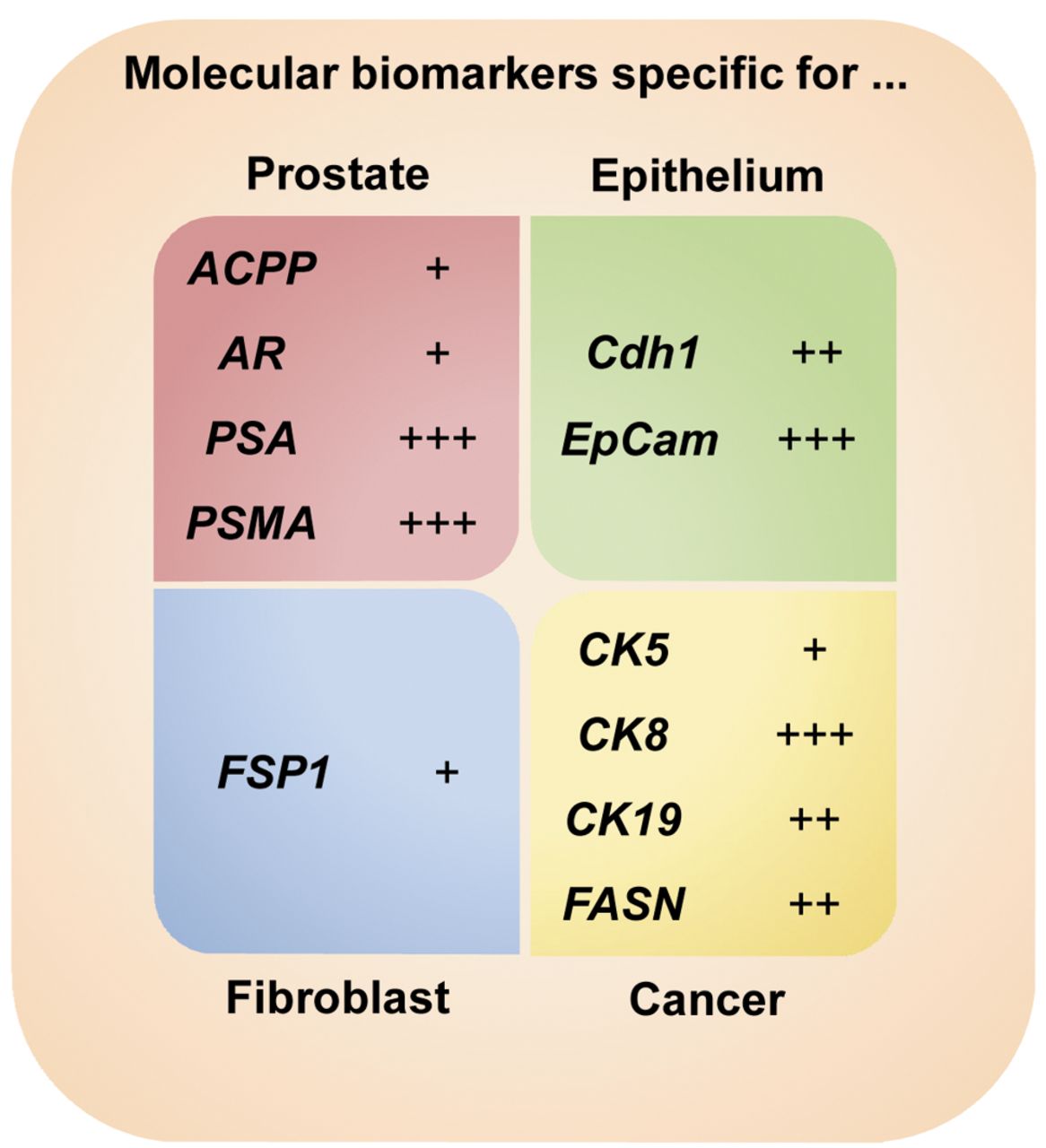
Detection of biomarker mRNA specific for cells of prostate, epithelium, fibroblast and cancer origin. mRNA amounts were normalized to AR transcript concentration and expressed as increased up to 10-fold (+), up to 100-fold (++) and up to 1,000-fold (+++).
Transcript steady-state levels of FASN and PSA have been detected highly variable in all patients with no clear tendency observed. Our examinations demonstrated mRNA concentration alterations from 0.1-fold (FASN; patient no. 3) and <0.1-fold (PSA; patient no. 4) decrease up to 16,158.4-fold (FASN; patient no. 1) and 596.3-fold (PSA; patient no. 5) increase, respectively. Finally, all of the samples were AR mRNA-negative.
Discussion
In the present study we examined the expression pattern of cancer-, prostate- and cell type-specific biomarker mRNA in primary PC cells during cultivation over a period time of up to 84 days. Overall, our data implicate a highly variable expression of well-known biomarkers at the mRNA level when comparing biomarker expression of the individual tumor tissue and analysing biomarker expression immediately after surgery in comparison to expression after cultivation of extracted primary cells. Our findings (i) put the suitability of the used biomarkers into question and (ii) shed further light on the progression-dependent, as well as the individual, biomarker expression in PC cells.
Patients' characteristics and related clinical data of PC patients undergoing radical prostatectomy at the Department of Urology, University Medicine Greifswald, Germany.
In general, RT-PCR is a highly acknowledged method of choice when it comes to high-throughput screening of biological samples. One important prerequisite is the fast and efficient purification of total RNA under denaturing conditions to strongly inactivate potent cellular and environmental RNases. However, the likelihood of partial degradation increases with length of the targeted mRNA, while the use of short amplicons increases the possibility of proper amplification from the obtained cDNA. In comparison to enzyme immunoassays, RT-PCR is simple to establish and very robust and can be used solely or in combination with, e.g., enzyme-linked immunosorbent assay (ELISA). Therefore, the validity of biomarker analyses should be taken into consideration when characterizing cells prepared from tumor tissue. For instance, FSP1 mRNA was detectable in LNCaP epithelial cell line preparations. This observation may cast into doubt the use of FSP1 as a specific biomarker for fibroblasts, particularly in the context of PC cells derived from metastatic tumor tissue (27). However, although it is frequently assumed that gene expression is correlated at the level of mRNA and protein, this is not always the case and transfer of biomarkers validated at the protein level to mRNA and vice versa has to be done with care. In our case, ACPP mRNA was used as a marker specific for PC. The corresponding protein is primarily used for immunohistochemical staining of PC and for differentiation of PC and bladder cancer (17, 28). This might indicate its limited benefit as a biomarker for PC at the level of mRNA. However, we were able to detect ACPP in the tissue samples of all five patients. The fact that ACPP was not detectable at all after cultivation of primary cells might be explained by the altered gene expression due to limitations during PC primary cell cultivation. The microenvironment appears important in cancer cell specification. From this, it follows that the use of artificial culture conditions may lead to a loss of environmental signals and a subsequent shift in cellular properties (29). Even by applying optimized and conditioned medium, cellular growth of non-malignant cells from the tumor environment, e.g. fibroblasts and blood cells, may weaken biomarker's significance.
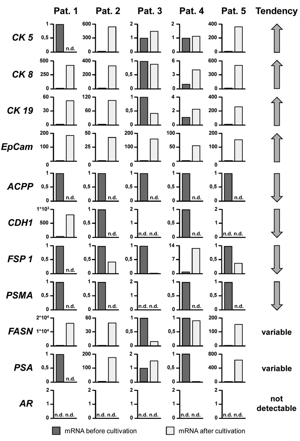
Transcript steady-state levels of 11 commonly accepted biomarkers for primary PC cell cultivation in preparations of prostate samples from 5 PC patients (pat). mRNA levels of biomarkers were relatively expressed before and after cultivation of primary PC cells and evaluated as increased (⇑), decreased (⇓), variable and not detectable (n.d.).
Furthermore, despite the controversy about PSA as a valid PC marker (30), PSA protein is expressed in the epithelial layer of the prostate and may be spread, e.g., during PC or inflammation upon epithelial disruption into the blood stream. Thus, expression of PSA is not specific for PC. However, since it is found in large amounts in prostatic tissue, PSA may be a valid indicator for cells with prostate origin. The observed high relative variability of expression between tissue sample of radical prostatectomy and cultured primary PC cells may, thus, reflect differences in the proportion of PC cells and fibroblast per donor, respectively.
In our study, the fibroblast-specific biomarker FSP1 has been detected in all 5 primary preparations suggesting fibroblasts in the PC preparation. However, FSP1 expression sank after primary cell propagation, whereas the epithelial biomarker EpCam increased. This may reflect a shift in epithelial cell type/fibroblast cell type ratio to more epithelial cells. Additionally, the presence of FPS1, as well as the loss of CDH1 during primary cell culture, may be regarded as the suppression of epithelial cell properties due to EMT during primary cell propagation (31, 32).
Consequently, temporal changes in biomarker expression need to be taken into account. Presumably, due to the in vitro conditions during primary PC culture, our analysis displayed highly variable expression rates of the 11 biomarkers examined in this study. For instance, alterability of the FASN expression varied from a 1.6×10⁴-fold increase (patient 1) to a decrease to 0.1-fold expression (patient 3). There are also some other biomarkers demonstrating a variability in expression over two magnitudes (CK5, CK8, PSA). Notably, ACPP and PSMA expression disappeared during propagation and, thus, demonstrating both of these factors as early biomarkers of PC. Some studies affirm highly variable expression of cancer-specific biomarkers in PC cells and PC patients (33-36), as well as in other malignancies, including cancer of lung, breast and ovary (35, 37, 38). However, there are indications that even anticancer treatment may cause variations in biomarker expression rates (37-39). Although cultivation of primary PC cells varied, no correlation between incubation time and expression levels was observable.
In conclusion, our study evaluated the 11 most commonly accepted and widely used biomarkers for primary PC cell characterization and revealed a temporal and highly individual course of biomarker expression at the mRNA level. Since the molecular characterization is prerequisite for a primary PC cell model, application of valid biomarker candidates combined with suitable detection methods is of great importance. Literature data, as well as our data presented here, suggest that some of the generally accepted biomarkers in PC research fail to be specific enough to distinguish significantly between type, origin and malignancy of primary PC cells. Particularly with regard to biomarkers, which have been defined long ago, the validity may be limited and should be reproved under application of recent techniques. In the near future, some new appropriate biomarkers, e.g. microRNAs, may be additionally included into the panel of cancer-specific and PC-specific factors. Finally, as a practical conclusion, experimental design, as well as proper data evaluation, may need to take under consideration the high variability of biomarker expression in primary PC cells.
Acknowledgements
The Authors thank Katja Wittig for excellent technical assistance and they are grateful to Dr. Matthias Saar for fruitful discussion.
Footnotes
Conflicts of Interests
The Authors have declared that no competing interests exist.
- Received April 25, 2016.
- Revision received May 31, 2016.
- Accepted June 1, 2016.
- Copyright © 2016 International Institute of Anticancer Research (Dr. John G. Delinassios), All rights reserved


