


Abstract
Although a large number of Kampo, Japanese traditional medicines, have been used for the treatment of oral diseases, little is known on their relative potency and endotoxin contamination. In order to obtain basic data for clinical applications, 10 Kampo, and 25 constituent plant extracts were tested for the contamination of lipopolysaccharide (LPS)-like substances, and anti-inflammatory activity. Human gingival (HGF) and periodontal ligament fibroblasts (HPLF) were cultured in 10% fetal bovine serum supplemented with Dulbecco's modified Eagle's medium. Viable cell number was measured by 3-(4, 5-dimethylthiazol-2-yl)-2,5-diphenyltetrazolium bromide method. Prostaglandin (PGE2) was determined by enzyme immunoassay. Cyclo-oxygenase (COX)-1 and COX-2 protein expressions were determined by western blot. COX activity was measured using Cox Inhibitor Screening Assay Kit. LPS, quantified by Endotoxin assay kit, was undetectable or relatively low in the test samples except for rikkosan and unseiin. Hangeshashinto potently inhibited PGE2 production by interleukin (IL)-1β-stimulated HPLFs and HGFs. Hangeshashinto suppressed the expression of COX-2 protein, but not that of COX-1 protein in IL-1β-induced HGF cells. Hangeshashinto slightly, but not significantly, inhibited both COX-1 and COX-2 activity. The present study provides the basis for clinical application of hangeshashinto for the treatment of stomatitis.
- Anti-inflammatory activity
- human fibroblasts
- LPS contamination
- Kampo medicines
- constituent plant extracts
- hangeshashinto
Kampo, Japanese traditional medicines are defined as drugs that are produced by mixing multiple numbers of “seeds, leaves, rhizomes, roots and shells of natural kingdom, and insects” that exhibit medicinal effects under the constant law. Kampo medicine prescriptions were systematically defined through the thousand years' investigations of the combination effects of many herbal drugs and hazardous effects. The number of species of constituent plant extracts (usually prepared by hot-water extraction or appropriate solvent and lyophilized) presently used in Kampo medicines nearly totals 300. More than 80 reports have investigated the anti-inflammatory effect of Kampo medicines [reviewed in (1) and (2)]. However, basic research and clinical studies for the treatment of oral diseases is much less, including our studies (3-19). Furthermore, little is known on the relative potency of Kampo medicines and their active ingredients. As far as we know, there is no report on the contamination of Kampo medicine by bacterial products derived from the soil such as endotoxin (lipopolysaccharide, LPS).
In the present study, we first investigated LPS contamination in 10 Kampo medicines and 25 constituent plant extracts (supplied as lyophilized materials of hot-water extracts). It was recently reported that orento (7), shosaikoto (8) and hangeshashinto (9) prevented LPS-stimulated inflammation of human gingival fibroblasts (HGFs). However, we found that interleukin (IL)-1β stimulated the prosta-glandin (PGE2), IL-6, IL-8 and monocyte chemoattractant protein-1 (MCP-1) production by HGFs to much higher extent than that achieved by LPS prepared from Escherichia coli and Porphyromonas gingivalis (20). Therefore, we investigated whether hangeshashinto also inhibits PGE2 production by IL-1β-stimulated HGFs as well as human periodontal ligament fibroblasts (HPLFs), in order to seek more broader application to oral diseases.
Materials and Methods
Materials. The following chemicals and reagents were obtained from the indicated companies: Glycyrrhizin, Wako Pure Chem. Ind. (Osaka, Japan); Dulbecco's modified Eagle's medium (DMEM), Invitrogen Corp (Carlsbad, CA, USA); fetal bovine serum (FBS), Gemini Bio-Products (Woodland, CA, USA); 3-[4,5-dimethylthiazol-2-yl]-2, 5-diphenyltetrazolium bromide (MTT) and dimethyl sulfoxide (DMSO), Sigma Chem. Ind. (St. Louis, MO, USA); Alisma rhizoma and Asiasarum root, Astragalus root, Atractylodes lancea rhizoma, Bupleurum root, Cimicifuga rhizoma, Cinnamon bark, Cnidium rhizoma, Coptis rhizoma, Gardenia fruit, ginger, ginseng, Glycyrrhiza, Japanese Angelica root, Japanese Gentian, Jujube, Peony root, Phellodendron bark, Pinellia tuber, Platycodon root, Polyporus sclerotium, Poria sclerotium, Rehmannia root, Saposhnikovia root, Scutellaria root, byakkokaninjinto, hangeshashinto, hotyuekkito, juzentaihoto, kikyoto, ninjinyoeito, rikkosan, saireito, shosaikoto and unseiin were obtained from TSUMURA & CO. (Tokyo, Japan). Kampo, Japanese traditional medicines were supplied as dried powers of hot-water extracts, and dissolved in phosphate-buffered saline without calcium and magnesium [PBS (−)] prior to the experiments. Enzyme immunoassay (EIA) kit was from Cayman Chemical Co. (Ann Arbor, MI, USA), Endotoxin assay kit (ToxinSensor™ Chromogenic LAL) from GenScript USA (Piscataway, NJ, USA), and IL-1β from RD Systems (Minneapolis, MN, USA).
Cell culture. HGF and HPLF cells, established from the first premolar tooth extracted from the lower jaw of a 12-year-old girl under the guideline of the Intramural Ethics Committee (21) were cultured at 37°C in DMEM supplemented with 10% heat-inactivated FBS, 100 U/ml penicillin G and 100 μg/ml streptomycin sulfate under a humidified 5% CO2 atmosphere. HGF and HPLF cells at a population doubling level of 10-20 were used in the present study.
Assay for cytotoxic activity. Cells were treated as described below, and the relative viable cell number was then determined by the MTT method. In brief, the culture medium was replaced with MTT (0.2 mg/ml) dissolved in DMEM, and cells were incubated for 2 h at 37°C. After replacing the medium, the formazan product was dissolved with DMSO, and the absorbance of the lysate at 540 nm was determined by using a microplate reader (Multiskan; Biochromatic Labsystem, Osaka, Japan). From the dose–response, the concentration that reduced the viable cell number by 50% (CC50) was determined. Three independent experiments were carried out.
Measurement of PGE2 production. HPLF and HGF cells were cultured in 24-well plates and incubated with different concentrations of hangeshashinto, in the presence or absence of 5 ng/ml IL-1β. The culture supernatant was collected by centrifugation, and the PGE2 concentration determined by EIA kit (Cayman Chemical Co.). To determine the PGE2 concentration in the cells, cells were washed twice with cold PBS(-) and lysed by sonication in PBS(-) containing 1% Triton X-100. PGE2 in the cell lysate was determined as described above. From the dose–response curve, the 50% cytotoxic concentration (CC50) and the concentration that reduced IL-1β-stimulated PGE2 production to 50% (EC50) was determined. Three independent experiments were carried out. The selectivity index (SI) was determined by the following equation: SI=CC50/EC50.
Measurement of COX-1 and COX-2 protein expression. The cell pellets were suspended in PBS(-) and mixed with an equal volume of 2× sodium dodecyle sulfate (SDS) sample buffer (0.1 M Tris-HCl, pH 6.8, 20% glycerol, 4% SDS, 0.01% bromophenol blue, 1.2% 2-mercaptoethanol) and were boiled for 10 min. The protein content of the cell lysate was determined by Protein Assay Kit (Bio Rad, Hercules, CA, USA) or BCA Protein Assay Reagent (Thermo Fisher Scientific, Rockford, IL USA) and aliquots equivalent to 30 or 50 μg protein were applied to 8% SDS polyacrylamide gel electrophoresis and then transferred to polyvinylidene difluoride membrane (Immobilon-P; Millipore Corp, Bedford, MA, USA). The membranes were then blocked with 5% skim milk in Tris-HCl-buffered saline plus TBS-T buffer overnight at 4°C and were incubated with rabbit polyclonal antibodies to human COX-1 and COX-2 (1:1000; BD Biosciences, Pharmingen, San Diego, CA, USA) or mouse monoclonal antibody to human β-actin 12000; Santa Cruz Biotechnology, Santa Cruz, CA, USA) for 90 min at room temperature and then incubated with second rabbit anti-mouse IgG antibody (1:2,000; Cell Signaling Technology, Boston, MA USA) for 60 min at room temperature. Immunoblots were washed with TBS-T buffer, developed with Western Lightning Chemiluminescence Reagent Plus (Perkin Elmer Life Sciences, Boston, MA, USA) and analyzed using an Imaging Analyzer (ChemiDoc MP System; Bio Rad).
Inhibition of purified COX activity. Purified COX-1 (ovine) or COX-2 (human recombinant) was incubated with different concentrations of hangeshashinto and indomethacin (20 μg/ml), and the enzyme activity was determined by measuring the concentration of PGF2α [quantified by EIA kit; Cayman Chemical], a reaction product, according to the manufacturer's instructions. Briefly, the sample (20 μl), heme (10 μl) and purified COX-1 or COX-2 (10 μl) were added to the reaction buffer (950 μl), and incubated for 2 min at 37°C to generate PGH2. The reaction was stopped by the addition of 50 μl HCl. SnCl2 (100 μl) was added and the mixture incubated for 5 min to reduce PGH2, to PGF2α. PGF2α (50 μl) thus produced was reacted for 18 h with Acetylcholinesterase Tracer (50 μl) and antiserum (50 μl), and then mixed with Ellman's Reagent (200 μl). After 60-90 min, the absorbance at 405 nm was measured. The inhibition of COX activity (%) was measured by the decrease in the absorbance.
Measurement of endotoxin contamination. The content of LPS-like substance in the samples was measured using Endotoxin assay kit (ToxinSensor™ Chromogenic LAL; GenScript USA). In brief, the sample was incubated for 30 min at 37°C with the LAL reagent, and the change in the absorbance at 540 nm was measured. Assuming that 1 endotoxin unit (EU) is equivalent to 0.1 ng LPS, LPS contamination in 1 g sample was determined.
Statistical analysis. The experimental data are expressed as the mean±SD of triplicate experiments. The statistical differences between control and treated groups were evaluated by Student's t-test. Comparison between multiple groups was done by Dunnett's test. Differences were considered significant at p<0.05.
Results
Assay for viable cell number. Hangeshashinto at concentrations up to 2 mg/ml showed no cytotoxicity against HPLF (CC50=4.27 mg/ml) (Figure 1A). Hangeshashinto at concentrations up to 1 mg/ml showed no cytotoxicity against HGF cells (CC50=5.52 mg/ml) (Figure 2A). Based on these data, the subsequent experiments were carried out with hangeshashinto of 1 or 2 mg/ml for HGFs and HPLFs, respectively.
Measurement of PGE2 production. We first confirmed that IL-1β significantly enhanced the production of PGE2 in both HGFs and HPLFs (Figure 1B and 2B). Hangeshashinto at a concentration above 0.0625 mg/ml significantly inhibited PGE2 production by IL-1β-stimulated HPLFs, with a 50% inhibitory concentration (IC50) of 0.015 mg/ml. The SI was calculated to be 285 (CC50/IC50=4.27/0.015) (Figure 1B).

A: Effect of hangeshashinto on the viability of unstimulated and interleukin (IL)-1β-stimulated human periodontal ligament fibroblasts (HPLFs). HPLFs were incubated for 24 h without (control) or with 5 ng/ml IL-1β in the presence of the indicated concentrations of hangeshashinto, and the relative viable cell number was determined by 3-[4,5-dimethylthiazol-2-yl]-2, 5-diphenyltetrazolium bromide method. B: Concentration-dependent effect of hangeshashinto on prostaglandin (PG)E2 production by (IL)-1β-stimulated HPLFs. HPLFs were treated for 24 h with the indicated concentrations of hangeshashinto in the presence or absence of 5 ng/ml IL-1β. The concentration of PGE2 in the medium was then determined. Each value represents the mean±SD of triplicate assays. Comparison between multiple groups used Dunnett's test. *Significant difference at p<0.01.
Similarly, hangeshashinto above 0.125 mg/ml significantly inhibited PGE2 production by IL-1β-stimulated HGF, with IC50 of 0.055 mg/ml. The SI value was calculated to be 100 (CC50/IC50=5.52/0.055) (Figure 2B).
Measurement of COX-1 and COX-2 protein expression. Hangeshashinto at 1 mg/ml inhibited the expression of IL-1β-induced COX-2 protein, but did not affect the expression of COX-1 protein in HGFs, whereas hangeshashinto affected neither COX-1 nor COX-2 protein expression in HPLFs (Figure 3).
Inhibition of purified COX activity. Hangeshashinto inhibited COX-1 activity slightly but not significantly. Hangeshashinto reduced COX-2 activity by approximately 50%, but also not significantly (Figure 4).
Measurement of endotoxin contamination. Glycyrrhizin (1 g) contained undetectable amount of LPS-like substances (less than 0.7 ng) (estimated by Endotoxin assay kit). Two Kampo medicines (rikkosan and unseiin) contained much higher concentrations of LPS-like substances (>200 ng/g) (Table I). On the other hand, four constituent plant extracts (Alisma rhizome, Scutellaria root, Pinellia tuber, Poria sclerotium) contained undetectable amounts of LPS contamination (below 2 ng/g). Another seven 7 Kampo medicines (hotyuekkito, byakkokaninjinto, ninjinyoeito, shosaikoto, juzentaihoto, saireito, kikyoto) and 21 plant constituents (Glycyrrhiza, Cimicifuga rhizome, Japanese Gentian, Asiasarum root, Saposhnikovia root, Coptis rhizome, Phellodendron bark, Polyporus sclerotium, Ginseng, Gardenia fruit, Japanese Angelica root, Platycodon root, Peony root, Astragalus root, Bupleurum root, Jujube, Cnidium rhizome, ginger, Cinnamon bark, Rehmannia root, Atractylodes lancea rhizome) contained 2.1-18.8 ng LPS/g (Table I).

A: Effect of hangeshashinto on the viability of unstimulated and interleukin (IL)-1β-stimulated human gingival fibroblasts(HGFs). HGFs were incubated for 24 h without (control) or with 5 ng/ml IL-1β in the presence of the indicated concentrations of hangeshashinto, and the relative viable cell number was determined by 3-[4,5-dimethylthiazol-2-yl]-2, 5-diphenyltetrazolium bromide method. B: Concentration-dependent effect of hangeshashinto on prostaglandin (PG)E2 production by IL-1β-stimulated HGFs. HGF cells were treated for 24 h with the indicated concentrations of hangeshashinto in the presence or absence of 5 ng/ml IL-1β. The concentration of PGE2 in the medium was then determined. Cells (−): PGE2 production without cells. HS+CM: Hangeshashinto (1 mg/ml) was added to conditioned medium of IL-1β-stimulated HGF cells at the ratio of 1:1. Each value represents the mean±SD of triplicate assays. Comparison between multiple groups used Dunnett's test. *Significant difference at p<0.01.
Discussion
The present study demonstrated for the first time to our knowledge that 10 Kampo, Japanese traditional medicine formulations and 25 constitutional plant extracts were contaminated with different amounts of LPS-like substances. Glycyrrhizin, the main component of Glycyrrhiza, and four constituent plant extracts (Alisma rhizome, Scutellaria root, Pinellia tuber, Poria sclerotium) contained undetectable amounts of LPS-like substances (below 0.7 and 2 ng/g, respectively). Asiasarum root also contained a very low concentration of LPS (2.1 ng/g). Hangeshashinto contained a slightly higher amount of LPS-like substances (less than 8.7 ng/g). Thus, when hangeshashinto was added to culture medium at 10-1000 μg/ml, LPS contamination would be expected to be 0.087-8.7 pg/ml, which would not significantly affect the experimental data. Most other Kampo medicines and constituent plant extracts contained a relatively low concentration of LPS contamination (10.4-18.8 ng/g) (Table I). On the other hand, rikkosan and unseiin contained unexpectedly high concentrations of LPS-like substances (>200 ng/g), which may counteract their anti-inflammatory activity.
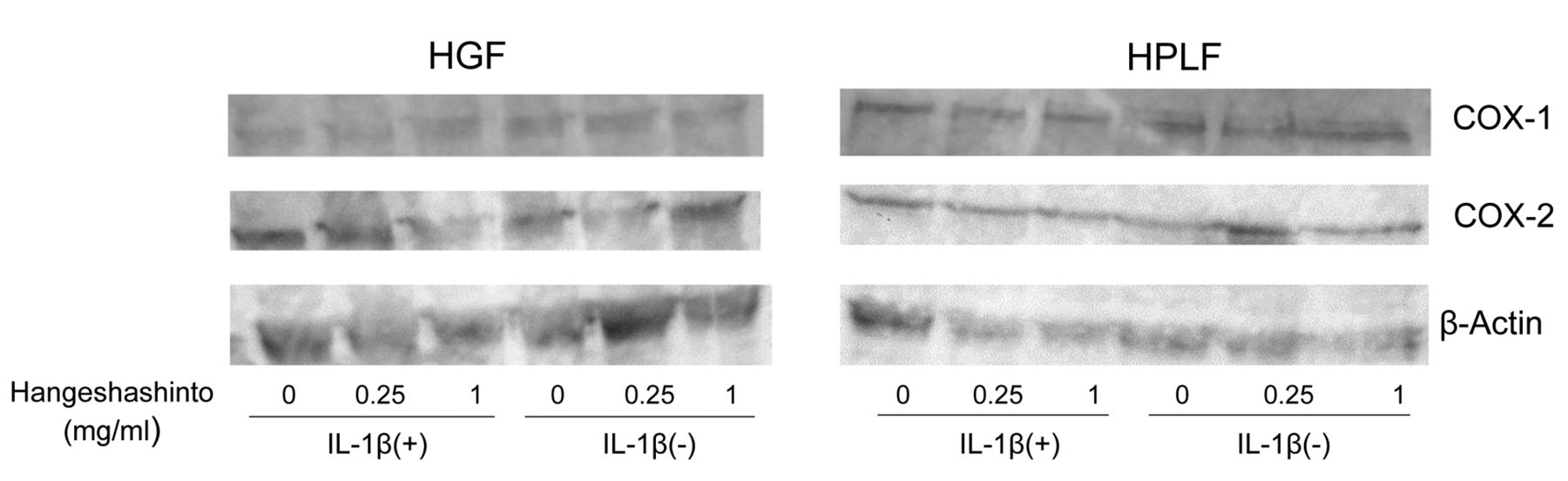
Effect of hangeshashinto on the intracellular concentration of cyclooxygenase (COX)-1 and COX-2 protein in unstimulated interleukin (IL)-1β-stimulated human gingival fibroblasts (HGFs) and human periodontal ligament fibroblasts (HPLFs). HGFs and HPLFs were treated for 24 h with 0, 0.25 or 1 mg/ml hangeshashinto in the absence or presence of 5 ng/ml IL-1β. COX protein expression in the cells was assayed by western blot analysis.

Effect of hangeshashinto on the enzymatic activity of purified cyclooxygenase (COX)-1 (A) and COX-2 (B) in vitro. COX-1 and COX-2 activity was determined by the amount of product [prostaglandin (PG)F2α] of enzymatic reaction. Each value represents the mean±SD from three independent experiments.
Contamination by lipopolysaccharide (LPS)-like substances of Kampo, Japanese traditional medicines (bold) and constituent plant extracts (supplied as lyophilized material of hot-water extracts), and glycyrrhizin (bold).
We next investigated the anti-inflammatory effect of hangeshashinto in IL-1β-stimulated HGFs and HPLFs, model systems of gingivitis and periodontitis. We previously reported that IL-1β treatment of HGFs resulted in two orders higher production of IL-6, IL-8, MCP-1 and PGE2, without induction of nitric oxide (NO) and tumor necrosis factor (TNF-α), in contrast to activated macrophages (20). The present study demonstrated for the first time that IL-1β treatment also stimulated HPLFs to produce two-order higher amounts of PGE2. We found that hangeshashinto inhibited PGE2 production by both IL-1β-stimulated HGFs and HPLFs, at SI values of 100 and 285, respectively. There was a possibility that hangeshashinto may have interfered with the coloring reaction of the PGE2 assay kit. We first confirmed that hangeshashinto did not induce PGE2 production without cells [indicated by cell (−), Figure 2B]. We also found that even when we added hangeshashinto to the culture medium of IL-1β-treated cells, the amount of PGE2 production did not change (indicated by HF+CM in Figure 2B). These experimental data eliminated this possibility, suggesting that the inhibition by hangeshashinto was not due simply to the interference of the PGE2 assay system. We also found that more than 80% of PGE2 produced by IL-1β-stimulated cells was found in the medium fraction (data not shown), indicating that IL-1β stimulated the actual production of PGE2, rather than stimulating its release.
The present study suggests that the inhibition of PGE2 production by hangeshashinto is mainly due to its inhibition of the expression of COX-2 protein, rather than that of COX-1 protein, or of COX-1 and COX-2 activity. This is in agreement with a previous finding that hangeshashinto inhibited PGE2 production via COX-2 protein expression in HGFs and human oral keratinocytes (22). We previously reported that the SI of rikkosan was low (SI=4) (10), possibly due to the higher background level of PGE2 production by contaminating LPS. It should be noted that the SI value (SI=100) for hangeshashinto was 25 times higher than rikkosan, substantiating its efficacy against stomatitis. This may explain why among 150 Kampo medicines, only hangeshashinto, ourento and inchinkouto are used for the treatment of stomatitis (23). A single pack (1.875 g) of hangeshashinto is administered to adult patients (over 15 years of age) in the clinic. Assuming the bioavailability of hangeshashinto to be 100%, its serum concentration is estimated to be 0.47 mg/ml. This concentration is lower than the cytotoxicity concentrations (1 mg/ml), and 9-31-times higher the 50% effective dose (0.015-0.055 mg/ml), which may be sufficient to exert its pharmacological effects.
As far as we know, this is the first report to present SI values for the comparison of relative anti-inflammatory activity against HGFs and HPLFs. A further comparative study using this index may identify the most appropriate plant extracts for the treatment of stomatitis, and elucidate its pharmaceutical action.
In conclusion, the present study demonstrated that most Kampo medicines contain undetectable to relatively low concentrations of LPS-like substances, except for rikkosan and unseiin, and hageshashinto had relatively higher anti-inflammatory activity against both human stomatitis and periodontitis model systems, further substantiating its therapeutic potential.
- Received February 9, 2016.
- Revision received March 24, 2016.
- Accepted March 28, 2016.
- Copyright © 2016 International Institute of Anticancer Research (Dr. John G. Delinassios), All rights reserved


