
Main menu

  • Home
  • Current Issue
  • Archive
  • Info for
    • Authors
    • Advertisers
    • Editorial Board
  • Other Publications
    • Anticancer Research
    • Cancer Genomics & Proteomics
    • Cancer Diagnosis & Prognosis
  • More
    • IIAR
    • Conferences
  • About Us
    • General Policy
    • Contact
  • Other Publications
    • In Vivo
    • Anticancer Research
    • Cancer Genomics & Proteomics

User menu

  • Register
  • Subscribe
  • My alerts
  • Log in
  • My Cart

Search

  • Advanced search
In Vivo
  • Other Publications
    • In Vivo
    • Anticancer Research
    • Cancer Genomics & Proteomics
  • Register
  • Subscribe
  • My alerts
  • Log in
  • My Cart
In Vivo

Advanced Search

  • Home
  • Current Issue
  • Archive
  • Info for
    • Authors
    • Advertisers
    • Editorial Board
  • Other Publications
    • Anticancer Research
    • Cancer Genomics & Proteomics
    • Cancer Diagnosis & Prognosis
  • More
    • IIAR
    • Conferences
  • About Us
    • General Policy
    • Contact
  • Visit iiar on Facebook
  • Follow us on Linkedin
Review ArticleReviewsR

The Prolonged QT Interval: Role of Pro-inflammatory Cytokines, Reactive Oxygen Species and the Ceramide and Sphingosine-1 Phosphate Pathways

PETER P. SORDILLO, DIANA C. SORDILLO and LAWRENCE HELSON
In Vivo November 2015, 29 (6) 619-636;
PETER P. SORDILLO
SignPath Pharma, Inc., Quakertown, PA, U.S.A.
  • Find this author on Google Scholar
  • Find this author on PubMed
  • Search for this author on this site
DIANA C. SORDILLO
SignPath Pharma, Inc., Quakertown, PA, U.S.A.
  • Find this author on Google Scholar
  • Find this author on PubMed
  • Search for this author on this site
LAWRENCE HELSON
SignPath Pharma, Inc., Quakertown, PA, U.S.A.
  • Find this author on Google Scholar
  • Find this author on PubMed
  • Search for this author on this site
  • For correspondence: lhelson@comcast.net
  • Article
  • Figures & Data
  • Info & Metrics
  • PDF
Loading

Abstract

Patients with QT prolongation have delayed cardiac repolarization and may suffer fatal ventricular arrhythmias. To determine the role of cytokines in causing this syndrome, we reviewed reports on patients with rheumatoid arthritis, psoriasis and other inflammatory conditions. These patients frequently have prolonged QT, which correlates with increases in tumor necrosis factor alpha, and interleukin-1β and 6. Studies in experimental models have shown that these cytokines act through stimulation of reactive oxygen species. Our review of data on phospholipidosis and on QT-shortening agents suggests a key role in QT prolongation for the ceramide/sphingosine-1-phosphate rheostat. We conclude that the cause of prolonged QT in inflammatory conditions is cytokine induction of reactive oxygen species and then ceramides, and believe that QT-prolonging agents bypass initial steps of this pathway and directly affect ceramides. Since both pro-inflammatory cytokines and numerous medications cause QT prolongation and ventricular arrhythmias by this mechanism, extra caution is needed when using these agents in patients with inflammatory conditions.

  • Prolonged QT
  • repolarization
  • ventricular arrhythmia
  • hERG
  • potassium channel
  • inflammation
  • cytokines
  • TNFα
  • interleukin-1β
  • interleukin-6
  • reactive oxygen species
  • ceramide
  • sphingosine-1 phosphate
  • review

An increase in the levels of pro-inflammatory cytokines plays a role in the morbidity of numerous diseases. Marked elevations in levels of tumor necrosis factor-alpha (TNFα), and interleukins IL1β and IL6, in particular, have been found in diseases as diverse as diabetes (1), Alzheimer's disease (2), inflammatory bowel disease (3) and cancer (4). When elevation of these cytokines becomes massive, a ‘cytokine storm’ may develop and cause death of the patient (5-7). These pro-inflammatory cytokines are also found to be increased in autoimmune diseases, and blockers of these cytokines are now standard therapies for patients with rheumatoid arthritis (RA) and many other diseases (8-10).

In long QT syndrome, there is delayed repolarization secondary to abnormalities in the potassium, sodium and calcium ion channels in myocardial cells. This may be caused by a genetic abnormality, coexisting cardiac diseases, hypokalemia or hypomagnesemia, or by one of numerous pharmaceutical agents, including commonly used antihistamines, sedatives, antibiotics, antiarrhythmics and psychiatric drugs. Ventricular tachycardia and sudden death secondary to delay in repolarization may occur (11-15). There is now increasing evidence suggesting that overexpression of IL1β, IL6, TNFα, and other cytokines plays a role in the pathogenesis of the long QT syndrome.

Data from Patient Trials

It has been observed that patients with rheumatoid arthritis (RA) have a markedly increased incidence of QT prolongation and an increased mortality secondary to cardiac arrhythmias (16-19). Patients with RA are thought to have twice the risk of death from ventricular arrhythmias compared to the normal population (17), and a 50 ms increase in QT in patients with RA doubles the risk for all-cause mortality (18). This increase in mortality risk also correlates with increases in the levels of inflammatory markers (17). Patients with other autoimmune diseases, such as psoriasis (20), systemic lupus erythematosus (21-24) and inflammatory bowel disease (25), have also been shown to have an increased incidence of QT prolongation. Prolonged QT from myocarditis secondary to infectious diseases is also well-described (26-31).

In all of these inflammatory conditions, marked increases in IL1β, IL6, TNFα, in particular, are seen. Adlan et al. have shown that the levels of these three cytokines and of the anti-inflammatory cytokine IL10 in patients with RA correlate directly with the extent of prolongation of the QT interval (32); and that elevation of these cytokines correlates with increased QT prolongation even in patients without disease (33). In a study of 1200 patients with heart failure, Deswal et al. showed the chance of dying could be predicted by the extent of elevations of TNFα and IL6 (34). The ‘electrical storm’ of recurrent ventricular arrhythmia seen with cardiac disease is also associated with elevated levels of cytokines and other inflammatory markers (35, 36). Furthermore, prolongation of the QT interval has been reported as a side-effect of therapy in anticancer trials of the pro-inflammatory cytokines, IL18 and interferon-γ (IFNγ) (37, 38).

In a recent study, Lazzerini et al. noted that patients with RA treated with tocilizumab, an IL6 blocker, had shortening of their previously prolonged QT interval, and this shortening was associated with concomitant decreases in inflammatory markers. In this trial of 17 patients with severe RA, 76% (13/17) had QT intervals of greater than 440 ms (mean±SD=452.3±35.8 ms) before starting therapy. After six months of therapy, only 29% (5/17) had a QT interval greater than 440 ms (mean±SD=428.1±34.3 ms). Decreases in the levels of TNFα and C-reactive protein correlated with the decrease in QT (39).

Cytokines: Data from Experimental Models

Transgenic mice which overexpress TNFα have a markedly prolonged action potential duration and experience recurrent ventricular arrhythmias (40). Studies in animal models or in cultured cardiomyocytes support the hypothesis that elevations in cytokines can cause prolonged QT. Wang et al. studied the effects of TNFα on canine cardiomyocytes using whole-cell patch clamp techniques. Marked inhibition of the rapid delayed-rectifier K+ current (IKr) was seen. TNFα was also found to reduce the human ether-à-go-go-related gene (hERG) current in HEK 293 cells expressing hERG. The suppressive effect of TNFα was concentration-dependent, and could be blocked by co-administration of an antibody to TNFα (41). The effect was thought to be due to TNFα stimulation of reactive oxygen species (ROS) since it was also blocked by vitamin E and by the superoxide dismutase mimic manganese (III) tetrakis (4-benzoic acid) porphyrin (MnTRAP). Kawada et al. reported that rat cardiomyocytes incubated with TNFα showed a decreased transient outward potassium current (Ito), and reductions in Kv4.2 and K+ channel-interacting protein-2 (42). Fernandez-Velasco found similar results, and also suggested that TNFα affects cardiac K+ channels through stimulation of ROS. Again, the effect of TNFα on QT prolongation was blocked by vitamin E and by MnTRAP (43). Petkova-Kirova et al. studied mice overexpressing TNFα in the heart, using whole-cell voltage-clamp recording of K+ current, and observed a 50% decrease in Ito, as well as a major decrease in the slow component of the delayed rectifier potassium current (IKs) (44). In another study, Grandy and Fiset reported that ventricular myocytes from mice chronically treated with TNFα to achieve blood levels equivalent to those seen in patients with congestive heart failure or HIV exhibited significantly reduced Ito and IKr (45). Li and Rozanski reported that IL1 significantly increased the duration of the action potential and the effective refractory period in excised papillary muscle, and increased the L-type calcium current (ICaL) in isolated cardiomyocytes. The increase in action potential duration in papillary muscle was blocked by aspirin and by indomethacin (46). Hagiwara et al. used whole-cell patch clamp recordings of the ventricles of mice exposed to IL6, and found a marked increase in ICaL (47).

Phospholipidosis and Ceramides

Another clue to the mechanism by which prolonged QT may occur is the similarity between the list of medications that cause QT prolongation and the list of those that cause phospolipidosis (PLD), the excess accumulation of intracellular phospholipids due to abnormalities in lysosomal lipid metabolism. Sun et al. screened 4,090 approved and investigational agents and found that 209 induced PLD in Hep G2 cells. Seventy-seven percent of these compounds were hERG channel blockers, and when steroidal drugs were excluded, 87% of PLD-inducing drugs were found to be hERG channel blockers (48). Abnormalities in cytokine production have been seen in phospholipidotic cells. We noted a study by Reasor et al. that showed marked enhancement of IL6 and TNFα release by rat phospholipidotic alveolar macrophages after lipopolysaccharide (LPS) stimulation (49). Likewise, Reinhart et al. reported that amiodarone, an agent which causes both PLD and prolonged QT, stimulated the release of TNFα and transforming growth factor (TGF)-β from pulmonary alveolar macrophages (50). Similar results have been reported by Masubuchi et al. (51). On the other hand, Munic et al. reported that the phospholipidotic potential of various macrolide antibiotics correlated with a reduction in IL6 levels in LPS-stimulated cells. Small increases in TNFα levels were seen with increasing PLD (52).

More importantly, although the exact biochemical mechanism by which PLD occurs is not known, it is clear that abnormalities in ceramide metabolism play a role in this disease entity. Several investigators have shown that PLD is associated with marked abnormalities in ceramide metabolism (53, 54). Ceramides are lipid molecules consisting of sphingosine and fatty acids. They are formed by hydrolysis of sphingomyelin by sphingomyelinases, but may also be synthesized through other pathways (55). It has been suggested that PLD results in increased levels of ceramides within cells and that this is part of the mechanism of drug-induced PLD (56). Other investigations support the hypothesis that ceramide metabolism plays a key role in PLD. Exposure of human macrophages to oxidized low-density lipoprotein results in both PLD and an increased content of acid sphingomyelinase and ceramides (57). The potential importance of the ceramide transacylase lysosomal phospholipase A2 (LPLA2) in alternate pathways of ceramide metabolism has been emphasized by Shayman et al. (53). Hiraoka et al. studied mice made deficient in LPLA2 by deletion of exon 5 of the (Lpla2) gene. These mice develop increases in ceramides and PLD of alveolar macrophages, peritoneal macrophages and spleen (58). Abe et al. studied the effects of three compounds: D-threo-1-phenyl-2-decanoylamino-3-morpholino-propanol (PDMP), a glucosylceramide synthase inhibitor and PLD-inducing agent; the strongly phospholipidotic agent, amiodarone; and tetracycline, which does not cause PLD, on Madin Darby canine kidney cells. Both PDMP and amiodarone inhibited LPLA2 and the transacylation of ceramide. This effect was concentration-dependent, and no effect was seen with tetracycline (59). Lecommandeur studied rat HEP G2 liver cells after chloroquine-induced PLD and found changes in the fatty acyl chain lengths of the ceramides, as well as changes in ceramide synthases, and suggested these changes might affect membrane morphology (60).

It is known that ceramides are important in the induction of multiple diseases, including atherosclerosis (61), diabetes (62) and cancer (63), as well as in growth arrest and cell apoptosis (64). It is also known that ceramides play a key role in TNFα and IL1 signal transduction (65-68). Studies have shown that IL1, IFNγ and TNFα can stimulate sphingomyelinase activity, resulting in ceramide accumulation (61, 69-72), and ceramide has frequently been referred to as the mimic of TNFα because of its similar effects on tumor-cell apoptosis and proliferation, and its role in many other disease processes (73-77). Lopez-Marure et al. showed that treatment with C8-ceramide had identical effects to those of TNFα on endothelial-cell proliferation and DNA synthesis (74). Similarly, Dbaibo et al. showed that TNFα and C2-ceramide had identical effects on the growth of Jurkat T-cells (76). Raines et al. showed that C2-ceramide and C6-ceramide mimicked the effects of TNFα on mitogen-activated protein kinase (MAPK) enzymatic activity, and on tyrosine phosphorylation (77). It is known that ceramides down-regulate the hERG K+ channel (78-80). Ceramides are metabolized to sphingosine and fatty acids, and sphingosine is phosphorylated by sphingosine kinases to form sphingosine-1 phosphate. The ceramide and sphingosine-1 pathways have opposite effects, the ceramide pathway causing cell death, and the sphingosine-1 phosphate pathway promoting cell survival (81-83). It is the balance of these two pathways that determines the fate of the cell (81). Since it has been postulated that ceramide metabolism is the common pathway to the development of atherosclerosis, perhaps through stimulation by TNFα (61), it is not unreasonable to ask whether the ceramide pathway might also be key to the mechanism of QT prolongation.

Agents that Reduce the Prolonged QT Interval

It has been long known that female sex is a risk factor for the long QT syndrome (84). However, QT intervals are shorter in the luteal phase of the menstrual cycle, when progesterone levels are higher, than in the follicular phase (85). This suggests that progestins have a protective effect against long QT (86). Kadish et al. studied 34,378 post-menopausal women who used unopposed estrogen, combined estrogen and progesterone, or used no therapy. A mild QT-prolonging effect was seen with estrogen, which was reversed with progesterone (87). Nakamura et al. investigated the effects of progesterone on guinea pig ventricular myocytes using patch clamp techniques. Progesterone shortened action potentials by lengthening IKs and inhibiting ICaL (88). Odening et al. studied ovariectomized transgenic LQT2 rabbits, which were exposed to estradiol, progesterone or dihydrotestosterone. In the estrogen-treated rabbits, ICaL increased, while in the progesterone-treated animals, ICaL decreased. Major cardiac ventricular events occurred in five out of seven estradiol-treated rabbits, but in none of the progesterone- or dihydrotestosterone-treated animals (89). Progesterone is known to have anti-inflammatory actions in many diseases. Aisenberg et al. showed that the anti-inflammatory effects of progesterone were essential in preventing pregnancy loss after LPS administration in mice (90). Loudon emphasized the importance of progesterone suppression of IL8 and cyclooxygenase-2 in maintaining myometrial quiescence in pregnancy and avoiding premature labor (91). Multiple studies have suggested that progesterone can reduce secondary damage from inflammation after traumatic brain injury by reducing levels of TNFα, IL1β and IL6 (92-94). Giannoni et al. showed that progesterone dramatically reduced secretion of TNFα and IL6 from mononuclear cells in umbilical cord blood from newborn children (95). Progesterone is known to induce sphingosine kinase, and the S1P pathway, the counteracting pathway to ceramides (96). Recent studies have suggested that non-genomic progesterone signaling may be a counteracting force to the effects of ceramides (97-103). Kupchak et al. have shown that non-genomic progesterone receptor activity can be antagonized by TNFα, and by 1(S),2(R)-D-erythro-2-(N-myristoylamino)-1-phenyl-1-propanol, a ceramidase inhibitor (101). Moussatche and Lyons have suggested that progesterone receptors act as the ‘fulcrum’ of the ceramide/S1P rheostat, determining whether ceramide synthase or ceramidase activity is greater, and that progestins and TNFα play opposing roles in the actions of these enzymes (98). Thomas and Pang suggested the neuroprotective effects of progestins are mediated by this mechanism (100). This suggests that the QT-shortening action of these agents may occur because of their opposing actions to TNFα and ceramides. It is also interesting that both estrogens and anti-estrogens, such as tamoxifen and toremifene, cause QT prolongation, and that both estrogens and anti-estrogens increase production of ceramides, although by different mechanisms. Tamoxifen is a potent inhibitor of glucosylceramide synthase and of acid ceramidase (104, 105). Estrogens increase the activity of ceramide synthases (106, 107).

3-Hydroxy-3-methylglutaryl coenzyme A reductase inhibitors (statins) may reduce ceramides by a number of mechanisms. Statins have also been shown to reduce levels of pro-inflammatory cytokines in many conditions, and discontinuation of these medications in patients being treated for hypercholesterolemia may result in a marked increase of cytokines and other inflammatory markers (108-110). In animal models of traumatic brain injury, the anti-cholesterol agents, atorvastatin, simvastatin and lovastatin, reduce brain and cerebrospinal fluid levels of IL1β, IL6 and TNFα (111-113). Iwata et al. showed that statins reduced IL6 and IL8 expression from LPS-stimulated human bronchial epithelial cells (114), and statin use is associated with decreased mortality in patients with pneumonia (115-117) and sepsis (118, 119). Thomsen et al., for example, reviewed the case histories of 29,900 patients hospitalized with pneumonia. Mortality among statin users was 10.3% at 30 days and 16.8% at 90 days, compared to 15.7% and 22.4% among non-statin users. These investigators also noted that statin users had lower blood levels of the inflammatory marker, C-reactive protein (116). Statins are known to reduce the QT interval (120, 121), and to reduce the chance of ventricular arrhythmia (122-125), and the reduction in ventricular arrhythmias has been linked to decreased inflammation (121, 124). Several investigators have argued that the primary underlying mechanism for the pleiotropic effects of statins is activation of sphingosine-1 phosphate-1 (S1P1) signaling (126, 127). Sugiura et al. showed that treatment with rosuvastatin led to an increase in the plasma level of S1P1 (128), and it has been reported that both atorvastatin and pitavastatin cause an up-regulation of S1P1 receptors (127). Furthermore, addition of pravastatin has been shown to prevent ceramide-induced death of mouse cerebral endothelial cells (129). Wei et al. showed that pravastatin and simuvastatin blocked oxidized LDL-induced acid sphingomyelinase activity and ceramide production in coronary artery endothelial cells (130).

Other agents have been shown to shorten the QT. We showed that liposomal curcumin, as well as empty liposomes, can attenuate the prolongation of the QT interval caused by crizotinib, nilotinib and other QT-prolonging agents (131-133). Liposomal curcumin and empty liposomes suppress TNFα, IL1β, IL6, monocyte chemoattractant protein 1 (MCP1), macrophage inflammatory protein 1-alpha (MIP1α) and Rantes, in both in vitro and in vivo models (134). Curcumin, which has both pro- and antioxidant effects, has been reported to protect against amiodarone pulmonary toxicity [a precursor to pulmonary PLD (135)] by reducing TNFα and TGFβ (136). Curcumin itself may cause prolonged QT because of its pro-oxidant effects, or because it can stimulate de novo ceramide synthesis (104), yet QT prolongation is blocked by liposomes. It has been suggested that liposomes, besides their anti-inflammatory activities, may compete with sphingomyelin for the enzyme sphingomyelinase, thus reducing the production of ceramides, or act by direct interaction with the cell membrane (137-139).

We have noted that in experimental models, the inducing effect of TNFα on QT prolongation can be blocked by antioxidants (41, 43), and other studies in animal models (140) and in patients (141, 142) have confirmed this. Zhang et al. showed that antioxidants restored IKr and hERG in the diabetic rabbit model of diabetes (140). Bednnarz et al. showed antioxidant vitamins corrected QT dispersion after exercise in patients post myocardial infarction (141). Kuklinski et al. reported that in patients treated with coenzyme Q and selenium, prolonged QT intervals were normalized in all, while 40% of controls continued to demonstrate prolonged QT. None of the treated patients died of re-infarction, while 20% of control patients had died of re-infarction by one year (142). Prolonged QT in diabetic patients has been associated with reduced plasma concentrations of vitamin C (143). Numerous studies have shown that ROS cause activation of sphingomyelinases (144-150). Vitamin E has been shown to reduce neutral sphingomyelinase activity and prevent neuronal death in rats after cerebral ischemia (151), although it may have contrary effects in cancer cells (152). Vitamin E has also been shown to prevent the induction of PLD in U937 cells exposed to 7-ketocholesterol (153). Navas et al. reported that coenzyme Q regulates the release of ceramides by non-competitive inhibition of neutral sphingomyelinase (154). The antioxidants 4,5-dihydroxy-1,3-benzenedisulfonic acid disodium salt monohydrate (Tiron) and N-acetylcysteine have been shown to block acid sphingomyelinase activity and the formation of ceramide-enriched membrane platforms that are stimulated by TNF-related apoptosis-inducing ligand (104, 155).

Cytokine-induced Prolongation of QT Likely Occurs Through ROS and Ceramides

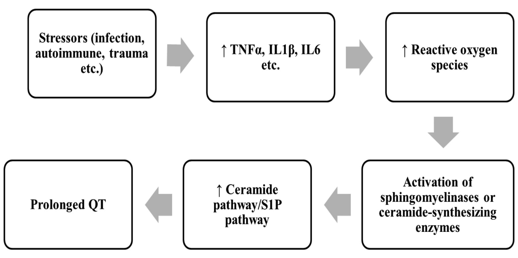
The information from these studies suggests the pathway by which cytokines cause prolonged QT. After stress, cytokines are released, and in turn, stimulate production of ROS. ROS cause activation of sphingomyelinases, which stimulate the ceramide pathway, and alter the ceramide pathway/sphingosine-1-phosphate pathway balance (Figure 1). The first step, concerning the role of stressors in the induction of pro-inflammatory cytokines, is well documented. As noted above, Wang et al. (41) and Fernandez Velasco et al. (43) both found that TNFα caused IKr, IKs and Ito suppression in experimental models, and that this effect was reversible by antioxidants such as vitamin E, and by a superoxide dismutase mimic, suggesting that TNFα caused this suppression by stimulating ROS. Pro-inflammatory cytokines (TNFα, IL1β, IL6, and IFNγ) have been shown to stimulate ROS in other experimental models (156-159). As noted, studies have shown that ROS can cause the activation of sphingomyelinases, causing an increase of ceramide levels (144-150). The mechanism of histone deacetylase inhibitor-induced leukemia cell death, for example, is thought to be secondary to acid sphingomyelinase-dependent generation of ceramides. This process does not occur in the absence of ROS stimulation of sphingomyelinase, and is blocked by antioxidants (146). ROS can also stimulate the salvage and de novo pathways of ceramide synthesis (160, 161), and this is likely the mechanism of TNFα stimulus of the de novo pathway (162). An increase in ROS appears to be the major mechanism of the cytokine effect on these enzymes, particularly on sphingomyelinases, although ROS-independent pathways may also play a role (163). ROS also inhibit sphingosine kinase-1, and thus further tilt the balance between the ceramide pathway (destructive) and the S1P pathway (protective) (81, 164, 165). The ceramide pathway is known to regulate numerous potassium channels, including hERG (78-80, 166-169). Ganapathi et al. studied the effects of C6-ceramide on HEK-293 cells and found that ceramide inhibited hERG channel current, and recruited hERG channels within caveolin-enriched lipid rafts (169). Chapman et al. reported an up to 30% decrease in hERG current with C6-ceramide, which they attributed to a reduction in the total number of hERG channels (78). Bai et al. found that C2-ceramide reduced hERG current, an effect they concluded was moderated by ROS (80). Wu et al. showed that C2-ceramide inhibited IKr in neuroblastoma IMR-32 cells and in rat pituitary GH(3) cells, and that this effect could be duplicated by TNFα (79).

Figure 1.
  • Download figure
  • Open in new tab
  • Download powerpoint
Figure 1.

A simplified model: Cytokine causation of the prolonged QT syndrome.

Drug-induced Prolonged QT

We have argued that pro-inflammatory cytokines cause prolonged QT by stimulating the ceramide pathway. If this hypothesis is accurate, we must account for the mechanism of action of QT-prolonging drugs, many of which are actively associated with the suppression of cytokines. We believe that although elevated levels of TNFα, IL1β and IL6 cause QT prolongation through the ceramide pathway and that this is the explanation for the high incidence of prolonged QT in inflammatory conditions, agents inducing QT prolongation may bypass the initial steps in this model and cause prolonged QT by directly affecting the ceramide pathway and disrupting the ceramide pathway/S1P pathway balance.

An important class of agents that cause QT prolongation is the tyrosine kinase inhibitors (TKIs), which are now used extensively for the treatment of chronic myelogenous leukemia (CML), renal cell carcinoma, hepatoma, gastrointestinal stromal tumors and many other types of cancer. One of the most prominent toxicities of nilotinib, a second-generation TKI which targets the breakpoint cluster region–ABL proto-oncogene 1 (BCR–ABL) receptor on CML cells, is QT prolongation. It has been shown that nilotinib induces apoptosis of CML cells by up-regulating ceramide synthase and down-regulating sphingosine kinase (170, 171). Addition of exogenous ceramides increased the apoptotic effect of nilotinib. In another study, El-Agamy showed that nilotinib caused a decrease in inflammatory cytokines and ROS generation and ameliorated LPS-induced acute lung injury in rats, presumably secondary to its stimulatory effect on ceramides and subsequent reduction in TNFα (172). Similarly, dasatinib, another second-generation TKI which is similar to nilotinib and which also causes QT prolongation, also causes apoptosis of CML cells by up-regulating ceramide synthase and down-regulating sphingosine kinase, thus shifting the ceramide/S1P balance toward ceramide (173). Likewise, alterations in the ceramide/S1P rheostat toward excess S1P have been associated with resistance to imatinib in patients with CML. The efficacy of imatinib has been linked to its generation of ceramides, particularly C18-ceramide, whereas resistance is thought to be caused by increased sphingosine kinase-1 activity and increased levels of S1P (174). Resistance has also been correlated with increased activity of glucosylceramide synthase, which converts ceramide to glucosylceramide. PDMP, a glucosylceramide synthase inhibitor and inducer of PLD, increased CML cell kill after treatment with imatinib (175). Huang et al. showed PDMP sensitizes resistant T315I mutant CML cells to a BCR–ABL inhibitor (176). Gao et al. reported that resistance to sunitinib, a tyrosine kinase inhibitor which causes prolonged QT and which is commonly used for treatment of renal carcinoma, gastrointestinal stromal tumors and pancreatic neuroendocrine tumors, is caused by activation of sphingosine kinase-1 and stimulation of the S1P pathway (177). Salas et al. showed that sphingosine kinase-1 and S1P mediate BCR–ABL stability and cause resistance to therapy, and that the efficacy of nilotinib depended on suppression of S1P, consistent with the view that all these agents work by tilting the ceramide/S1P balance toward ceramide (178). Many other investigators have found similar results (179-181).

While it is well-known that pro-inflammatory cytokines can stimulate the production of ceramides, it is also well-documented that exogenous ceramides, and the stimulation of the ceramide pathway, can cause a feedback inhibition of cytokines (182-188). For example, Jozefowski et al. showed that treatment with C8-ceramide reduced production of TNFα and MIP2 in murine peritoneal macrophages and in J774A.1 macrophage-like cells after LPS stimulation, and concluded that ceramide acted as a negative regulator of cytokine production. Inhibition of sphingomyelinase increased cytokine production (182). Rozenova et al. found similar results. They studied acid sphingomyelinase-deficient mice and peritoneal macrophages derived from the animals, and found that both the animals and the isolated macrophages produced 10- to 15-fold higher levels of TNFα when stimulated with LPS (183). Walton et al. reported that C6-ceramide inhibited LPS-induced IL8 synthesis in human aortic endothelial cells (184). The negative regulation of cytokines by ceramides may explain why medications which bypass the initial steps in the inflammatory pathway to ceramides and cause prolonged QT directly, may also cause cytokine suppression (189).

Other agents that cause QT prolongation are known to stimulate aberrant ceramide metabolism. For example, chlorpromazine, a commonly used antipsychotic drug, has been shown to reduce the level of acid ceramidase, which degrades ceramides to sphingosine and fatty acids, causing increased levels of ceramides (190-192). It is also well-known that chlorpromazine can induce inflammatory conditions, such as drug-induced lupus (193, 194). Hieronymus et al. have shown that the addition of S1P, which shifts the ceramide/S1P balance away from ceramide, can block chlorpromazine-induced apoptosis of peripheral blood mononuclear cells, and have suggested that aberrations of the ceramide pathway are the cause of drug-induced lupus (195).

Tricyclic antidepressants have been shown to have effects on ceramide/S1P balance. Desipramine inhibits acid ceramidase (190-192), although it also has an inhibitory effect on sphingomyelinase (196-198). Because of their inhibitory effects on sphingomyelinase, tricyclic antidepressants have been studied extensively as possible suppressors of ceramides, and it has been speculated that the antidepressant effects of these agents may be due to their effects on this enzyme. However, Elojeimy et al. treated DU145 prostate carcinoma cells with desipramine and found an increase in ceramide levels, suggesting that in some cases, the effects of desipramine on acid ceramidase may outweigh its effects on sphingomyelinase (192). That sphingomyelin hydrolysis and ceramidase activity may produce ceramides with different fatty acid chain lengths, which is discussed below, may be another explanation why tricyclic antidepressants cause prolonged QT despite inhibiting sphingomyelinase.

Antimalarials, such as mefloquine and chloroquine, have profound effects on the ceramide pathway. Mefloquine inhibits sphingomyelin synthase and glucosylceramide synthase, leading to an increased intracellular ceramide concentration, and this is thought to be its mechanism of action against Plasmodium falciparum (199). Mefloquine also increases sphingomyelinase (200) and reduces sphingosine kinase (201). Chloroquine inhibits acid ceramidase (190).

Cyclooxygenase-2 inhibitors, such as celecoxib, are known to cause prolonged QT (202), and to increase ceramide levels by activating sphingomyelinase (203-205). In one study, addition of C6-ceramide duplicated the effects of celecoxib on the cell cycle and cell growth inhibition (204). Schiffmann et al. showed that the toxic effects of celecoxib occur by activation of ceramide synthase 6 and the salvage pathway (206).

Fingolimod, a sphingosine analog with both agonist and antagonist effects on the S1P1 and other S1P receptors, and which sequesters lymphocytes in lymph nodes, is used to treat patients with relapsing multiple sclerosis. Fingolimod causes QT prolongation and inhibition of the hERG current (207-209), and sometimes fatal arrhythmia (210). This drug also can cause profound suppression of cytokines, including TNFα, IL1β, MCP1, interferon γ, chemokine (C-X-C motif) ligand 1 and TGFβ (211, 212).

In the mouse model of influenza-induced cytokine storm, modulation of the S1P1 receptor was identified as the primary pathway for initiation of the storm (213). The storm was reversed in this model through feedback inhibition by a S1P1 receptor modulator, with marked reductions in TNFα, MCP1, IFNγ, MIP1α, IL6 and other cytokines (214, 215). In this study, the S1P1 receptor modulator was a much more effective therapy than the antiviral agent, Tamiflu: 82% of mice treated with the receptor modulator survived compared to 50% with Tamiflu and to 20% of controls, 96% (27/28) of mice treated with both the receptor modulator and Tamiflu survived (213). Ceramides are composed of sphingosine and fatty acids. Sphingosine kinase, the enzyme which catalyzes sphingosine to S1P, is the crucial enzyme in the balance between the opposing ceramide to S1P pathways, and sphingosine has important ceramide-like effects (216). In addition, fingolimod, like tricyclic antidepressants, appears to inhibit acid ceramidase (217). Other sphingosine analogs, such as siponimod, have also been reported to cause prolonged QT (207).

Many other agents that are known to cause prolonged QT have been shown to affect the ceramide pathway and disrupt the ceramide/S1P balance, including aminoglycosides (218-221), antiestrogens such as tamoxifen (222), quinolones (221-224), monoclonal antibodies such as rituximab (225), platinum-containing antineoplastics (226-228), calcium channel blockers (229, 230), anthracyclines (231, 232), arsenic trioxide (233, 234), antihistamines (229), taxanes (235), cyclosporines (235) and imidazole antifungals such as ketoconazole (235). A list of QT-prolonging drugs and their effects on ceramide signaling is given in Table I.

Ceramide Fatty Acid Chain Length

The production of specific ceramides may be as important as the total amount of cytokines in the causation of the prolonged QT interval (236-238). Senkal et al. showed that treatment with the anthracycline doxorubin, in combination with gemcitabine, against human head and neck carcinoma cells, resulted in a 3.5-fold increase in ceramide synthase activity. When these cells were heterotransplanted into mice, a 7-fold increase in C-18 ceramide was seen after treatment, despite no increase in total ceramide levels (236). Veret et al. showed that treatment of pancreatic β-cells in culture with high doses of glucose resulted in apoptosis through stimulation of ceramide synthase-4, and increases in the levels of C18:0, C22:0 and C24:1 ceramides, without an increase in total ceramides. Down-regulation of ceramide synthase-4 by short-interfering RNA reduced apoptosis (238). C16:0 ceramide appears to have an important role in the induction of glucose intolerance. Turpin et al. reported that obese patients had elevated levels of ceramide synthase-6 mRNA and C16:0 ceramide in their adipose tissue. In addition, they showed that ceramide synthase-6-deficient mice had reduced levels of C16:0 ceramide and did not develop glucose intolerance or obesity after a high-fat diet (239). In patients with Parkinson's disease, ceramide levels are generally elevated. However, levels of C16:0, C18:0, C20:0, C22:0 and C24:1 ceramides are especially high, particularly in those who have significant cognitive impairment (240). There is also a great deal of data showing that elevated levels of C18:0 and C24:0 ceramides contribute to disease severity in patients with Alzheimer's disease (241). Such patients have greatly increased levels of C24:0 ceramide in the middle frontal gyrus, and the levels of C18:0 and C24:0 ceramides there correlate with disease severity. Addition of amyloid β-peptide-42 to cultured hippocampal neurons has been shown to result in increased C18:0 and C24:0 ceramide levels, and this effect can be reversed by an inhibitor of serine palmitoyltransferase (242). Numerous investigators have shown that C16:0, in particular, is crucial for apoptosis, both of malignant and normal cells (241-245). In cancer, and other diseases, it has been suggested that very long chain ceramides interfere with the toxic effects of C16:0 ceramide, and that what is crucial in many diseases is the balance between C16:0 and C24:0 ceramides, with C16:0 having toxic and C24:0 having protective effects (241, 242, 247).

The role of ceramides with specific fatty acid lengths in the pathogenesis of the prolonged QT syndrome and other abnormalities of cardiac function is not known. However, as noted, the production of different cytokines by different enzymes in the various ceramide pathways could be part of the reason why tricyclic antidepressants cause prolonged QT despite their tendency to inhibit sphingomyelinase. Di Paola et al. showed that C2-ceramide stimulated, while C16-ceramide inhibited, cytochrome c oxidase activity in rat heart mitochondria. Short-chain ceramides inhibited the generation of ROS associated with membrane potential-dependent reverse electron flow from succinate to complex 1, while long-chain ceramides had no effect. The investigators in this study concluded that short-chain ceramides caused a collapse in membrane potential, leading to an increase in permeability (248). Monette et al. reported that the impaired electron transport and increase in ROS in the aging rat heart after chronic inflammation was caused by increased levels of C16:0, C18:0 and C24:1 ceramides. The increased ceramide levels and mitochondrial abnormalities were reversed by (R)-α-lipoic acid (249). Russo et al. reported that mice which were fed a diet high in myristate (C14:0) developed marked increases in ceramide synthase-5 and C14-ceramide through the de novo pathway, resulting in cardiac hypertrophy and dysfunction (250). No effect was seen with a high palmitate (C16:0) diet.

View this table:
  • View inline
  • View popup
  • Download powerpoint
Table I.

QT-Prolonging agents and their effects on ceramide/S1P signaling

The significance of these findings is unclear, but there are hints that C16:0 ceramide may be especially important in the causation of QT prolongation, just as it is in other diseases. In a series of elegant experiments, Schiffmann et al. reported that celecoxib activation of ceramide synthase-6 induced C16:0 ceramide selectively, and showed that the toxic effects of celecoxib on cell growth were solely due to C16:0 ceramide production (206). As noted, Lecommandeur reported that drug-induced PLD is characterized by an increase in C16:0 ceramide, even when the total ceramide content is not changed (60). Bock et al. showed that treatment of Jurkat T-lymphocytes with C16:0 ceramide causes clustering of the voltage-gated potassium channels Kv1.3 within ceramide-enriched membranes, resulting in inhibition of Kv1.3 activity (251). It is also known that many of the toxic effects of TNFα occur specifically through generation of C16:0 ceramide, and that these toxic effects can be blocked by reduction of this ceramide (252, 253). Likewise, it has been shown that the toxic effects of IFNγ in models of autoimmune disease occur specifically through ceramide synthase-6 and C16:0 ceramides, and that these effects are reversed by inhibition of this ceramide (254).

Conclusion

We conclude that excess levels of pro-inflammatory cytokines play a role in causing prolongation of the QT interval, and that the likely mechanism of action is through ROS and the ceramide pathway. We also believe that QT-prolonging drugs may bypass the initial steps and cause prolonged QT by direct effects on the ceramide and S1P pathways and alterations of the ceramide/S1P rheostat. Elevated levels of cytokines may predispose a patient to QT prolongation, and this may be the reason that the QT-prolonging potential of these agents may not be observed in initial studies in human volunteers. Recent studies have suggested that it may be the stimulation of ceramides with particular fatty acid chain lengths, as much as the increase in total ceramides, which is important in the pathogenesis of some diseases. The potential role of individual ceramides, particularly C16:0 ceramide, in the causation of QT prolongation needs to be investigated. That pro-inflammatory cytokines and QT-prolonging medications may cause QT prolongation by the same mechanism means extra caution is needed when using these agents in patients with inflammatory conditions.

Footnotes

  • This article is freely accessible online.

  • Conflicts of Interest

    The Authors acknowledge a potential conflict of interest. Dr. Peter Sordillo is a member of the Scientific Advisory Board of SignPath Pharma, which makes several formulations of intravenous curcumin. Dr. Helson is CEO of SignPath Pharma.

  • Received July 28, 2015.
  • Revision received September 9, 2015.
  • Accepted September 25, 2015.
  • Copyright © 2015 International Institute of Anticancer Research (Dr. John G. Delinassios), All rights reserved

References

  1. ↵
    1. Mirza S,
    2. Hossain M,
    3. Mathews C,
    4. Martinez P,
    5. Pino P,
    6. Gay JL,
    7. Rentfro A,
    8. McCormick JB,
    9. Fisher-Hoch SP
    : Type-2 diabetes is associated with elevated levels of TNFα, IL6 and adiponectin and low levels of leptin in a population of Mexican Americans: A cross-sectional study. Cytokine 57: 136-142, 2012.
    OpenUrlCrossRefPubMed
  2. ↵
    1. Swardfager W,
    2. Lanctot K,
    3. Rothenburg L,
    4. Wong A,
    5. Cappell J,
    6. Hermann N
    : A meta-analysis of cytokines in Alzheimer's disease. Biol Psychiatry 68: 930-941, 2010.
    OpenUrlCrossRefPubMed
  3. ↵
    1. Nielsen OH,
    2. Ainsworth MA
    : Tumor necrosis factor inhibitors for inflammatory bowel disease. N Engl J Med 360: 754-762, 2013.
    OpenUrl
  4. ↵
    1. Sordillo PP,
    2. Helson L
    : Curcumin and cancer stem cells: Curcumin has asymmetrical effects on cancer and normal stem cells. Anticancer Res 35: 599-614, 2015.
    OpenUrlAbstract/FREE Full Text
  5. ↵
    1. Clark IA
    : The advent of the cytokine storm. Immunol Cell Biol 85: 271-273, 2007.
    OpenUrlCrossRefPubMed
    1. D'Elia RV,
    2. Harrison K,
    3. Oyston PC,
    4. Lukaszewski RA,
    5. Clark GC
    : Targeting the ‘cytokine storm’ for therapeutic benefit. Clin Vaccine Immunol 20: 319-327, 2013.
    OpenUrlAbstract/FREE Full Text
  6. ↵
    1. Sordillo PP,
    2. Helson L
    : Curcumin suppression of cytokine release and cytokine storm. A potential therapy for patients with Ebola and other severe viral infections. In Vivo 29: 1-4, 2015.
    OpenUrlAbstract/FREE Full Text
  7. ↵
    1. Genovese MC,
    2. Cohen S,
    3. Moreland L,
    4. Lium D,
    5. Robbins S,
    6. Newmark R,
    7. Bekker P
    : Combination therapy with etanercept and anakinra in the treatment of patients with rheumatoid arthritis who have been treated unsuccessfully with methotrexate. Arthritis Rheum 50: 1412-1419, 2004.
    OpenUrlCrossRefPubMed
    1. Kaneko A
    : Tocilizumab in rheumatoid arthritis: Efficacy, safety and its place in therapy. Ther Adv Chronic Dis 4: 15-21, 2013.
    OpenUrlAbstract/FREE Full Text
  8. ↵
    1. Tobin AM,
    2. Kirby B
    : TNFα inhibitors in the treatment of psoriasis and psoriatic arthritis. BioDrugs 19: 47-57, 2005.
    OpenUrlCrossRefPubMed
  9. ↵
    1. Morita H,
    2. Wu J,
    3. Zipes DP
    : The QT syndromes: Long and short. Lancet 372: 750-763, 2008.
    OpenUrlCrossRefPubMed
    1. Hunter JD,
    2. Sharma P,
    3. Rathi S
    : Long QT syndrome. Contin Educ Anaesth Crit Care Pain 8: 67-70, 2008.
    OpenUrlFREE Full Text
    1. Hedley PL,
    2. Jorgensen P,
    3. Schlamowitz S,
    4. Wangari R,
    5. Moolman-Smook J,
    6. Brink PA,
    7. Kanters JK,
    8. Corfield VA,
    9. Christiansen M
    : The genetic basis of long QT and short QT syndromes: A mutation update. Human Mutat 30: 1486-1511, 2009.
    OpenUrlCrossRefPubMed
    1. Kallergis EM,
    2. Goudis CA,
    3. Simantirakis EN,
    4. Kochiadakis GE,
    5. Vardas PE
    : Mechanisms, risk factors, and management of acquired long QT syndrome: A comprehensive review. Sci World J 2012: 212178, 2012.
    OpenUrl
  10. ↵
    1. Kannankeril P,
    2. Roden DM,
    3. Darbar D
    : Drug-induced long QT syndrome. Pharmacol Rev 62: 760-781, 2010.
    OpenUrlAbstract/FREE Full Text
  11. ↵
    1. Lazzerini PE,
    2. Acampa M,
    3. Capecchi PL,
    4. Hammoud M,
    5. Maffei S,
    6. Bisogno S,
    7. Barreca C,
    8. Galeazzi M,
    9. Laghi-Pasini F
    : Association between high sensitivity C-reactive protein, heart rate variability and corrected QT interval in patients with chronic inflammatory arthritis. Eur J Intern Med 24: 368-374, 2013.
    OpenUrlCrossRefPubMed
  12. ↵
    1. Lazzerini PE,
    2. Capecchi PL,
    3. Acampa M,
    4. Galeazzi M,
    5. Laghi-Pasini F
    : Arrhythmic risk in rheumatoid arthritis: The driving role of systemic inflammation. Autoimmun Rev 13: 936-944, 2014.
    OpenUrlCrossRefPubMed
  13. ↵
    1. Panoulas VF,
    2. Toms TE,
    3. Douglas KM,
    4. Sandoo A,
    5. Metsios GS,
    6. Stavropoulos-Kalinoglou A,
    7. Kitas GD
    : Prolonged QTc interval predicts all-cause mortality in patients with rheumatoid arthritis: An association driven by high inflammatory burden. Rheumatol 53: 131-137, 2014.
    OpenUrlAbstract/FREE Full Text
  14. ↵
    1. Chauhan K,
    2. Ackerman M,
    3. Crowson CS,
    4. Matteson EL,
    5. Gabriel SE
    : Population-based study of QT interval prolongation in patients with rheumatoid arthritis. Clin Exp Rheumatol 33: 84-89, 2015.
    OpenUrlPubMed
  15. ↵
    1. Simsek H,
    2. Sahin M,
    3. Akyol A,
    4. Akdag S,
    5. Ozkol HU,
    6. Gumrukcuoglu HA,
    7. Gunes Y
    : Increased risk of atrial and ventricular arrhythmia in long-lasting psoriasis patients. Sci World J 2013: 901215, 2013.
    OpenUrl
  16. ↵
    1. Bourre-Tessier J,
    2. Clarke AE,
    3. Huynh T,
    4. Bernatsky S,
    5. Joseph L,
    6. Belisle P,
    7. Pineau CA
    : Prolonged corrected QT interval in anti-Ro/SSA-positive adults with systemic lupus erythematosus. Arthritis Care Res 63: 1031-1037, 2011.
    OpenUrl
    1. Cardoso CR,
    2. Sales MA,
    3. Papi JA,
    4. Salles GF
    : QT-interval parameters are increased in systemic lupus erythematosus patients. Lupus 14: 846-852, 2005.
    OpenUrlAbstract/FREE Full Text
    1. Bourre-Tessier J,
    2. Urowitz MB,
    3. Clarke AE,
    4. Bernatsky S,
    5. Krantz MJ,
    6. Huynh T,
    7. Joseph L,
    8. Belisle P,
    9. Bae SC,
    10. Hanly JG,
    11. Wallace DJ,
    12. Gordon C,
    13. Isenberg D,
    14. Rahman A,
    15. Gladman DD,
    16. Fortin PR,
    17. Merill JT,
    18. Romero-Diaz J,
    19. Sanchez-Guerrero J,
    20. Fessler B,
    21. Alarcon GS,
    22. Steinsson K,
    23. Bruce IN,
    24. Ginzler E,
    25. Dooley MA,
    26. Nived O,
    27. Sturfelt G,
    28. Kalunian K,
    29. Ramos-Casals M,
    30. Petri M,
    31. Zoma A
    : Electrocardiographic findings in systemic lupus erythematosus: Data from an international inception cohort. Arthritis Care Res 67: 128-135, 2015.
    OpenUrl
  17. ↵
    1. Milovanovic B,
    2. Stojanovic L,
    3. Milicevik N,
    4. Vasic K,
    5. Bjelakovic B,
    6. Krotin M
    : Cardiac autonomic dysfunction in patients with systemic lupus, rheumatoid arthritis and sudden death risk. Srp Arh Celok Lek 138: 26-32, 2010.
    OpenUrlCrossRefPubMed
  18. ↵
    1. Curione M,
    2. Aratari A,
    3. Amato S,
    4. Colotto M,
    5. Barbato M,
    6. Leone S,
    7. Tego A,
    8. Panetti D,
    9. Parlapiano C
    : A study on QT interval in patients affected with inflammatory bowel disease without cardiac involvement. Intern Emerg Med 5: 307-310, 2010.
    OpenUrlPubMed
  19. ↵
    1. Sareli P,
    2. Schamroth CL,
    3. Passias J,
    4. Schamroth L
    : Torsade de pointes due to coxsackie B3 myocarditis. Clin Cardiol 10: 361-362.
    1. Jensen TB,
    2. Dalsgaard D,
    3. Johansen JB
    : Cardiac arrest due to torsades de pointes ventricular tachycardia in a patient with Lyme carditis. Ugeskr Laeger 176, 2014.
    1. Mitamura E,
    2. Mifune J,
    3. Kanamori K,
    4. Hifumi N,
    5. Shimizu O,
    6. Yamamura I,
    7. Takahashi Y,
    8. Oonaka M,
    9. Tanaka T,
    10. Nunoda S
    : A case of torsade de pointes tachycardia complicating diphtheria. Kokyu To Junkan 33: 223-228, 1985.
    OpenUrlPubMed
    1. Badorff C,
    2. Zeiher AM,
    3. Hohnloser SH
    : Torsade de pointes tachycardia as a rare manifestation of acute enteroviral myocarditis. Heart 86: 489-490, 2001.
    OpenUrlAbstract/FREE Full Text
    1. Gowani SA,
    2. Kumar A,
    3. Arora S,
    4. Lahiri B
    : Legionella pneumonia complicated by myocarditis and torsades de pointes: A case report and review of literature. Conn Med 77: 331-334, 2014.
    OpenUrl
  20. ↵
    1. Sayar N,
    2. Terzi S,
    3. Yilmaz HY,
    4. Atmaca H,
    5. Kocak F,
    6. Dayi SU,
    7. Cakmak N,
    8. Tarhan A,
    9. Ozler A,
    10. Yesilcimen K
    : A case of prosthetic mitral valve Brucella endocarditis complicated with torsades de pointes. Heart Vessels 21: 331-333, 2006.
    OpenUrlPubMed
  21. ↵
    1. Adlan AM,
    2. Panoulas VF,
    3. Smith JP,
    4. Fisher JP,
    5. Kitas GD
    : Association between corrected QT interval and inflammatory cytokines in rheumatoid arthritis. J Rheumatol 42: 421-428, 2015.
    OpenUrlAbstract/FREE Full Text
  22. ↵
    1. Medenwald D,
    2. Kors JA,
    3. Loppnow H,
    4. Thiery J,
    5. Kluttig A,
    6. Nuding S,
    7. Tiller D,
    8. Greiser KH,
    9. Werdan K,
    10. Haerting J
    : Inflammation and prolonged QT time: Results from the cardiovascular disease, living and ageing in Halle (CARLA) study. PLoS ONE 9: e95994, 2014.
    OpenUrlCrossRefPubMed
  23. ↵
    1. Deswal A,
    2. Petersen NJ,
    3. Feldman AM,
    4. Young JB,
    5. White BG,
    6. Mann DL
    : Cytokines and cytokine receptors in advanced heart failure: An analysis of the cytokine database from the vesnarinone trial (VEST). Circulation 103: 2055-2059, 2001.
    OpenUrlAbstract/FREE Full Text
  24. ↵
    1. Streitner F,
    2. Kuschyk J,
    3. Veltmann C,
    4. Ratay D,
    5. Schoene N,
    6. Streitner I,
    7. Brueckmann M,
    8. Schumacher B,
    9. Borggrefe M,
    10. Wolpert C
    : Role of proinflammatory markers and NT-proBNP in patients with an implantable cardioverter-defibrillator and an electrical storm. Cytokine 47: 166-172, 2009.
    OpenUrlCrossRefPubMed
  25. ↵
    1. Gupalo EM,
    2. Kostyukevich MV,
    3. Mironova NA,
    4. Shlevkov NB,
    5. Kiktev VG,
    6. Bakalov SA,
    7. Sharf TV,
    8. Efremov EE,
    9. Chumachenko PV,
    10. Kotina ED,
    11. Ostroumov EN,
    12. Shumakov DV
    : Electrical storm due to myocarditis in post-infarct patient: When two diseases meet. Cor et Vasa doi:10.1016/j.crvasa.2015.05.006.
  26. ↵
    1. Robertson MJ,
    2. Kline J,
    3. Struemper H,
    4. Koch KM,
    5. Bauman JW,
    6. Gardner OS,
    7. Murray SC,
    8. Germaschewski F,
    9. Weisenbach J,
    10. Jonak Z,
    11. Toso JF
    : A dose-escalation study of recombinant human interleukin-18 in combination with rituximab in patients with non-Hodgkin's lymphoma. J Immunother 36: 331-341, 2013.
    OpenUrlCrossRef
  27. ↵
    1. Mattson K,
    2. Niiranen A,
    3. Pyrhonen S,
    4. Farkkila M,
    5. Cantell K
    : Recombinant interferon-γ treatment in non-small cell lung cancer. Antitumor effect and cardiotoxicity. Acta Oncol 30: 607-610, 1991.
    OpenUrlPubMed
  28. ↵
    1. Lazzerini PE,
    2. Acampa M,
    3. Capecchi PL,
    4. Fineshi I,
    5. Selvi E,
    6. Moscadelli V,
    7. Zimbone S,
    8. Gentile D,
    9. Galeazzi M,
    10. Laghi-Pasini F
    : Antiarrhythmic potential of anticytokine therapy in rheumatoid arthritis: Tocilizumab reduces corrected QT interval by controlling systemic inflammation. Arthritis Care Res 67: 332-339, 2015.
    OpenUrl
  29. ↵
    1. London B,
    2. Baker LC,
    3. Lee JS,
    4. Shusterman V,
    5. Choi BR,
    6. Kubota T,
    7. McTiernan CF,
    8. Feldman AM,
    9. Salama G
    : Calcium-dependent arrhythmias in transgenic mice with heart failure. Am J Physiol Heart Circ Physiol 284: H431-H441, 2003.
    OpenUrlAbstract/FREE Full Text
  30. ↵
    1. Wang J,
    2. Wang H,
    3. Zhang Y,
    4. Gao H,
    5. Nattel S,
    6. Wang Z
    : Impairment of HERG K(+) channel function by tumor necrosis factor-alpha: Role of reactive oxygen species as a mediator. J Biol Chem 279: 13289-13292, 2004.
    OpenUrlAbstract/FREE Full Text
  31. ↵
    1. Kawada H,
    2. Niwano S,
    3. Niwano H,
    4. Yumoto Y,
    5. Wakisaka Y,
    6. Yuge M,
    7. Kawahara K,
    8. Izumi T
    : Tumor necrosis factor-α downregulates the voltage gated outward K+ current in cultured neonatal rat cardiomyocytes: A possible cause of electrical remodeling in diseased hearts. Circulation 70: 605-609, 2006.
    OpenUrl
  32. ↵
    1. Fernandez-Velasco M,
    2. Ruiz-Hurtado G,
    3. Hurtado O,
    4. Moro MA,
    5. Delgado C
    : TNFα down-regulates transient outward potassium current in rat ventricular myocytes through iNOS overexpression and oxidant species generation. Am J Physiol Heart Circ Physiol 293: H238-H245, 2007.
    OpenUrlAbstract/FREE Full Text
  33. ↵
    1. Petkova-Kirova PS,
    2. Gursoy E,
    3. Mehdi H,
    4. McTiernan CF,
    5. London B,
    6. Salama G
    : Electrical remodeling of cardiac myocytes from mice with heart failure due to the overexpression of tumor necrosis factor-alpha. Am J Physiol Heart Circ Physiol 290: H2098-H2107, 2006.
    OpenUrlAbstract/FREE Full Text
  34. ↵
    1. Grandy SA,
    2. Fiset C
    : Ventricular K+ currents are reduced in mice with elevated levels of serum TNFα. J Mol Cell Cardiol 47: 238-246, 2009.
    OpenUrlCrossRefPubMed
  35. ↵
    1. Li YH,
    2. Rozanski GJ
    : Effects of human recombinant interleukin-1 on electrical properties of guinea pig ventricular cells. Cardiovasc Res 27: 525-530, 1993.
    OpenUrlAbstract/FREE Full Text
  36. ↵
    1. Hagiwara Y,
    2. Miyoshi S,
    3. Fukuda K,
    4. Nishiyama N,
    5. Ikegami Y,
    6. Tanimoto K,
    7. Murata M,
    8. Takahashi E,
    9. Shimoda K,
    10. Hirano T,
    11. Mitamura H,
    12. Ogawa S
    : SHP2-mediated signaling cascade through GP130 is essential for LIF-dependent ICaL, [Ca2+]i transient, and APD increase in cardiomyocytes. J Mol Cell Cardiol 43: 710-716, 2007.
    OpenUrlCrossRefPubMed
  37. ↵
    1. Sun H,
    2. Xia M,
    3. Shahane SA,
    4. Jadhav A,
    5. Austin CP,
    6. Huang R
    : Are hERG channel blockers also phospholipidosis inducers? Bioorg Med Chen Lett 23: 4587-4590, 2013.
    OpenUrl
  38. ↵
    1. Reasor MJ,
    2. McCloud CM,
    3. DiMatteo M,
    4. Schafer R,
    5. Ima A,
    6. Lemaire I
    : Effects of amiodarone-induced phospholipidosis on pulmonary host defense functions in rats. Proc Soc Exp Biol Med 211: 346-352, 1996.
    OpenUrlAbstract/FREE Full Text
  39. ↵
    1. Reinhart PG,
    2. Gairola CG
    : Amiodarone-induced pulmonary toxicity in Fischer rats: Release of tumor necrosis factor alpha and transforming growth factor beta by pulmonary alveolar macrophages. J Toxicol Environ Health 52: 353-365, 1997.
    OpenUrlCrossRefPubMed
  40. ↵
    1. Masubuchi H,
    2. Ueno M,
    3. Maeno T,
    4. Hara K,
    5. Suzuki M,
    6. Kono S,
    7. Yamaguchi A,
    8. Yamaguchi K,
    9. Kanbe M,
    10. Kitahara S,
    11. Orii M,
    12. Aoki N,
    13. Aoki F,
    14. Suga T,
    15. Kurabayashi M
    : Pathological analysis of amiodarone-induced lung injury. Am J Respir Crit Care Med 189: A5734, 2014.
    OpenUrl
  41. ↵
    1. Munic V,
    2. Banjanac M,
    3. Kostrun S,
    4. Nujic K,
    5. Bosnar M,
    6. Marjanovic N,
    7. Ralic J,
    8. Martijasic M,
    9. Hlevnjak M,
    10. Haber VE
    : Intensity of macrolide anti-inflammatory activity in J774A.1 cells positively correlates with cellular accumulation and phospholipidosis. Pharmocol Res 64: 298-307, 2011.
    OpenUrl
  42. ↵
    1. Shayman JA,
    2. Abe A
    : Drug induced phospholipidosis: An acquired lysosomal storage disorder. Biochem Biophys Acta 1831: 602-611, 2013.
    OpenUrlCrossRef
  43. ↵
    1. Anderson N,
    2. Borlak J
    : Drug-induced phospholipidosis. FEBS Lett 580: 5533-5540, 2006.
    OpenUrlCrossRefPubMed
  44. ↵
    1. Kitatani K,
    2. Idkowiak-Baldys J,
    3. Hannun YA
    : The sphingolipid salvage pathway in ceramide metabolism and signaling. Cell Signal 20: 1010-1018, 2008.
    OpenUrlCrossRefPubMed
  45. ↵
    1. Nioi P,
    2. Perry BK,
    3. Wang EJ,
    4. Gu YZ,
    5. Snyder RD
    : In vitro detection of drug-induced phospholipidosis using gene expression and fluorescent phospholipid-based methodologies. Toxicol Sci 99: 162-173, 2007.
    OpenUrlAbstract/FREE Full Text
  46. ↵
    1. Grandl M,
    2. Liebisch G,
    3. Robenek H,
    4. Rubsamen K,
    5. Boettcher A,
    6. Englmaier M,
    7. Schmitz G
    : Ox-LDL induces endolysosomal phospholipidosis and ceramide species while E-LDL induces lipid droplets and cholesterylester in human macrophages. Chem Phys Lipids 163s: s40, 2010.
    OpenUrl
  47. ↵
    1. Hiraoka M,
    2. Abe A,
    3. Lu Y,
    4. Yang K,
    5. Han X,
    6. Gross RW,
    7. Shayman JA
    : Lysosomal phospholipase A2 and phospholipidosis. Mol Cell Biol 26: 6139-6148, 2006.
    OpenUrlAbstract/FREE Full Text
  48. ↵
    1. Abe A,
    2. Hiraoka M,
    3. Shayman JA
    : A role for lysosomal phospholipase A2 in drug induced phospholipidosis. Drug Metab Lett 1: 49-53, 2007.
    OpenUrlCrossRefPubMed
  49. ↵
    1. Lecommandeur E
    : Ceramide metabolism in the liver and its role in two lysosomal lipidoses: Drug-induced phospholipidosis and Sandhoff disease. Metabomeeting, 10-12 September, London 2014.
  50. ↵
    1. Bismuth J,
    2. Lin P,
    3. Yao Q,
    4. Chen C
    : Ceramide: A common pathway for atherosclerosis? Atherosclerosis 196: 497-504, 2008.
    OpenUrlCrossRefPubMed
  51. ↵
    1. Galadari S,
    2. Rahman A,
    3. Pallichankandy S,
    4. Galadari A,
    5. Thayyullathil F
    : Role of ceramide in diabetes mellitus: Evidence and mechanisms. Lipids Health Dis 12: 98, 2013.
    OpenUrlCrossRefPubMed
  52. ↵
    1. Saddoughi SA,
    2. Ogretmen B
    : Diverse function of ceramide in cancer cell death and proliferation. Adv Cancer Res 117: 37-58, 2013.
    OpenUrlCrossRefPubMed
  53. ↵
    1. Ogretman B,
    2. Hannun YA
    : Biologically active sphingolipids in cancer pathogenesis and treatment. Nature Rev Cancer 4: 604-616, 2004.
    OpenUrlCrossRefPubMed
  54. ↵
    1. Schutze S,
    2. Machleidt T,
    3. Kronke M
    : The role of diacylglycerol and ceramide in tumor necrosis factor and interleukin-1 signal transduction. J Leukoc Biol 56: 533-541, 1994.
    OpenUrlAbstract
    1. Wiegmann K,
    2. Schultz S,
    3. Machleidt T,
    4. Witte D,
    5. Kronke M
    : Functional dichotomy of neutral and acidic sphingomyelinases in tumor necrosis factor signaling. Cell 78: 1005-1015, 1994.
    OpenUrlCrossRefPubMed
    1. Dressler KA,
    2. Mathias S,
    3. Kolesnick RN
    : Tumor necrosis factor-alpha activates the sphingomyelin signal transduction pathway in a cell-free system. Science 255: 1715-1718, 1992.
    OpenUrlAbstract/FREE Full Text
  55. ↵
    1. Kuno K,
    2. Matsushima K
    : The IL1 receptor signaling pathway. J Leukoc Biol 56: 542-547, 1994.
    OpenUrlAbstract
  56. ↵
    1. Xu J,
    2. Yeh CH,
    3. Chen S,
    4. He L,
    5. Sensi SL,
    6. Canzoniero LM,
    7. Choi DW,
    8. Hsu CY
    : Involvement of de novo ceramide biosynthesis in tumor necrosis factor-alpha/cycloheximide-induced cerebral endothelial cell death. J Biol Chem 273: 16521-16526, 1998.
    OpenUrlAbstract/FREE Full Text
    1. Modur V,
    2. Zimmerman GA,
    3. Prescott SM,
    4. McIntyre TM
    : Endothelial cell inflammatory responses to tumor necrosis factor alpha. Ceramide-dependent and -independent mitogen-activated protein kinase cascades. J Biol Chem 271: 13094-13102, 1996.
    OpenUrlAbstract/FREE Full Text
    1. Krown KA,
    2. Page MT,
    3. Nguyen C,
    4. Zechner D,
    5. Gutierrez V,
    6. Comstock KL,
    7. Glembotski CC,
    8. Quintana PJ,
    9. Sabbadini RA
    : Tumor necrosis factor alpha-induced apoptosis in cardiac myocytes. Involvement of the sphingolipid signaling cascade in cardiac cell death. J Clin Invest 98: 2854-2865, 1996.
    OpenUrlCrossRefPubMed
  57. ↵
    1. Ruvolo PP
    : Ceramide regulates cellular homeostasis via diverse stress signaling pathways. Leukemia 15: 1153-1160, 2001.
    OpenUrlCrossRefPubMed
  58. ↵
    1. Hanna AN,
    2. Chan EY,
    3. Xu J,
    4. Stone JC,
    5. Brindley DN
    : A novel pathway for tumor necrosis factor-alpha and ceramide signaling involving sequential activation of tyrosine kinase, p21(ras), and phosphatidylinositol 3-kinase. J Biol Chem 274: 12722-12729, 1999.
    OpenUrlAbstract/FREE Full Text
  59. ↵
    1. Lopez-Marure R,
    2. Ventura JL,
    3. Sanchez L,
    4. Montano LF,
    5. Zentella A
    : Ceramide mimics tumour necrosis factor-alpha in the induction of cell-cycle arrest in endothelial cells. Induction of the tumour suppressor p53 with decrease in retinoblastoma/protein levels. Eur J Biochem 267: 4325-4333, 2000.
    OpenUrlPubMed
    1. Michael JM,
    2. Lavin MF,
    3. Watters DJ
    : Resistance to radiation-induced apoptosis in Burkitt's lymphoma cells is associated with defective ceramide signaling. Cancer Res 57: 3600-3605, 1997.
    OpenUrlAbstract/FREE Full Text
  60. ↵
    1. Dbaibo GS,
    2. Obeid LM,
    3. Hannun YA
    : Tumor necrosis factor-α (TNFα) signal transduction through ceramide. J Biol Chem 268: 17762-17766, 1993.
    OpenUrlAbstract/FREE Full Text
  61. ↵
    1. Raines MA,
    2. Kolesnick RN,
    3. Golde DW
    : Sphingomyelinase and ceramide activate mitogen-activated protein kinase in myeloid HL-60 cells. J Biol Chem 268: 14572-14575, 1993.
    OpenUrlAbstract/FREE Full Text
  62. ↵
    1. Chapman H,
    2. Ramstrom C,
    3. Korhonen L,
    4. Laine M,
    5. Wann KT,
    6. Lindholm D,
    7. Pasternack M,
    8. Tornquist K
    : Downregulation of the HERG (KCNH2) K+ channel by ceramide: Evidence for ubiquitin-mediated lysosomal degradation. J Cell Sci 118: 5325-5334, 2005.
    OpenUrlAbstract/FREE Full Text
  63. ↵
    1. Wu SN,
    2. Lo YK,
    3. Kuo BI,
    4. Chiang HT
    : Ceramide inhibits the inwardly rectifying potassium current in GH(3) lactotrophs. Endocrinology 142: 4785-4794, 2001.
    OpenUrlCrossRefPubMed
  64. ↵
    1. Bai Y,
    2. Wang J,
    3. Shan H,
    4. Lu Y,
    5. Zhang Y,
    6. Luo X,
    7. Yang B,
    8. Wang Z
    : Sphingolipid metabolite ceramide causes metabolic perturbation contributing to HERG K+ channel dysfunction. Cell Physiol Biochem 20: 429-440, 2007.
    OpenUrlCrossRefPubMed
  65. ↵
    1. Cuvillier O,
    2. Pirianov G,
    3. Kleuser B,
    4. Vanek PG,
    5. Coso OA,
    6. Gutkind S,
    7. Spiegel S
    : Suppression of ceramide-mediated programmed cell death by sphingosine-1-phosphate. Nature 381: 800-803, 1996.
    OpenUrlCrossRefPubMed
    1. Maceka M,
    2. Payne SG,
    3. Milstien S,
    4. Spiegel S
    : Sphingosine kinase, sphingosine-1-phosphate and apoptosis. Biochim Biophys Acta 1585: 193-201, 2002.
    OpenUrlCrossRefPubMed
  66. ↵
    1. Spiegel S,
    2. Milstien S
    : Sphingosine-1-phosphate: An enigmatic signaling lipid. Nature Rev Mol Cell Biol 4: 397-407, 2003.
    OpenUrlCrossRefPubMed
  67. ↵
    1. Conrath CE,
    2. Wilde AAM,
    3. Jongbloed RJE,
    4. Alders M,
    5. van Langen IM,
    6. van Tintelen JP,
    7. Doevendans PA,
    8. Opthof T
    : Gender differences in the long QT syndrome: Effects of β-adrenoceptor blockade. Cardiovasc Res 53: 770-776, 2002.
    OpenUrlAbstract/FREE Full Text
  68. ↵
    1. Nakagawa M,
    2. Ooie T,
    3. Takahashi N,
    4. Taniguchi Y,
    5. Anan F,
    6. Yonemochi H,
    7. Saikawa T
    : Influence of menstrual cycle on QT interval dynamics. Pacing Clin Electrophysiol 29: 607-613, 2006.
    OpenUrlCrossRefPubMed
  69. ↵
    1. Odening KE,
    2. Choi BR,
    3. Koren G
    : Sex hormones and cardic arrest in long QT syndrome: Does progesterone represent a potential new antiarrhythmic therapy? Heart Rhythm 9: 1150-1152, 2012.
    OpenUrlPubMed
  70. ↵
    1. Kadish AH,
    2. Greenland P,
    3. Limacher MC,
    4. Frishman WH,
    5. Daugherty SA,
    6. Schwartz JB
    : Estrogen and progestin use and the QT interval in postmenopausal women. Ann Noninvasive Electrocardiol 9: 366-374, 2004.
    OpenUrlCrossRefPubMed
  71. ↵
    1. Nakamura H,
    2. Kurokawa J,
    3. Bai CX,
    4. Asada K,
    5. Xu J,
    6. Oren RV,
    7. Zhu ZI,
    8. Clancy CE,
    9. Isobe M,
    10. Furukawa T
    : Progesterone regulates cardiac repolarization through a nongenomic pathway: An in vitro patch-clamp and computational modeling study. Circulation 116: 2913-2922, 2007.
    OpenUrlAbstract/FREE Full Text
  72. ↵
    1. Odening KE,
    2. Choi BR,
    3. Liu GX,
    4. Hartmann K,
    5. Ziv O,
    6. Chaves L,
    7. Schofield L,
    8. Centracchio J,
    9. Zehender M,
    10. Peng X,
    11. Brunner M,
    12. Koren G
    : Estradiol promotes sudden cardiac death in transgenic long QT type 2 rabbits while progesterone is protective. Heart Rhythm 9: 823-832, 2012.
    OpenUrlCrossRefPubMed
  73. ↵
    1. Aisemberg J,
    2. Vercelli CA,
    3. Bariani MV,
    4. Billi SC,
    5. Wolfson ML,
    6. Franchi AM
    : Progesterone is essential for protecting against LPS-induced pregnancy loss. LIF as a potential mediator of the anti-inflammatory effect of progesterone. PLoS ONE 8: e56161, 2013 doi:10.1371/journal.pone.0056161.
    OpenUrlPubMed
  74. ↵
    1. Loudon JAZ,
    2. Elliot CL,
    3. Hills F,
    4. Bennett PR
    : Progesterone represses interleukin-8 and cyclo-oxygenase-2 in human lower segment fibroblast cells and amnion epithelial cells. Biol Reprod 69: 331-337, 2003.
    OpenUrlAbstract/FREE Full Text
  75. ↵
    1. He J,
    2. Evans CO,
    3. Hoffman SW,
    4. Oyesiku NM,
    5. Stein DG
    : Progesterone and allopregnanolone reduce inflammatory cytokines after traumatic brain injury. Exp Neurol 189: 404-412, 2004.
    OpenUrlCrossRefPubMed
    1. Chen G,
    2. Shi J,
    3. Jin W,
    4. Wang L,
    5. Xie W,
    6. Sun J,
    7. Hang C
    : Progesterone administration modulates TLRs/NF-κB signaling pathway in rat brain after cortical contusion. Ann Clin Lab Sci 38: 65-74, 2008.
    OpenUrlAbstract/FREE Full Text
  76. ↵
    1. Pan DS,
    2. Liu WG,
    3. Yang XF,
    4. Cao F
    : Inhibitory effect of progesterone on inflammatory factors after experimental traumatic brain injury. Biomed Environ Sci 20: 432-438, 2007.
    OpenUrlPubMed
  77. ↵
    1. Giannoni E,
    2. Guignard L,
    3. Knaup Reymond M,
    4. Perreau M,
    5. Roth-Kleiner M,
    6. Calandra T,
    7. Roger T
    : Estradiol and progesterone strongly inhibit the innate immune response of newborn mononuclear cells. Infect Immun doi:10.1128/IAI.00076-11, 2011.
  78. ↵
    1. Jeng YJ,
    2. Suarez VR,
    3. Izban MG,
    4. Wang HQ,
    5. Soloff MS
    : Progesterone-induced sphingosine kinase-1 expression in the rat uterus during pregnancy and signaling consequences. Am J Physiol Endocrinol Metab 292: E1110-E1121, 2007.
    OpenUrlAbstract/FREE Full Text
  79. ↵
    1. Holland WL,
    2. Scherer PE
    : PAQRs: A counteracting force to ceramides? Mol Pharmacol 75: 740-743, 2009.
    OpenUrlAbstract/FREE Full Text
  80. ↵
    1. Moussatche P,
    2. Lyons TJ
    : Non-genomic progesterone signaling and its non-canonical receptor. Biochem Soc Trans 40: 200-204, 2012.
    OpenUrlAbstract/FREE Full Text
    1. Wang Y,
    2. Wang X,
    3. Lau WB,
    4. Yuan Y,
    5. Booth D,
    6. Li JJ,
    7. Scalia R,
    8. Preston K,
    9. Gao E,
    10. Koch W,
    11. Ma XL
    : Adiponectin inhibits tumor necrosis factor-α induced vascular inflammatory response via caveolin-mediated ceramidase recruitment and activation. Circ Res 114: 792-805, 2014.
    OpenUrlAbstract/FREE Full Text
  81. ↵
    1. Thomas P,
    2. Pang Y
    : Membrane progesterone receptors: Evidence for neuroprotective, neurosteroid signaling and neuroendocrine functions in neuronal cells. Neuroendorinology 96: 162-171, 2012.
    OpenUrl
  82. ↵
    1. Kupchak BR,
    2. Garitaonandia I,
    3. Villa NY,
    4. Smith JL,
    5. Lyons TJ
    : Antagonism of human adiponectin receptors and their membrane progesterone receptor paralogs by TNFα and a ceramidase inhibitor. Biochemistry 48: 5504-5506, 2009.
    OpenUrlCrossRefPubMed
    1. Buechler C,
    2. Wanninger J,
    3. Neumeier M
    : Adiponectin receptor binding proteins – recent advances in elucidating adiponectin signaling pathways. FEBS Lett 584: 4280-4286, 2010.
    OpenUrlCrossRefPubMed
  83. ↵
    1. Holland WL,
    2. Miller RA,
    3. Wang ZV,
    4. Sun K,
    5. Barth BM,
    6. Bui HH,
    7. Davis KE,
    8. Bikman BT,
    9. Halberg N,
    10. Rutkowski JM,
    11. Wade MR,
    12. Tenorio VM,
    13. Kuo MS,
    14. Brozinick JT,
    15. Zhang BB,
    16. Birnbaum MJ,
    17. Summers SA,
    18. Scherer PE
    : Receptor-mediated activation of ceramidase activity initiates the pleiotropic actions of adiponectin. Nature Med 17: 55-63, 2011.
    OpenUrlCrossRefPubMed
  84. ↵
    1. Morad SAF,
    2. Cabot MC
    : Ceramide-orchestrated signaling in cancer cells. Nature Rev Cancer 13: 51-65, 2013.
    OpenUrlCrossRefPubMed
  85. ↵
    1. Furlong SJ,
    2. Mader JS,
    3. Hoskin DW
    : Lactoferricin-induced apoptosis in estrogen-nonresponsive MDA-MB-435 breast cancer cells is enhanced by C6 ceramide or tamoxifen. Oncol Rep 15: 1385-1390, 2006.
    OpenUrlPubMed
  86. ↵
    1. Wegner MS,
    2. Wanger RA,
    3. Oertel S,
    4. Brachtendorf S,
    5. Hartmann D,
    6. Schiffmann S,
    7. Marschalek R,
    8. Schreiber Y,
    9. Ferreiros N,
    10. Geisslinger G,
    11. Grosch S
    : Ceramide synthases CerS4 and CerS5 are upregulated by 17β-estradiol and GPER1 via AP-1 in human breast cancer cells. Biochem Pharmacol 92: 577-589, 2014.
    OpenUrlPubMed
  87. ↵
    1. Schiffmann S,
    2. Sandner J,
    3. Birod K,
    4. Wobst I,
    5. Angioni C,
    6. Ruckhaberle E,
    7. Kauffmann M,
    8. Ackermann H,
    9. Lotsch J,
    10. Schmidt H,
    11. Geisslinger G,
    12. Grosch S
    : Ceramide synthases and ceramide levels are increased in breast cancer tissue. Carcinogenesis 30: 745-752, 2009.
    OpenUrlAbstract/FREE Full Text
  88. ↵
    1. Li JJ,
    2. Li YS,
    3. Chu JM,
    4. Zhang CY,
    5. Wang Y,
    6. Huang Y,
    7. Chen J,
    8. Yuan JQ,
    9. Huang YL
    : Changes of plasma inflammatory markers after withdrawal of statin therapy in patients with hyperlipidemia. Clin Chim Acta 366: 269-273, 2006.
    OpenUrlPubMed
    1. Lai WT,
    2. Lee KT,
    3. Chu CS,
    4. Voon WC,
    5. Yen HW,
    6. Tsai LY,
    7. Sheu SH
    : Influence of withdrawal of statin treatment on proinflammatory response and fibrinolytic activity in humans: An effect independent on cholesterol elevation. Int J Cardiol 98: 459-464, 2005.
    OpenUrlCrossRefPubMed
  89. ↵
    1. Lee KT,
    2. Lai WT,
    3. Chu CS,
    4. Tsai LY,
    5. Yen HW,
    6. Voon WC,
    7. Sheu SH
    : Effect of withdrawal of statin on C-reactive protein. Cardiology 102: 166-170, 2004.
    OpenUrlCrossRefPubMed
  90. ↵
    1. Li B,
    2. Mahmood A,
    3. Lu D,
    4. Wu H,
    5. Xiong Y,
    6. Qu C,
    7. Chopp M
    : Simvastatin attenuates microglia, astrocyte activation and decreases IL1β level following traumatic brain injury. Neurosurgery 65: 179-186, 2009.
    OpenUrlCrossRefPubMed
    1. Chen SF,
    2. Hung TH,
    3. Chen CC,
    4. Lin KH,
    5. Huang YN,
    6. Tsai HC,
    7. Wang JY
    : Lovastatin improves histological and functional outcomes and reduces inflammation after experimental traumatic brain injury. Life Sci 81: 288-298, 2007.
    OpenUrlCrossRefPubMed
  91. ↵
    1. Wang H,
    2. Lynch JR,
    3. Song P,
    4. Yang HJ,
    5. Yates RB,
    6. Mace B,
    7. Warner DS,
    8. Guyton JR,
    9. Laskowitz DT
    : Simvastatin and atorvastatin improve behavioral outcome, reduce hippocampal degeneration, and improve cerebral blood flow after experimental traumatic brain injury. Exp Neurol 206: 59-69, 2007.
    OpenUrlCrossRefPubMed
  92. ↵
    1. Iwata A,
    2. Shirai R,
    3. Ishii H,
    4. Kushima H,
    5. Otani S,
    6. Hashinaga K,
    7. Umeki K,
    8. Kishi K,
    9. Tokimatsu I,
    10. Hiramatsu K,
    11. Kadota J
    : Inhibitory effect of statins on inflammatory cytokine production from human bronchial epithelial cells. Clin Exp Immunol 168: 234-240, 2012.
    OpenUrlCrossRefPubMed
  93. ↵
    1. Mortensen EM,
    2. Pugh MJ,
    3. Copeland LA,
    4. Restrepo MI,
    5. Cornell JE,
    6. Anzueto A,
    7. Pugh JA
    : Impact of statins and angiotensin-converting enzyme inhibitors on mortality of subjects hospitalized with pneumonia. Eur Respir J 31: 611-617, 2008.
    OpenUrlAbstract/FREE Full Text
  94. ↵
    1. Thomsen RW,
    2. Riis A,
    3. Kornum JB,
    4. Christensen S,
    5. Johnsen SP,
    6. Sorensen HT
    : Pre-admission use of statin and outcomes after hospitalization with pneumonia: population-based cohort study of 29,900 patients. Arch Intern Med 168: 2081-2087, 2008.
    OpenUrlCrossRefPubMed
  95. ↵
    1. Chalmers JD,
    2. Singanayagam A,
    3. Murray MP,
    4. Hill AT
    : Prior statin use is associated with improved outcomes in community-acquired pneumonia. Am J Med 121: 1002-1007, 2008.
    OpenUrlCrossRefPubMed
  96. ↵
    1. Dobesh PP,
    2. Klepser DG,
    3. McGuire TR,
    4. Morgan CW,
    5. Olsen KM
    : Reduction in mortality associated with statin therapy in patients with severe sepsis. Pharmacotherapy 29: 621-630, 2000.
    OpenUrl
  97. ↵
    1. Almog Y,
    2. Shefer A,
    3. Novak V,
    4. Maimon N,
    5. Barski L,
    6. Eizinger M,
    7. Friger M,
    8. Zeller L,
    9. Danon A
    : Prior statin therapy is associated with a decreased rate of severe sepsis. Circulation 110: 880-885, 2004.
    OpenUrlAbstract/FREE Full Text
  98. ↵
    1. Vrtovec B,
    2. Okrajsek R,
    3. Golicnik A,
    4. Ferjan M,
    5. Starc V,
    6. Radovancevic B
    : Atorvastatin therapy increases heart rate variability, decreases QT variability, and shortens QTc interval duration in patients with advanced chronic heart failure. J Cardiac Failure 11: 684-690, 2005.
    OpenUrlCrossRefPubMed
  99. ↵
    1. Mark L,
    2. Katona A
    : Effect of fluvastatin on QT dispersion: A new pleiotropic effect? Am J Cardiol 85: 919-920, 2000.
    OpenUrlPubMed
  100. ↵
    1. Chiu JH,
    2. Abdelhadi RH,
    3. Chung MK,
    4. Gurm HS,
    5. Marrouche NF,
    6. Saliba WJ,
    7. Natale A,
    8. Martin DO
    : Effect of statin therapy on risk of ventricular arrhythmia among patients with coronary artery disease and an implantable cardioverter-defibrillator. Am J Cardiol 95: 490-491, 2005.
    OpenUrlCrossRefPubMed
    1. Kostapanos MS,
    2. Liberopoulos EN,
    3. Goudevenos JA,
    4. Mikhailidis DP,
    5. Elisaf MS
    : Do statins have an antiarrhythmic activity? Cardiovasc Res 75: 10-20, 2007.
    OpenUrlAbstract/FREE Full Text
  101. ↵
    1. Buber J,
    2. Goldenberg I,
    3. Moss AJ,
    4. Wang PJ,
    5. McNitt S,
    6. Hall J,
    7. Eldar M,
    8. Barsheshet A,
    9. Schecter M
    : Reduction in life-threatening ventricular tachyarrhythmias in statin-treated patients with nonischemic cardiomyopathy enrolled in the MADIT-CRT (multicenter automatic defibrillator implantation trial with cardiac resynchronization therapy). J Am Coll Cardiol 60: 749-755, 2012.
    OpenUrlCrossRefPubMed
  102. ↵
    1. Barsheshet A,
    2. Wang PJ,
    3. Moss AJ
    : MADIT-CRT investigators reverse remodeling and the risk of ventricular tachyarrhythmias in MADIT-CRT. J Am Coll Cardiol 57: 2416-2423, 2011.
    OpenUrlCrossRefPubMed
  103. ↵
    1. Egom EE,
    2. Rose RA,
    3. Neyses L,
    4. Soran H,
    5. Cleland JG,
    6. Mamas MA
    : Activation of sphingosine-1-phosphate signaling as a potential underlying mechanism of the pleiotropic effects of statin therapy. Crit Rev Clin Lab Sci 50: 79-89, 2013.
    OpenUrlCrossRefPubMed
  104. ↵
    1. Igarashi J,
    2. Miyoshi M,
    3. Hashimoto T,
    4. Kubota Y,
    5. Kosaka H
    : Statins induce S1P1 receptors and enhance endothelial nitric oxide production in response to high-density lipoproteins. Br J Pharmacol 150: 470-479, 2007.
    OpenUrlCrossRefPubMed
  105. ↵
    1. Sugiura T,
    2. Dohi Y,
    3. Yamashita S,
    4. Ohte N,
    5. Ito S,
    6. Iwaki S,
    7. Hirowatari Y,
    8. Ohkawa R,
    9. Mishima Y,
    10. Yatomi Y,
    11. Kimura G,
    12. Fujii S
    : Analytical evaluation of plasma serotonin and sphingosine 1-phosphate and their clinical assessment in early atherosclerosis. Coron Artery Dis 23: 234-238, 2012.
    OpenUrlCrossRefPubMed
  106. ↵
    1. Chen SD,
    2. Hu CJ,
    3. Yang DI,
    4. Nassief A,
    5. Chen H,
    6. Yin K,
    7. Xu J,
    8. Hsu CY
    : Pravastatin attenuates ceramide-induced cytotoxicity in mouse cerebral endothelial cells with HIF1 activation and VEGF up-regulation. Ann NY Acad Sci 1042: 357-364, 2005.
    OpenUrlCrossRefPubMed
  107. ↵
    1. Wei YM,
    2. Li X,
    3. Xiong J,
    4. Abais JM,
    5. Xia M,
    6. Boini KM,
    7. Zhang Y,
    8. Li PL
    : Attenuation by statins of membrane raft-redox signaling in coronary arterial endothelium. J Pharmacol Exp Ther 345: 170-179, 2013.
    OpenUrlAbstract/FREE Full Text
  108. ↵
    1. Shopp G,
    2. Helson L,
    3. Bouchard A,
    4. Salvail,
    5. Majeed M
    : Liposomes ameliorate crizotinib- and nilotinib-induced inhibition of the cardiac IKr channel and QTc prolongation. Anticancer Res 34: 4733-4740, 2014.
    OpenUrlAbstract/FREE Full Text
    1. Helson L,
    2. Shopp G,
    3. Bouchard A,
    4. Majeed M
    : Liposome mitigation of curcumin inhibition of cardiac potassium delayed-rectifier current. J Recept Ligand Channel Res 5: 1-8, 2012.
    OpenUrl
  109. ↵
    1. Ranjan AP,
    2. Mukerjee A,
    3. Helson L,
    4. Vishwanatha JK
    : Mitigating prolonged QT interval in cancer nanodrug development for accelerated clinical translation. J Nanobiotechnol 11: 40, 2013.
    OpenUrl
  110. ↵
    1. Helson L,
    2. Sordillo PP,
    3. Schopp G,
    4. Savail D,
    5. Bouchard A,
    6. Shaw WA,
    7. Burgess SW,
    8. Kloesch B,
    9. Majeed M
    : Liposomal curcumin (Lipocurc) and in vitro/in vivo surrogates for cytokine storm associated with uncontrolled Ebola infection. Targeting Ebola Paris 2015, Recent Advances and Strategies, May 28-29, 2015.
  111. ↵
    1. Martin WJ,
    2. Kachel DL,
    3. Vilen T,
    4. Natarajan V
    : Mechanism of phospholipidosis in amiodarone pulmonary toxicity. J Pharmacol Exp Ther 251: 272-278, 1989.
    OpenUrlAbstract/FREE Full Text
  112. ↵
    1. Punithavathi D,
    2. Venkatesan N,
    3. Babu M
    : Protective effects of curcumin against amiodarone-induced pulmonary fibrosis in rats. Br J Pharmacol 139: 1342-1350, 2003.
    OpenUrlCrossRefPubMed
  113. ↵
    1. Stein O,
    2. Oette K,
    3. Dabach Y,
    4. Hollander G,
    5. Ben Naim M,
    6. Stein Y
    : Persistence of increased cholesteryl ester in human skin fibroblasts is caused by residual exogenous sphingomyelinase and is reversed by phospholipid liposomes. Biochim Biophys Acta 1165: 153-159, 1992.
    OpenUrlPubMed
    1. Gupta AK,
    2. Rudney H
    : Plasma membrane sphingomyelin and the regulation of HMG-CoA reductase activity and cholesterol biosynthesis in cell cultures. J Lipid Res 32: 125-136, 1991.
    OpenUrlAbstract
  114. ↵
    1. Liu JJ,
    2. Nilsson A,
    3. Duan RD
    : Effects of phospholipids on sphingomyelin hydrolysis induced by intestinal alkaline sphingomyelinase: An in vitro study. J Nutr Biochem 11: 192-197, 2000.
    OpenUrlCrossRefPubMed
  115. ↵
    1. Zhang Y,
    2. Sun X,
    3. Zhang Y,
    4. Wang J,
    5. Lu Y,
    6. Yang B,
    7. Wang Z
    : Potential therapeutic value of antioxidants for abnormal prolongation of QT interval and the associated arrhythmias in a rabbit model of diabetes. Cell Physiol Biochem 28: 97-102, 2011.
    OpenUrlCrossRefPubMed
  116. ↵
    1. Bednarz B,
    2. Chamiec T,
    3. Ceremuzynski L
    : Antioxidant vitamins decrease exercise-induced QT dispersion after myocardial infarction. Kardiol Pol 58: 375-379, 2003.
    OpenUrlPubMed
  117. ↵
    1. Kuklinski B,
    2. Weissenbacher E,
    3. Fahnrich A
    : Coenzyme Q10 and antioxidants in acute myocardial infarction. Mol Aspects Med 15: S143-S147, 1994.
    OpenUrlPubMed
  118. ↵
    1. Odermarsky M,
    2. Lykkesfeldt J,
    3. Liuba P
    : Poor vitamin C status is associated with increased carotid intima-media thickness, decreased microvascular function, and delayed myocardial repolarization in young patients with type 1 diabetes. Am J Clin Nutr 90: 447-452, 2009.
    OpenUrlAbstract/FREE Full Text
  119. ↵
    1. Bezombes C,
    2. Plo I,
    3. Mansat-De Mas V,
    4. Quillet-Mary A,
    5. Negre-Salvayre A,
    6. Laurent G,
    7. Jaffrezou JP
    : Oxidative stress-induced activation of Lyn recruits sphingomyelinase and is requisite for its stimulation by Ara-C. FASEB J doi.10.1096/fj.00-0787fje, 2001.
    1. Sawada M,
    2. Nakashima S,
    3. Kiyono T,
    4. Nakagawa M,
    5. Yamada J,
    6. Yamakawa H,
    7. Banno Y,
    8. Shinoda J,
    9. Nishimura Y,
    10. Nozawa Y,
    11. Sakai N
    : p53 regulates ceramide formation by neutral sphingomyelinase through reactive oxygen species in human glioma cells. Oncogene 20: 1368-1378, 2001.
    OpenUrlCrossRefPubMed
  120. ↵
    1. Rosato RR,
    2. Maggio SC,
    3. Almenara JA,
    4. Payne SG,
    5. Atadja P,
    6. Spiegel S,
    7. Dent P,
    8. Grant S
    : The histone deacetylase inhibitor LAQ824 induces human leukemia cell death through a process involving XIAP down-regulation, oxidative injury, and the acid sphingomyelinase-dependent generation of ceramide. Molec Pharmacol 69: 216-225, 2006.
    OpenUrlAbstract/FREE Full Text
    1. Manago A,
    2. Becker KA,
    3. Carpinteiro A,
    4. Wilker B,
    5. Soddemann M,
    6. Seitz AP,
    7. Edwards MJ,
    8. Grassme H,
    9. Szabo I,
    10. Gulbins E
    : Pseudomonas aeruginosa pyocyanin induces neutrophil death via mitochondrial reactive oxygen species and mitochondrial acid sphingomyelinase. Antioxid Redox Signal 22: 1097-1110, 2015.
    OpenUrlPubMed
    1. Hatanaka Y,
    2. Fujii J,
    3. Fukutomi T,
    4. Watanabe T,
    5. Che W,
    6. Sanada Y,
    7. Igarashi Y,
    8. Taniguchi N
    : Reactive oxygen species enhances the induction of inducible nitric oxide synthase by sphingomyelinase in RAW264.7 cells. Biochim Biophys Acta 1393: 203-210, 1998.
    OpenUrlPubMed
    1. Castillo SS,
    2. Levy M,
    3. Thaikoottathil JV,
    4. Goldkorn T
    : Reactive nitrogen and oxygen species activate different sphingomyelinases to induce apoptosis in airway epithelial cells. Exp Cell Res 313: 2680-2686, 2007.
    OpenUrlCrossRefPubMed
  121. ↵
    1. Hernandez OM,
    2. Discher DJ,
    3. Bishopric NH,
    4. Webster KA
    : Rapid activation of neutral sphingomyelinase by hypoxia-reoxygenation of cardiac myocytes. Circ Res 86: 198-204, 2000.
    OpenUrlAbstract/FREE Full Text
  122. ↵
    1. Yamagata K,
    2. Ichinose S,
    3. Tagawa C,
    4. Tagami M
    : Vitamin E regulates SMase activity, GSH levels, and inhibit neuronal death in stroke-prone spontaneously hypertensive rats during hypoxia and reoxygenation. J Exp Stroke Trans Med 2: 41-48, 2009.
    OpenUrl
  123. ↵
    1. Gu X,
    2. Song X,
    3. Dong Y,
    4. Cai H,
    5. Walters E,
    6. Zhang R,
    7. Pang X,
    8. Xie T,
    9. Guo Y,
    10. Sridhar R,
    11. Califano JA
    : Vitamin E succinate induces ceramide-mediated apoptosis in head and neck squamous cell carcinoma in vitro and in vivo. Clin Cancer Res 14: 1840-1848, 2008.
    OpenUrlAbstract/FREE Full Text
  124. ↵
    1. Vejux A,
    2. Guyot S,
    3. Montange T,
    4. Riedinger JM,
    5. Kahn E,
    6. Lizard G
    : Phospholipidosis and down-regulation of the PI3-K/PDK-1/AKT signaling pathway are vitamin E inhibitable events associated with 7-ketocholesterol-induced apoptosis. J Nutr Biochem 20: 45-61, 2009.
    OpenUrlCrossRefPubMed
  125. ↵
    1. Navas P,
    2. Villalba JM,
    3. de Cabo R
    : The importance of plasma membrane coenzyme Q in aging and stress responses. Mitochondrion 7: S34-S40, 2007.
    OpenUrlCrossRefPubMed
  126. ↵
    1. Dumitru CA,
    2. Gulbins E
    : TRAIL activates acid sphingomyelinase via a redox mechanism and releases ceramide to trigger apoptosis. Oncogene 25: 5612-5625, 2006.
    OpenUrlCrossRefPubMed
  127. ↵
    1. Woo CH,
    2. Eom YW,
    3. Yoo MH,
    4. You MH,
    5. Han HJ,
    6. Song WK,
    7. Yoo YJ,
    8. Chun JS,
    9. Kim JH
    : Tumor necrosis factor-alpha generates reactive oxygen species via a cytosolic phospholipase A2-linked cascade. J Biol Chem 275: 32357-32362, 2000.
    OpenUrlAbstract/FREE Full Text
    1. Kim JJ,
    2. Lee SB,
    3. Park JK,
    4. Yoo YD
    : TNFα-induced ROS production triggering apoptosis is directly linked to ROMO1 and BCL-X(L). Cell Death Differ 17: 1420-1434, 2010.
    OpenUrlCrossRefPubMed
    1. Yang D,
    2. Elner SG,
    3. Bian ZM,
    4. Till GO,
    5. Petty HR,
    6. Elner VM
    : Pro-inflammatory cytokines increase reactive oxygen species through mitochondria and NADPH oxidase in cultured RPE cells. Exp Eye Res 85: 462-472, 2007.
    OpenUrlCrossRefPubMed
  128. ↵
    1. Lo YYC,
    2. Cruz TF
    : Involvement of reactive oxygen species in cytokine and growth factor induction of c-fos expression in chondrocytes. J Biol Chem 270: 11727-11730, 1995.
    OpenUrlAbstract/FREE Full Text
  129. ↵
    1. Sultan I,
    2. Senkal CE,
    3. Ponnusamy S,
    4. Bielawski J,
    5. Szulc Z,
    6. Bielawska A,
    7. Hannun YA,
    8. Ogretman B
    : Regulation of the sphingosine-recycling pathway for ceramide generation by oxidative stress, and its role in controlling c-Myc/Max function. Biochem J 393: 513-521, 2006.
    OpenUrlAbstract/FREE Full Text
  130. ↵
    1. Son JH,
    2. Yoo HH,
    3. Kim DH
    : Activation of de novo synthetic pathway of ceramides is responsible for the initiation of hydrogen peroxide-induced apoptosis in HL-60 cells. J Toxicol Environ Health A 70: 1310-1318, 2007.
    OpenUrlCrossRefPubMed
  131. ↵
    1. Meyer SGE,
    2. de Groot H
    : Cycloserine and threo-dihydrosphingosine inhibit TNFα-induced cytotoxicity: Evidence for the importance of de novo ceramide synthesis in TNFα signaling. Biochimica et Biophysica Acta 1643: 1-4, 2003.
    OpenUrlPubMed
  132. ↵
    1. Sawada M,
    2. Kiyono T,
    3. Nakashima S,
    4. Shinoda J,
    5. Naganawa T,
    6. Hara S,
    7. Iwama T,
    8. Sakai N
    : Molecular mechanisms of TNFα-induced ceramide formation in human glioma cells: P53-mediated oxidant stress-dependent and-independent pathways. Cell Death Differ 11: 997-1008, 2004.
    OpenUrlCrossRefPubMed
  133. ↵
    1. Maceyka M,
    2. Milstien S,
    3. Spiegel S
    : Shooting the messenger: Oxidative stress regulates sphingosine-1-phosphate. Circ Res 100: 7-9, 2007.
    OpenUrlFREE Full Text
  134. ↵
    1. Pchejetski D,
    2. Kunduzova O,
    3. Dayon A,
    4. Calise D,
    5. Seguelas MH,
    6. Leducq N,
    7. Seif I,
    8. Parini A,
    9. Cuvillier O
    : Oxidative stress-dependent sphingosine kinase-1 inhibition mediates monoamine oxidase A-associated cardiac cell apoptosis. Circ Res 100: 41-49, 2007.
    OpenUrlAbstract/FREE Full Text
  135. ↵
    1. Volpi S,
    2. Heaton C,
    3. Mack K,
    4. Hamilton JB,
    5. Lannan R,
    6. Wolfgang CD,
    7. Licamele L,
    8. Polymeropoulos H,
    9. Lavedan C
    : Whole-genome association study identifies polymorphisms associated with QT prolongation during iloperidone treatment of schizophrenia. Mol Psychiatry 14: 1024-1031, 2009.
    OpenUrlCrossRefPubMed
    1. Hida H,
    2. Takeda M,
    3. Soliven B
    : Ceramide inhibits inwardly rectifying K+ currents via a Ras- and Raf-1-dependent pathway in cultured oligodendrocytes. J Neurosci 18: 8712-8719, 1998.
    OpenUrlAbstract/FREE Full Text
    1. Chik CL,
    2. Li B,
    3. Karpinski E,
    4. Ho AK
    : Ceramide inhibits the outward potassium current in rat pinealocytes. J Neurochem 79: 339-348, 2001.
    OpenUrlCrossRefPubMed
  136. ↵
    1. Ganapathi SB,
    2. Fox TE,
    3. Kester M,
    4. Elmslie KS
    : Ceramide modulates HERG potassium channel gating by translocation into lipid rafts. Am J Physiol 299: C74-C86, 2010.
    OpenUrl
  137. ↵
    1. Camgoz A,
    2. Gencer EB,
    3. Ural AU,
    4. Avcu F,
    5. Baran Y
    : Roles of ceramide synthase and ceramide clearance genes in nilotinib-induced cell death in chronic myeloid leukemia cells. Leuk Lymphoma 52: 1574-1584, 2011.
    OpenUrlCrossRefPubMed
  138. ↵
    1. Baran Y,
    2. Camgoz A,
    3. Gencer E,
    4. Ural A,
    5. Avcu F
    : A novel mechanism of nilotinib-induced apoptosis, bioactive sphingolipids. Haematologica 95: 528, 2010.
    OpenUrl
  139. ↵
    1. El-Algamy DS
    : Nilotinib ameliorates lipopolysaccharide-induced acute lung injury in rats. Toxicol Appl Pharmacol 253: 153-160, 2011.
    OpenUrlPubMed
  140. ↵
    1. Gencer EB,
    2. Ural AU,
    3. Avcu F,
    4. Baran Y
    : A novel mechanism of dasatinib-induced apoptosis in chronic myeloid leukemia; ceramide synthase and ceramide clearance genes. Ann Hematol 90: 1265-1275, 2011.
    OpenUrlPubMed
  141. ↵
    1. Baran Y,
    2. Salas A,
    3. Senkal CE,
    4. Gunduz U,
    5. Bielawski J,
    6. Obeid LM,
    7. Ogretmen B
    : Alterations of ceramide/sphingosine 1-phosphate rheostat involved in the regulation of resistance to imatinib-induced apoptosis in K562 human chronic myeloid leukemia cells. J Biol Chem 282: 10922-10934, 2007.
    OpenUrlAbstract/FREE Full Text
  142. ↵
    1. Baran Y,
    2. Bielawski J,
    3. Gunduz U,
    4. Ogretmen B
    : Targeting glucosylceramide synthase sensitizes imatinib-resistant chronic myeloid leukemia cells via endogenous ceramide accumulation. J Cancer Res Clin Oncol 137: 1535-1544, 2011.
    OpenUrlCrossRefPubMed
  143. ↵
    1. Huang WC,
    2. Tsai CC,
    3. Chen CL,
    4. Chen TY,
    5. Chen YP,
    6. Lin YS,
    7. Lu PJ,
    8. Lin CM,
    9. Wang SH,
    10. Tsao CW
    : Glucosylceramide synthesis inhibitor PDMP sensitizes chronic myeloid leukemia T315I mutant to BCR–ABL inhibitor and cooperatively induces glycogen synthase kinase-3 regulated apoptosis. FASEB J 25: 3661-3673, 2011.
    OpenUrlAbstract/FREE Full Text
  144. ↵
    1. Gao H,
    2. Deng L
    : Sphingosine kinase-1 activation causes acquired resistance against sunitinib in renal cell carcinoma cells. Cell Biochem Biophys 68: 419-425, 2014.
    OpenUrlPubMed
  145. ↵
    1. Salas A,
    2. Ponnusamy S,
    3. Senkal CE,
    4. Meyers-Needham M,
    5. Selvam SP,
    6. Saddoughi SA,
    7. Apohan E,
    8. Sentelle RD,
    9. Smith C,
    10. Gault CR
    : Sphingosine kinase-1 and sphingosine 1-phosphate receptor 2 mediate Bcr–Abl stability and drug resistance by modulation of protein phosphatase 2A. Blood 117: 5941-5952, 2011.
    OpenUrlAbstract/FREE Full Text
  146. ↵
    1. Bonhoure E,
    2. Lauret A,
    3. Barnes DJ,
    4. Martin C,
    5. Malavaud B,
    6. Kohama T,
    7. Melo JV,
    8. Cuvillier O
    : Sphingosine kinase-1 is a downstream regulator of imatinib-induced apoptosis in chronic myeloid leukemia cells. Leukemia 22: 971-979, 2008.
    OpenUrlCrossRefPubMed
    1. Ekiz HA,
    2. Baran Y
    : Therapeutic applications of bioactive sphingolipids in hematological malignancies. Int J Cancer 127: 1497-1506, 2010.
    OpenUrlCrossRefPubMed
  147. ↵
    1. Unlu M,
    2. Kiraz Y,
    3. Kaci FN,
    4. Ozcan MA,
    5. Baran Y
    : Multidrug resistance in chronic myeloid leukemia. Turk J Biol 38: 806-816, 2014.
    OpenUrl
  148. ↵
    1. Jozefowski S,
    2. Czerkies M,
    3. Lukasik A,
    4. Bielawski A,
    5. Bielawski J,
    6. Kwaitkowska K,
    7. Sobota A
    : Ceramide and ceramide 1-phosphate are negative regulators of TNFα production induced by lipopolysaccharide. J Immunol 185: 6960-6973, 2010.
    OpenUrlAbstract/FREE Full Text
  149. ↵
    1. Rozenova KA,
    2. Deevska GM,
    3. Karakashian AA,
    4. Nikolova-Karakashian MN
    : Studies on the role of acid sphingomyelinase and ceramide in the regulation of tumor necrosis factor alpha (TNFα)-converting enzyme activity and TNFα secretion in macrophages. J Biol Chem 285: 21103-21113, 2010.
    OpenUrlAbstract/FREE Full Text
  150. ↵
    1. Walton KA,
    2. Gugiu BG,
    3. Thomas M,
    4. Basseri RJ,
    5. Eliav DR,
    6. Salomon RG,
    7. Berliner JA
    : A role for neutral sphingomyelinase activation in the inhibition of LPS action by phospholipid oxidation products. J Lipid Res 47: 1967-1974, 2006.
    OpenUrlAbstract/FREE Full Text
    1. Jung JS,
    2. Shin KO,
    3. Lee YM,
    4. Shin JA,
    5. Park EM,
    6. Jeong J,
    7. Kim DH,
    8. Choi JW,
    9. Kim HS
    : Anti-inflammatory mechanism of exogenous C2 ceramide in lipopolysaccharide-stimulated microglia. Biochim Biophys Acta 1831: 1016-1026, 2013.
    OpenUrl
    1. Goldsmith M,
    2. Avni D,
    3. Levy-Rimler G,
    4. Mashiach R,
    5. Ernst O,
    6. Levi M,
    7. Webb B,
    8. Meijler MM,
    9. Gray NS,
    10. Rosen H,
    11. Zor T
    : A ceramide-1-phosphate analogue, PCERA-1, simultaneously suppresses tumour necrosis factor-α and induces interleukin-10 production in activated macrophages. Immunology 127: 103-115, 2008.
    OpenUrl
    1. Abboushi N,
    2. El-Hed A,
    3. El-Assaad W,
    4. Kozhaya L,
    5. El-Sabban ME,
    6. Bazarbachi A,
    7. Badreddine R,
    8. Bielawski A,
    9. Usta J,
    10. Dbaibo GS
    : Ceramide inhibits IL2 production by preventing protein kinase C-dependent NF-κB activation: Possible role in protein kinase Cθ regulation. J Immunol 173: 3193-3200, 2004.
    OpenUrlAbstract/FREE Full Text
  151. ↵
    1. Sun Y,
    2. Fox T,
    3. Adhikary G,
    4. Kester M,
    5. Pearlman E
    : Inhibtion of corneal inflammation by liposomal delivery of short-chain, C-6 ceramide. J Leukoc Biol 83: 1512-1521, 2008.
    OpenUrlAbstract/FREE Full Text
  152. ↵
    1. Fujita H,
    2. Kitawaki T,
    3. Sato T,
    4. Maeda T,
    5. Kamihira S,
    6. Takaori-Kondo A,
    7. Kadowaki N
    : The tyrosine kinase inhibitor dasatinib suppresses cytokine production by plasmacytoid dendritic cells by targeting endosomal transport of CpG DNA. Eur J Immunol 43: 93-103, 2013.
    OpenUrlCrossRefPubMed
  153. ↵
    1. Canals D,
    2. Perry DM,
    3. Jenkins RW,
    4. Hannun YA
    : Drug targeting of sphingolipid metabolism: Sphingomyelinases and ceramidases. Br J Pharmacol 163: 694-712, 2011.
    OpenUrlCrossRefPubMed
    1. Kornhuber J,
    2. Muehlbacher M,
    3. Trapp S,
    4. Pechmann S,
    5. Friedl A,
    6. Reichel M,
    7. Muhle C,
    8. Terfloth L,
    9. Groemer TW,
    10. Spitzer GM,
    11. Liedl KR,
    12. Gulbins E,
    13. Tripal P
    : Identification of novel functional inhibitors of acid sphingomyelinase. PLoS One 6: e23852, 2011.
    OpenUrlCrossRefPubMed
  154. ↵
    1. Elojeimy S,
    2. Holman DH,
    3. Liu X,
    4. El-Zawahry A,
    5. Villani M,
    6. Cheng JC,
    7. Mahdy A,
    8. Zeidan Y,
    9. Bielwaska A,
    10. Hannun YA,
    11. Norris JS
    : New insights on the use of desipramine as an inhibitor for acid ceramidase. FEBS Lett 580: 4751-4756, 2006.
    OpenUrlCrossRefPubMed
  155. ↵
    1. Canoso RT,
    2. Sise HS
    : Chlorpromazine-induced lupus anticoagulant and associated immunologic abnormalities. Am J Hematol 13: 121-129, 1982.
    OpenUrlPubMed
  156. ↵
    1. Pretel M,
    2. Marques L,
    3. Espana A
    : Drug-induced lupus erythematosus. Actas Dermosifiliogr 105: 18-30, 2014.
    OpenUrlPubMed
  157. ↵
    1. Hieronymus T,
    2. Grotsch P,
    3. Blank N,
    4. Grunke M,
    5. Capraru D,
    6. Geiler T,
    7. Winkler S,
    8. Kalden JR,
    9. Lorenz HM
    : Chlorpromazine induces apoptosis in activated human lymphoblasts: A mechanism supporting the induction of drug-induced lupus erythematosus. Arthritis Rheum 43: 1994-2004, 2000.
    OpenUrlCrossRefPubMed
  158. ↵
    1. Delgado A,
    2. Casas J,
    3. Llebaria A,
    4. Abad JL,
    5. Fabrias G
    : Inhibitors of sphingolipid metabolism enzymes. Biochim Biophys Acta Biomembranes 1758: 1957-1977, 2006.
    OpenUrl
    1. Kolzer M,
    2. Werth N,
    3. Sandhoff K
    : Interactions of acid sphingomyelinase and lipid bilayers in the presence of the tricyclic antidepressant desipramine. FEBS Lett 559: 96-98, 2004.
    OpenUrlCrossRefPubMed
  159. ↵
    1. Hurwitz R,
    2. Ferlinz K,
    3. Sandhoff K
    : The tricyclic antidepressant desipramine causes proteolytic degradation of lysosomal sphingomyelinase in human fibroblasts. Biol Chem Hoppe Seyler 375: 447-450, 1994.
    OpenUrlCrossRefPubMed
  160. ↵
    1. Pankova-Kholmyansky I,
    2. Flescher E
    : Potential new antimalarial chemotherapeutics based of sphingolipid metabolism. Chemotherapy 52: 205-209, 2006.
    OpenUrlCrossRefPubMed
  161. ↵
    1. Labaied M,
    2. Dagan A,
    3. Dellinger M,
    4. Geze M,
    5. Egee S,
    6. Thomas SL,
    7. Wang C,
    8. Gatt S,
    9. Grellier P
    : Anti-plasmodium activity of ceramide analogs. Malaria J 3: 49, 2004.
    OpenUrl
  162. ↵
    1. Sharma N
    : Inhibiton of tumor cell growth by mefloquine via multimechanistic effects involving increased cellular stress, inhibition of autophagy, and impairment of cellular energy metabolism. Ph.D. Thesis, University of Southern California, 3564014, 2013.
  163. ↵
    1. Frolov RV,
    2. Ignatova II,
    3. Singh S
    : Inhibition of HERG potassium channels by celecoxib and its mechanism, PLoS One 6: e26344, 2011.
    OpenUrlPubMed
  164. ↵
    1. Hsu AL,
    2. Ching TT,
    3. Wang DS,
    4. Song X,
    5. Rangnekar VM,
    6. Chen CS
    : The cyclooxygenase-2 inhibitor celecoxib induces apoptosis by blocking AKT activation in human prostate cancer cells independently of BCL-2. J Biol Chem 275: 11397-11403, 2000.
    OpenUrlAbstract/FREE Full Text
  165. ↵
    1. Kundu N,
    2. Smyth MJ,
    3. Samsel L,
    4. Fulton AM
    : Cyclooxygenase inhibitors block cell growth, increase ceramide and inhibit cell cycle. Breast Cancer Res Treat 76: 57-64, 2002.
    OpenUrlCrossRefPubMed
  166. ↵
    1. Chan TA,
    2. Morin PJ,
    3. Vogelstein B,
    4. Kinzler KW
    : Mechanisms underlying nonsteroidal anti-inflammatory drug-mediated apoptosis. Proc Natl Acad Sci USA 95: 681-686, 1998.
    OpenUrlAbstract/FREE Full Text
  167. ↵
    1. Schiffmann S,
    2. Ziebell S,
    3. Sandner J,
    4. Birod K,
    5. Deckmann K,
    6. Hartmann D,
    7. Rode S,
    8. Schmidt H,
    9. Angioni C,
    10. Geisslinger G,
    11. Grosch S
    : Activation of ceramide synthase-6 by celecoxib leads to a selective induction of C16:0-ceramide. Biochem Pharmacol 80: 1632-1640, 2010.
    OpenUrlCrossRefPubMed
  168. ↵
    1. Yagi Y,
    2. Nakamura Y,
    3. Kitahara K,
    4. Harada T,
    5. Kato K,
    6. Ninomiya T,
    7. Cao X,
    8. Ohara H,
    9. Izumi-Nakaseko H,
    10. Suzuki K,
    11. Ando K,
    12. Sugiyama A
    : Analysis of onset mechanisms of a sphingosine-1-phosphate receptor modulator fingolimod-induced atrioventricular conduction block and QT-interval prolongation. Toxicol Appl Pharmacol 281: 39-47, 2014.
    OpenUrlPubMed
    1. Camm J,
    2. Hla T,
    3. Bakshi R,
    4. Brinkmann V
    : Cardiac and vascular effects of fingolimod: Mechanistic basis and clinical implications. Amer Heart J 168: 632-644, 2014.
    OpenUrlCrossRefPubMed
  169. ↵
    1. Bermel RA,
    2. Hashmonay R,
    3. Meng X,
    4. Randhawa S,
    5. von Rosenstiel P,
    6. Sfikas N,
    7. Kantor D
    : Fingolimod first-dose effects in patients with relapsing multiple sclerosis concomitantly receiving selective serotonin-reuptake inhibitors. Mult Scler Relat Disord 4: 273-280, 2015.
    OpenUrlPubMed
  170. ↵
    1. Mori M
    : Lethal arrthymia due to fingolimod, a S1P receptor modulator: Are we overestimating or underestimating? J Neurol Neurosurg Psychiatry doi:10.1136/jnnp-2015-310451, 2015.
  171. ↵
    1. Thangada S,
    2. Shapiro LH,
    3. Silva C,
    4. Yamase H,
    5. Hla T,
    6. Ferrer FA
    : Treatment with the immunomodulator FTY720 (fingolimod) significantly reduces renal inflammation in murine unilateral ureteral obstruction. J Urol 191: 1508-1516, 2014.
    OpenUrlPubMed
  172. ↵
    1. Chiba K,
    2. Kataoka H,
    3. Seki N,
    4. Shimano K,
    5. Koyama M,
    6. Fukunari A,
    7. Sugahara K,
    8. Sugita T
    : Fingolimod (FTY720), sphingosine 1-phosphate receptor modulator, shows superior efficacy as compared with interferon-β in mouse experimental autoimmune encephalomyelitis. Int Immunopharmacol 11: 366-372, 2011.
    OpenUrlCrossRefPubMed
  173. ↵
    1. Oldstone MBA,
    2. Rosen H
    1. Oldstone MBA,
    2. Rosen H
    : Cytokine storm plays a direct role in the morbidity and mortality from influenza virus infection and is chemically treatable with a single sphingosine-1-phosphate agonist molecule. In: Sphingosine-1-Phosphate Signaling in Immunology and Infectious Diseases. Oldstone MBA, Rosen H (eds.). Switzerland, Springer, 2014.
  174. ↵
    1. Walsh KB,
    2. Teijaro JR,
    3. Wilker PR,
    4. Jatzek A,
    5. Fremgen DM,
    6. Das SC,
    7. Watanabe T,
    8. Hatta M,
    9. Shinya K,
    10. Suresh M,
    11. Kawaoka Y,
    12. Rosen H,
    13. Oldstone MBA
    : Suppression of cytokine storm with a sphingosine analog provides protection against pathogenic influenza virus. Proc Natl Acad Sci USA 108: 12018-12023, 2011.
    OpenUrlAbstract/FREE Full Text
  175. ↵
    1. Oldstone MB,
    2. Rosen H
    : Cytokine storm plays a direct role in the morbidity and mortality from influenza virus infection and is chemically treatable with a single sphingosine-1-phosphate agonist molecule. Curr Top Microbiol Immunol 378: 129-147, 2014.
    OpenUrlCrossRefPubMed
  176. ↵
    1. Van Brocklyn JR,
    2. Williams JB
    : The control of the balance between ceramide and sphingosine-1-phosphate by sphingosine kinase: Oxidative stress and the seesaw of cell survival and death. Comp Biochem Physiol B Biochem Mol Biol 163: 26-36, 2012.
    OpenUrlCrossRefPubMed
  177. ↵
    1. Dawson G,
    2. Qin J
    : Gilenya (FTY720) inhibits acid sphingomyelinase by a mechanism similar to tricyclic anti-depressants. Biochem Biophys Res Commun 404: 321-323, 2011.
    OpenUrlCrossRefPubMed
  178. ↵
    1. Codini M,
    2. Cataldi S,
    3. Ambesi-Impiombato FS,
    4. Lazzarini A,
    5. Floridi A,
    6. Lazzarini R,
    7. Curcio F,
    8. Beccari T,
    9. Albi E
    : Gentamicin arrests cancer cell growth: The intriguing involvement of nuclear sphingomyelin metabolism. Int J Mol Sci 16: 2307-2319, 2015.
    OpenUrlPubMed
    1. Ghosh P,
    2. Chatterjee S
    : Effects of gentamicin on sphingomyelinase activity in cultured human renal proximal tubular cells. J Biol Chem 262: 12550-12556, 1987.
    OpenUrlAbstract/FREE Full Text
    1. Naeem U,
    2. Waheed A,
    3. Bakhtiar S
    : Determination of the effects of ciprofloxacin in prevention of aminoglycoside induced nephrotoxicity in rabbits: A comparative study. Pakistan J Pharmacol 27: 1-10, 2010.
    OpenUrl
  179. ↵
    1. Beauchamp D,
    2. Laurent G,
    3. Grenier L,
    4. Gourde P,
    5. Zanen J,
    6. Heuson-Stiennon JA,
    7. Bergeron MG
    : Attenuation of gentamicin-induced nephrotoxicity in rats by fleroxacin. Antimicrob Agents Chemother 41: 1237-1245, 1997.
    OpenUrlAbstract/FREE Full Text
  180. ↵
    1. Morad SAF,
    2. Levin JC,
    3. Shanmugavelandy SS,
    4. Kester M,
    5. Fabrias G,
    6. Bedia C,
    7. Cabot MC
    : Ceramide-antiestrogen nanoliposomal combinations-Novel impact of hormonal therapy in hormone-insensitive breast cancer. Mol Cancer Ther 11: 2352-2361, 2012.
    OpenUrlAbstract/FREE Full Text
    1. Albi E,
    2. Magni MV
    : Sphingolipid metabolism inhibitors and cell function. Open Enzyme Inhibition J 1: 72-79, 2008.
    OpenUrl
  181. ↵
    1. Robert J
    : MS-209 Schering. Curr Opin Investig Drugs 5: 1340-1347, 2004.
    OpenUrlPubMed
  182. ↵
    1. Bezombes C,
    2. Grazide S,
    3. Garret C,
    4. Fabre C,
    5. Quillet-Mary A,
    6. Muller S,
    7. Jaffrezou JP,
    8. Laurent G
    : Rituximab antiproliferative effect in B-lymphoma cells is associated with acid-sphingomyelinase activation in raft microdomains. Blood 15: 1166-1173, 2004.
    OpenUrl
  183. ↵
    1. Zeidan YH,
    2. Jenkins RW,
    3. Hannun YA
    : Remodeling of cellular cytoskeleton by the acid sphingomyelinase/ceramide pathway. J Cell Biol 181: 335-350, 2008.
    OpenUrlAbstract/FREE Full Text
    1. Rebillard A,
    2. Rioux-Leclercq N,
    3. Muller C,
    4. Bellaud P,
    5. Jouan F,
    6. Meurette O,
    7. Jouan E,
    8. Vernhet L,
    9. Le Quement C,
    10. Carpinteiro A,
    11. Schenck M,
    12. Lagadic-Gossmann D,
    13. Gulbins E,
    14. Dimanche-Boitrel MT
    : Acid sphingomyelinase deficiency protects from cisplatin-induced gastrointestinal damage. Oncogene 27: 6590-6595, 2008.
    OpenUrlPubMed
  184. ↵
    1. Maurmann L,
    2. Belkacemi L,
    3. Adams NR,
    4. Majmudar PM,
    5. Moghaddas S,
    6. Bose RN
    : A novel cisplatin-mediated apoptosis pathway is associated with acid sphingomyelinase and FAS proapoptotic protein activation in ovarian cancer. Apoptosis 20: 960-974, 2015.
    OpenUrlPubMed
  185. ↵
    1. Petersen NHT,
    2. Olsen OD,
    3. Groth-Pedersen L,
    4. Ellegaard AM,
    5. Bilgin M,
    6. Redmer S,
    7. Ostenfeld MS,
    8. Ulanet D,
    9. Dovmark TH,
    10. Lonborg A,
    11. Vindelov SD,
    12. Hanahan D,
    13. Arenz C,
    14. Ejsing CS,
    15. Kirkegaard T,
    16. Rohde M,
    17. Nylandsted J,
    18. Jaattela M
    : Transformation-associated changes in sphingolipid metabolism sensitize cells to lysosomal cell death induced by inhibitors of acid sphingomyelinase. Cancer Cell 24: 379-393, 2013.
    OpenUrlCrossRefPubMed
  186. ↵
    1. Benaim G,
    2. Garcia-Marchan Y,
    3. Reyes C,
    4. Uzcanga G,
    5. Figarella K
    : Identification of a sphingosine-sensitve Ca2+ channel in the plasma membrane of Leishmania Mexicana. Biochem Biophys Res Commun 430: 1091-1096, 2013.
    OpenUrlCrossRefPubMed
  187. ↵
    1. Hazar-Rethinam M,
    2. de Long LM,
    3. Gannon OM,
    4. Topkas E,
    5. Boros S,
    6. Vargas AC,
    7. Dzienis M,
    8. Mukhopadhyay P,
    9. Simpson F,
    10. Endo-Munoz L,
    11. Saunders NA
    : A novel E2F/sphingosine kinase-1 axis regulates anthracycline response in squamous cell carcinoma. Clin Cancer Res 21: 417-427, 2015.
    OpenUrlAbstract/FREE Full Text
  188. ↵
    1. Mansat V,
    2. Bettaieb A,
    3. Levade T,
    4. Laurent G,
    5. Jaffrezou JP
    : Serine protease inhibitors block neutral sphingomyelinase activation, ceramide generation, and apoptosis triggered by daunorubicin. FASEB J 11: 695-702, 1997.
    OpenUrlAbstract
  189. ↵
    1. Straub AC,
    2. Klei LR,
    3. Stolz DB,
    4. Barchowsky A
    : Arsenic requires sphingosine-1-phosphate type 1 receptors to induce angiogenic genes and endothelial cell remodeling. Am J Pathol 174: 1949-1958, 2009.
    OpenUrlCrossRefPubMed
  190. ↵
    1. Park MT,
    2. Kang YH,
    3. Park IC,
    4. Kim CH,
    5. Lee YS,
    6. Chung HY,
    7. Lee SJ
    : Combination treatment with arsenic trioxide and phytosphingosine enhances apoptotic cell death in arsenic trioxide-resistant cancer cells. Mol Cancer Ther 6: 82-92, 2007.
    OpenUrlAbstract/FREE Full Text
  191. ↵
    1. Senchenkov A,
    2. Litvak DA,
    3. Cabot MC
    : Targeting ceramide metabolism – a strategy for overcoming drug resistance. J Natl Cancer Inst 93: 347-357, 2001.
    OpenUrlAbstract/FREE Full Text
  192. ↵
    1. Senkal CE,
    2. Ponnusamy S,
    3. Rossi MJ,
    4. Bialewski J,
    5. Sinha D,
    6. Jiang JC,
    7. Jazwinski SM,
    8. Hannun YA,
    9. Ogretmen B
    : Role of human longevity assurance gene-1 and C18-ceramide in chemotherapy-induced cell death in human head and neck squamous cell carcinomas. Mol Cancer Ther 6: 712-722, 2007.
    OpenUrlAbstract/FREE Full Text
    1. Kroesen BJ,
    2. Pettus B,
    3. Luberto C,
    4. Busman M,
    5. Siestma H,
    6. de Leij L,
    7. Hannun YA
    : Induction of apoptosis through B-cell receptor cross-linking occurs via de novo-generated C16-ceramide and involves mitochondria. J Biol Chem 276: 13606-13614, 2001.
    OpenUrlAbstract/FREE Full Text
  193. ↵
    1. Veret J,
    2. Coant N,
    3. Berdyshev EV,
    4. Skobeleva A,
    5. Therville N,
    6. Bailbe D,
    7. Gorshkova I,
    8. Natarajan V,
    9. Portha B,
    10. Le Stunff H
    ; Ceramide synthase 4 and de novo production of ceramides with specific N-acyl chain lengths are involved in glucolipotoxicity-induced apoptosis of INS-1 β-cells. Biochem J 438: 177-189, 2011.
    OpenUrlAbstract/FREE Full Text
  194. ↵
    1. Turpin SM,
    2. Nicholls HT,
    3. Willmes DM,
    4. Mourier A,
    5. Brodesser S,
    6. Wunderlich CM,
    7. Mauer J,
    8. Xu E,
    9. Hammerschmidt P,
    10. Bronneke HS,
    11. Trifunovic A,
    12. LoSasso G,
    13. Wunderlich FT,
    14. Kornfeld JW,
    15. Bluher M,
    16. Kronke M,
    17. Bruning JC
    : Obesity-induce CerS6-dependent C16:0 ceramide production promotes weight gain and glucose intolerance. Cell Metab 20: 678-686, 2014.
    OpenUrlCrossRefPubMed
  195. ↵
    1. Mielke MM,
    2. Maetzler W,
    3. Haughey NJ,
    4. Bandaru VVR,
    5. Savica R,
    6. Deuschle C,
    7. Gasser T,
    8. Hauser AK,
    9. Graber-Sultan S,
    10. Schleicher E,
    11. Berg D,
    12. Liepelt-Scarfone I
    : Plasma ceramide and glucosylceramide metabolism is altered in sporadic Parkinson's disease and associated with cognitive impairment: A pilot study. PLoS One 8: e73094, 2013.
    OpenUrlPubMed
  196. ↵
    1. Grosch S,
    2. Schiffman S,
    3. Geisslinger G
    : Chain length-specific properties of ceramides. Prog Lipid Res 51: 50-62, 2012.
    OpenUrlCrossRefPubMed
  197. ↵
    1. Cutler RG,
    2. Kelly J,
    3. Storie K,
    4. Pedersen WA,
    5. Tammara A,
    6. Hatanpaa K,
    7. Troncoso JC,
    8. Mattson MP
    : Involvement of oxidative stress-induced abnormalities in ceramide and cholesterol metabolism in brain aging and Alzheimer's disease. Proc Natl Acad Sci USA 101: 2070-2075, 2004.
    OpenUrlAbstract/FREE Full Text
    1. Paschall AV,
    2. Zimmerman MA,
    3. Torres CM,
    4. Yang D,
    5. Chen MR,
    6. Li X,
    7. Bieberich E,
    8. Bai A,
    9. Bielawski J,
    10. Bielawski A,
    11. Liu K
    : Ceramide targets xIAP and cIAP1 to sensitize metastatic colon and breast cancer cells to apoptosis induction to suppress tumor progression. BMC Cancer 14: 24, 2014.
    OpenUrlCrossRefPubMed
    1. Aflaki E,
    2. Doddapattar P,
    3. Radovic B,
    4. Povoden S,
    5. Kolb D,
    6. Vujic N,
    7. Wegscheider M,
    8. Koefeler H,
    9. Hornemann T,
    10. Graier WF,
    11. Malli R,
    12. Madeo F,
    13. Kratky D
    : C16 ceramide is crucial for triacylglycerol-induced apoptosis in macrophages. Cell Death Dis 3: e280, 2012 .
    OpenUrlCrossRefPubMed
  198. ↵
    1. Seumois G,
    2. Fillet M,
    3. Gillet L,
    4. Faccinetto C,
    5. Desmet C,
    6. Francois C,
    7. Dewals B,
    8. Oury C,
    9. Vanderplasschen A,
    10. Lekeux P,
    11. Bureau F
    : De novo C16- and C24-ceramide generation contributes to spontaneous neutrophil apoptosis. J Leukoc Biol 81: 1477-1486, 2007.
    OpenUrlAbstract/FREE Full Text
    1. Sassa T,
    2. Suto S,
    3. Okayasu Y,
    4. Kihara A
    : A shift in sphingolipid composition from C24 to C16 increases susceptibility to apoptosis in HeLa cells. Mol Cell Biol Lipids 1821: 1031-1037, 2012.
    OpenUrl
  199. ↵
    1. Stiban J,
    2. Perera M
    : Very long chain ceramides interfere with C16-ceramide-induced channel formation: A plausible mechanism for regulating the initiation of intrinsic apoptosis. Biochim Biophys Acta Biomembranes 1848: 561-567, 2015.
    OpenUrl
  200. ↵
    1. Di Paola M,
    2. Cocco T,
    3. Lorusso M
    : Ceramide interaction with the respiratory chain of heart mitochondria. Biochemistry 39: 6660-6668, 2000.
    OpenUrlCrossRefPubMed
  201. ↵
    1. Monette JS,
    2. Gomez LA,
    3. Moreau RF,
    4. Dunn KC,
    5. Butler JA,
    6. Finlay LA,
    7. Michels AJ,
    8. Shay KP,
    9. Smith EJ,
    10. Hagen TM
    : (R)-α-Lipoic acid treatment restores ceramide balance in aging rat cardiac mitochondria. Pharmacol Res 63: 23-29, 2011.
    OpenUrlCrossRefPubMed
  202. ↵
    1. Russo SB,
    2. Baicu CF,
    3. Van Laer A,
    4. Geng T,
    5. Kasiganesan H,
    6. Zile MR,
    7. Cowart LA
    : Ceramide synthase-5 mediates lipid-induced autophagy and hypertrophy in cardiomyocytes. J Clin Invest 122: 3919-3930, 2012.
    OpenUrlCrossRefPubMed
  203. ↵
    1. Bock J,
    2. Szabo I,
    3. Gamper N,
    4. Adams C,
    5. Gulbins E
    : Ceramide inhibits the potassium channel Kv1.3 by the formation of membrane platforms. Biochem Biophys Res Commun 305: 890-897, 2003.
    OpenUrlCrossRefPubMed
  204. ↵
    1. White-Gilbertson S,
    2. Mullen T,
    3. Senkal C,
    4. Lu P,
    5. Ogretmen B,
    6. Obeid L,
    7. Voelkel-Johnson C
    : Ceramide synthase-6 modulates TRAIL sensitivity and nuclear translocation of active caspase-3 in colon cancer cells. Oncogene 28: 1132-1141, 2009.
    OpenUrlCrossRefPubMed
  205. ↵
    1. Osawa Y,
    2. Uchinami H,
    3. Bielawski J,
    4. Schwabe RF,
    5. Hannun YA,
    6. Brenner DA
    : Mechanisms of signal transduction: Roles for C16-ceramide and sphingosine 1-phosphate in regulating hepatocyte apoptosis in response to tumor necrosis factor-α. J Biol Chem 280: 27879-27887, 2005.
    OpenUrlAbstract/FREE Full Text
  206. ↵
    1. Schiffmann S,
    2. Ferreiros N,
    3. Birod K,
    4. Eberle M,
    5. Schreiber Y,
    6. Pfeilschifter W,
    7. Ziemann U,
    8. Pierre S,
    9. Scholich K,
    10. Grosch S,
    11. Geisslinger G
    : Ceramide synthase-6 plays a critical role in the development of experimental autoimmune encephalomyelitis. J Immunol 188: 5727-5733, 2012.
    OpenUrl
    1. Futerman AH
    (Ed.): Ceramide Signaling. Kluwer Academic, Plenum Publishers 2002.
    1. Alemany R,
    2. Kleuser B,
    3. Ruwisch L,
    4. Danneberg K,
    5. Lass H,
    6. Hashemi R,
    7. Spiegel S,
    8. Jacobs KH,
    9. Meyer zu Heringdorf D
    : Depolarization induces rapid and transient formation of intracellular sphingosine-1-phosphate. FEBS Lett 509: 239-244, 2001.
    OpenUrlCrossRefPubMed
    1. Dbaibo GS,
    2. Kfoury Y,
    3. Darwiche N,
    4. Panjarian S,
    5. Kozhaya L,
    6. Nasr R,
    7. Abdallah M,
    8. Hermine O,
    9. El-Sabban M,
    10. de The H,
    11. Bazarbachi A
    : Arsenic trioxide induces accumulation of cytotoxic levels of ceramide in acute promyelocytic leukemia and adult T-cell leukemia/lymphoma cells through de novo ceramide synthesis and inhibition of glucosylceramide synthase activity. Haematologica 92: 753-762, 2007.
    OpenUrlAbstract/FREE Full Text
    1. Sun WY,
    2. Abeynaike LD,
    3. Escarbe S,
    4. Smith CD,
    5. Pitson SM,
    6. Hickey MJ,
    7. Bonder CS
    : Rapid histamine-induced neutrophil recruitment is sphingosine kinase-1 dependent. Am J Pathol 180: 1740-1750, 2012.
    OpenUrlCrossRefPubMed
    1. Cabot MC,
    2. Giuliano AE,
    3. Han TY,
    4. Liu YY
    : SDZ PSC 833, the cyclosporine A analogue and multidrug resistance modulator, activates ceramide synthesis and increases vinblastine sensitivity in drug-sensitive and drug-resistant cancer cells. Cancer Res 59: 880-885, 1999.
    OpenUrlAbstract/FREE Full Text
    1. Astarita G,
    2. Avanesian A,
    3. Grimaldi B,
    4. Realini N,
    5. Justinova Z,
    6. Panlilio LV,
    7. Basit A,
    8. Goldberg SR,
    9. Piomelli D
    : Methamphetamine accelerates cellular senescence through stimulation of de novo ceramide biosynthesis. PLoS One 10: e0116961, 2015.
    OpenUrlPubMed
    1. Kurita-Ochiai T,
    2. Amano S,
    3. Fukushima K,
    4. Ochiai K
    : Cellular events in butyric acid-induced T cell apoptosis. J Immunol 171: 3576-3584, 2003.
    OpenUrlAbstract/FREE Full Text
    1. Maggio SC,
    2. Rosato RR,
    3. Kramer LB,
    4. Dai Y,
    5. Rahmani M,
    6. Paik DS,
    7. Czarnik AC,
    8. Payne SG,
    9. Spiegel S,
    10. Grant S
    : The histone deacetylase inhibitor MS-275 interacts synergistically with fludarabine to induce apoptosis in human leukemia cells. Cancer Res 64: 2590-2600, 2004.
    OpenUrlAbstract/FREE Full Text
    1. Muscoli C,
    2. Doyle T,
    3. Dagostino C,
    4. Bryant L,
    5. Chen Z,
    6. Watkins L,
    7. Ryerse J,
    8. Bieberich E,
    9. Neumman W,
    10. Salvemini D
    : Counter-regulation of opioid analgesia by glial-derived bioactive sphingolipids. J Neurosci 30: 15400-15408, 2010.
    OpenUrlAbstract/FREE Full Text
    1. Ndengele MM,
    2. Cuzzocrea S,
    3. Masini E,
    4. Vinci MC,
    5. Esposito E,
    6. Muscoli C,
    7. Petrusca DN,
    8. Mollace V,
    9. Mazzon E,
    10. Li D,
    11. Petrache I,
    12. Matuschak GM,
    13. Salvemini D
    : Spinal ceramide modulates the development of morphine antinociceptive tolerance via peroxynitrite-mediated nitroxidative stress and neuroimmune activation. J Pharmacol Exp Ther 329: 64-75, 2009.
    OpenUrlAbstract/FREE Full Text
    1. Moussavi M,
    2. Assi K,
    3. Gomez-Munoz A,
    4. Salh B
    : Curcumin mediates ceramide generation via the de novo pathway in colon cancer cells. Carcinogenesis 27: 1636-1644, 2006.
    OpenUrlAbstract/FREE Full Text
    1. Gong L,
    2. Yang B,
    3. Xu M,
    4. Cheng B,
    5. Tang X,
    6. Zheng P,
    7. Jing Y,
    8. Wu GJ
    : Bortezomib-induced apoptosis in cultured pancreatic cancer cells is associated with ceramide production. Cancer Chemother Pharmacol 73: 69-77, 2014.
    OpenUrlPubMed
    1. Liu GY,
    2. Han F,
    3. Yang Y,
    4. Xie Y,
    5. Jiang H,
    6. Mao YY,
    7. Wang HP,
    8. Wang MM,
    9. Chen R,
    10. Yang J,
    11. Chen JH
    : Evaluation of sphingolipid metabolism in renal cortex of rats with streptozotocin-induced diabetes and the effects of rapamycin. Nephrol Dial Transplant 26: 1493-502, 2010.
    OpenUrlPubMed
    1. Linardic CM,
    2. Jayadev S,
    3. Hannun YA
    : Activation of the sphingomyelin cycle by brefeldin A: Effects of brefeldin A on differentiation and implications for a role for ceramide in regulation of protein trafficking. Cell Growth Differ 7: 765-774, 1996.
    OpenUrlAbstract
    1. Mandala SM,
    2. Thornton RA,
    3. Milligan J,
    4. Rosenbach M,
    5. Garcia-Calvo M,
    6. Bull HG,
    7. Harris G,
    8. Abruzzo GK,
    9. Flattery AM,
    10. Gill CJ,
    11. Bartizal K,
    12. Dreikorn S,
    13. Kurtz MB
    : Rustmicin, a potent antifungal agent, inhibits sphingolipid synthesis at inositol phosphoceramide synthase. J Biol Chem 273: 14942-14949, 1998.
    OpenUrlAbstract/FREE Full Text
    1. Zager RA
    : Polyene antibiotics: Relative degrees of in vitro cytotoxicity and potential effects on tubule phospholipid and ceramide content. Am J Kidney Dis 36: 238-249, 2000.
    OpenUrlPubMed
    1. Zollinger M,
    2. Gschwind HP,
    3. Jin Y,
    4. Sayer C,
    5. Zecri F,
    6. Hartmann S
    : Absorption and disposition of the sphingosine 1-phosphate receptor modulator fingolimod (FTY720) in healthy volunteers: A case of xenobiotic biotransformation following endogenous metabolic pathways. Drug Metab Dispos 39: 199-207, 2011.
    OpenUrlAbstract/FREE Full Text
PreviousNext
Back to top

In this issue

In Vivo: 29 (6)
In Vivo
Vol. 29, Issue 6
November-December 2015
  • Table of Contents
  • Table of Contents (PDF)
  • Index by author
  • Back Matter (PDF)
  • Ed Board (PDF)
  • Front Matter (PDF)
Print
Download PDF
Article Alerts
Sign In to Email Alerts with your Email Address
Email Article

Thank you for your interest in spreading the word on In Vivo.

NOTE: We only request your email address so that the person you are recommending the page to knows that you wanted them to see it, and that it is not junk mail. We do not capture any email address.

Enter multiple addresses on separate lines or separate them with commas.
The Prolonged QT Interval: Role of Pro-inflammatory Cytokines, Reactive Oxygen Species and the Ceramide and Sphingosine-1 Phosphate Pathways
(Your Name) has sent you a message from In Vivo
(Your Name) thought you would like to see the In Vivo web site.
CAPTCHA
This question is for testing whether or not you are a human visitor and to prevent automated spam submissions.
5 + 6 =
Solve this simple math problem and enter the result. E.g. for 1+3, enter 4.
Citation Tools
The Prolonged QT Interval: Role of Pro-inflammatory Cytokines, Reactive Oxygen Species and the Ceramide and Sphingosine-1 Phosphate Pathways
PETER P. SORDILLO, DIANA C. SORDILLO, LAWRENCE HELSON
In Vivo Nov 2015, 29 (6) 619-636;

Citation Manager Formats

  • BibTeX
  • Bookends
  • EasyBib
  • EndNote (tagged)
  • EndNote 8 (xml)
  • Medlars
  • Mendeley
  • Papers
  • RefWorks Tagged
  • Ref Manager
  • RIS
  • Zotero
Reprints and Permissions
Share
The Prolonged QT Interval: Role of Pro-inflammatory Cytokines, Reactive Oxygen Species and the Ceramide and Sphingosine-1 Phosphate Pathways
PETER P. SORDILLO, DIANA C. SORDILLO, LAWRENCE HELSON
In Vivo Nov 2015, 29 (6) 619-636;
del.icio.us logo Digg logo Reddit logo Twitter logo CiteULike logo Facebook logo Google logo Mendeley logo
  • Tweet Widget
  • Facebook Like
  • Google Plus One

Jump to section

  • Article
    • Abstract
    • Data from Patient Trials
    • Cytokines: Data from Experimental Models
    • Phospholipidosis and Ceramides
    • Agents that Reduce the Prolonged QT Interval
    • Cytokine-induced Prolongation of QT Likely Occurs Through ROS and Ceramides
    • Drug-induced Prolonged QT
    • Ceramide Fatty Acid Chain Length
    • Conclusion
    • Footnotes
    • References
  • Figures & Data
  • Info & Metrics
  • PDF

Related Articles

  • No related articles found.
  • PubMed
  • Google Scholar

Cited By...

  • Pirarubicin, an Anthracycline Anticancer Agent, Induces Apoptosis Through Generation of Hydrogen Peroxide
  • The Relationship Between Cardiac Conduction Times, Cardiovascular Risk Factors, and Inflammation in Patients with Early Arthritis
  • In Utero Particulate Matter Exposure Produces Heart Failure, Electrical Remodeling, and Epigenetic Changes at Adulthood
  • Sphingosine Kinase Inhibitors as Maintenance Therapy of Glioblastoma After Ceramide-Induced Response
  • Google Scholar

More in this TOC Section

  • Research Progress on the Microregulatory Mechanisms of Fertilization: A Review
  • Gastric Cancer Invading the Pancreas: A Review of the Role of Pancreatectomy
  • Circulating microRNAs and Clinicopathological Findings of Papillary Thyroid Cancer: A Systematic Review
Show more Reviews

Similar Articles

Keywords

  • Prolonged QT
  • repolarization
  • ventricular arrhythmia
  • hERG
  • potassium channel
  • inflammation
  • cytokines
  • TNFα
  • interleukin-1β
  • interleukin-6
  • reactive oxygen species
  • ceramide
  • sphingosine-1 phosphate
  • review
In Vivo

© 2023 In Vivo

Powered by HighWire