
Main menu

  • Home
  • Current Issue
  • Archive
  • Info for
    • Authors
    • Editorial Policies
    • Advertisers
    • Editorial Board
  • Other Publications
    • Anticancer Research
    • Cancer Genomics & Proteomics
    • Cancer Diagnosis & Prognosis
  • More
    • IIAR
    • Conferences
  • About Us
    • General Policy
    • Contact
  • Other Publications
    • In Vivo
    • Anticancer Research
    • Cancer Genomics & Proteomics

User menu

  • Register
  • Subscribe
  • My alerts
  • Log in
  • My Cart

Search

  • Advanced search
In Vivo
  • Other Publications
    • In Vivo
    • Anticancer Research
    • Cancer Genomics & Proteomics
  • Register
  • Subscribe
  • My alerts
  • Log in
  • My Cart
In Vivo

Advanced Search

  • Home
  • Current Issue
  • Archive
  • Info for
    • Authors
    • Editorial Policies
    • Advertisers
    • Editorial Board
  • Other Publications
    • Anticancer Research
    • Cancer Genomics & Proteomics
    • Cancer Diagnosis & Prognosis
  • More
    • IIAR
    • Conferences
  • About Us
    • General Policy
    • Contact
  • Visit iiar on Facebook
  • Follow us on Linkedin
Research ArticleClinical Studies

Interferon Gamma Release Assays and Tubercolin Skin Test Performance in Different Settings of HIV Immunodeficiency

ROBERTO PARRELLA, VINCENZO ESPOSITO, MIRELLA ONOFRIO, GIOVANNI PARRELLA, ROSARIA VIGLIETTI, VINCENZO SANGIOVANNI, MIRIAM GARGIULO, FILOMENA DI MARTINO, ANNALISA DEL GIUDICE, GIULIA SANTORO, MARIANO BERNARDO, MARIA AURORA CARLEO and ANTONIO CHIRIANNI
In Vivo January 2015, 29 (1) 137-140;
ROBERTO PARRELLA
1Third Department of Infectious Diseases- D. Cotugno Hospital- AORN dei Colli, Naples, Italy
  • Find this author on Google Scholar
  • Find this author on PubMed
  • Search for this author on this site
  • For correspondence: rob.parrella@gmail.com
VINCENZO ESPOSITO
1Third Department of Infectious Diseases- D. Cotugno Hospital- AORN dei Colli, Naples, Italy
  • Find this author on Google Scholar
  • Find this author on PubMed
  • Search for this author on this site
MIRELLA ONOFRIO
1Third Department of Infectious Diseases- D. Cotugno Hospital- AORN dei Colli, Naples, Italy
  • Find this author on Google Scholar
  • Find this author on PubMed
  • Search for this author on this site
GIOVANNI PARRELLA
1Third Department of Infectious Diseases- D. Cotugno Hospital- AORN dei Colli, Naples, Italy
  • Find this author on Google Scholar
  • Find this author on PubMed
  • Search for this author on this site
ROSARIA VIGLIETTI
1Third Department of Infectious Diseases- D. Cotugno Hospital- AORN dei Colli, Naples, Italy
  • Find this author on Google Scholar
  • Find this author on PubMed
  • Search for this author on this site
VINCENZO SANGIOVANNI
1Third Department of Infectious Diseases- D. Cotugno Hospital- AORN dei Colli, Naples, Italy
  • Find this author on Google Scholar
  • Find this author on PubMed
  • Search for this author on this site
MIRIAM GARGIULO
1Third Department of Infectious Diseases- D. Cotugno Hospital- AORN dei Colli, Naples, Italy
  • Find this author on Google Scholar
  • Find this author on PubMed
  • Search for this author on this site
FILOMENA DI MARTINO
1Third Department of Infectious Diseases- D. Cotugno Hospital- AORN dei Colli, Naples, Italy
  • Find this author on Google Scholar
  • Find this author on PubMed
  • Search for this author on this site
ANNALISA DEL GIUDICE
2Unit of Microbiology and Virology- D. Cotugno Hospital- AORN dei Colli, Naples, Italy
  • Find this author on Google Scholar
  • Find this author on PubMed
  • Search for this author on this site
GIULIA SANTORO
2Unit of Microbiology and Virology- D. Cotugno Hospital- AORN dei Colli, Naples, Italy
  • Find this author on Google Scholar
  • Find this author on PubMed
  • Search for this author on this site
MARIANO BERNARDO
2Unit of Microbiology and Virology- D. Cotugno Hospital- AORN dei Colli, Naples, Italy
  • Find this author on Google Scholar
  • Find this author on PubMed
  • Search for this author on this site
MARIA AURORA CARLEO
1Third Department of Infectious Diseases- D. Cotugno Hospital- AORN dei Colli, Naples, Italy
  • Find this author on Google Scholar
  • Find this author on PubMed
  • Search for this author on this site
ANTONIO CHIRIANNI
1Third Department of Infectious Diseases- D. Cotugno Hospital- AORN dei Colli, Naples, Italy
  • Find this author on Google Scholar
  • Find this author on PubMed
  • Search for this author on this site
  • Article
  • Figures & Data
  • Info & Metrics
  • PDF
Loading

Abstract

Background/Aim: HIV infection is a risk factor for re-activation of latent tubercolosis infection (LTBI). In recent years new blood tests for the detection of TB infection have been developed: Quantiferon TB Gold in Tube and TSPOT TB, which are interferon-γ releasing assays (IGRAs), have improved the identification of LTBI. In our study we have compared IGRAs and TST in HIV-positive patients with different settings of immunodeficiency. Patients and Methods: 98 consecutive HIV patients were recruited. They underwent a blood draw, a chest radiography and a tuberculin skin test. The HIV infection setting was detected and IGRAs were carried-out. Five patients showed a complete correspondence of TST, TSPOT-TB and QFT-IT. Discordant results were observed in patients testing positive to IGRAs but negative to TST. Only 2 patients showed positive TST and negative IGRAs. Conclusion: Our study showed a poor concordance between tuberculin skin test and IGRAs, mainly in patients with a low CD4 cell count.

  • Latent tubercolosis infection
  • HIV infection
  • interferon gamma releasing assays
  • tuberculin skin test

Tubercolosis (TB) is the leading cause of death in patients with HIV infection. According to data reported by the WHO, one -third of the 34 million people living with HIV worldwide are infected with latent tubercolosis and most of these subjects live in Sub-Saharian Africa (1, 2).

Latent Tubercolosis infection (LTBI) is characterized by the absence of symptoms and signs of disease, with a negative chest radiography and negative direct and cell culture tests, and by a positive tuberculin skin test (TST) or positive interferon-γ (IFNγ) release assays (IGRAs) (3).

Medical conditions associated with immunosuppression, such as HIV infection, treatments based on corticosteroids >15 mg/day or tumor necrosis factor antagonists or immunosuppressor drugs after organ transplant, represent a risk factor for progression to active TB and for development of reactivation. Other risk factors are age <5 years old, recent TB infection (in the last 2 years), smoke, drug addiction, chronic diseases such as diabetes and chronic kidney failure (4-6).

The risk of progression from latent to active TB is between 5% and 10% and this risk is greater in the first years after the development of latent infection (6, 7).

People with TB and HIV co-infection are estimated to be 11 million worldwide and in these subjects the risk of progression from latent TB infection to active disease seems to be about 10% annually, which is also the case in patients who receive highly active anti-retroviral therapy (HAART) (7).

Several studies have shown that isoniazid preventive therapy (IPT) as treatment of latent TB infection (LTBI) may reduce the risk of progression to active disease by about 36% and that this risk reduction is about 62% in patients with a positive tuberculin skin test (TST) (8, 10). In order to enhance TB control, it is necessary and urgent to identify and to treat LTBI in HIV patients.

Until recently, TST was the only test available for diagnosing latent TB infection, but this tool has some limitations such as: false-positive results due to exposure to non-tuberculous mycobacteria and to BCG vaccine; false negative results due to impaired cellular immunity; a reader related variability of the results (9).

Certain studies have shown that among HIV patients, the proportion of negative TST is higher than in people without HIV infection (10, 11).

In recent years new blood tests for the detection of TB infection have been developed: Quantiferon TB Gold in Tube (QFT-GIT) and TSPOT TB (T-Spot), that measure the amount of IFN-γ-released from activated T-cells after exposure to M. tubercolosis antigens. Quantiferon determines the level of soluble IFNγ produced, while TSPOT detects the number of IFNγ- producing cells (12, 13).

The antigens used in these tests seem to be more specific than the ones included in Purified Protein Derivative (PPD) and do not cross-react with BCG and M. avium complex. Moreover several studies have shown that IGRAs may be more sensitive than TST in HIV patients for diagnosis of M. tubercolosis infection and less affected by advanced immunosuppression (12-14).

In current literature the data about the performance of the IGRAs in HIV subjects are discordant and limited and few studies compare both IGRAs with TST for diagnosis of LTBI in patients with HIV infection and different grades of immunodeficiency (6, 13, 14).

The aim of the present study was to compare both IGRAs with TST for the diagnosis of latent tubercolosis infection in a cohort of patients with HIV infection and different settings of immunosuppression.

Patients and Methods

Patients and samples. Patients were recruited into at the 3rd Department of Infectious Diseases, Unit of Pneumology and Unit of Microbiology and Virology at D. Cotugno Hospital- AORN dei Colli in Naples.

All eligible patients were HIV-infected adults (age>20 years) treated from December 2011 to December 2013. At enrollment they gave their written informed consent. The subjects with signs and symptoms related to active TB were considered not eligible. Patients with opportunistic infections and pneumonia were also excluded from the study.

Biochemical, clinical and demographic data, such as age, gender, nationality, sexual attitudes, drug addiction, HIV transmission route, history of HIV infection, history of contacts with TB cases, incarceration, were recorded in a database and reported on a medical record.

Procedures. Blood samples were collected to evaluate immunological and viral status of HIV infection at enrollment (CD4 cell count, CD4 cell nadir and HIV-RNA viral load were detected) and to measure IGRAs tests (Quantiferon TB Gold in Tube- Cellestis Ltd, Australia and T-Spot TB- Oxford Immunotec Limited, UK).

After blood sampling, in the same day, each patient underwent a TST with an intradermal injection of PPD 5 UI (Tubertest- Sanofi Pasteur MSD, SNC). The diameter of induration was measured at 48-72 h and considered positive if equal or higher than 5 mm.

Patients with a positive TST or a positive result of Quantiferon or TSPOT-TB underwent chest radiography in order to exclude active TB.

All patients recruited into the study were divided into four groups on the basis of their immunological status:

  • Group 1: CD4≤ 100 cells/μL

  • Group 2: CD4≥ 101 and ≤ 200 cells/μL

  • Group 3: CD4≥ 201 and ≤300 cells/μL.

  • Group 4: CD4≥301 cells/μL

View this table:
  • View inline
  • View popup
  • Download powerpoint
Table I.

Demographical and biochemical characteristics of patients included in the study.

To perform Quantiferon, blood samples from each patient were inoculated in three-1 mL heparinized tubes, one containing a negative control, one containing a positive control (phytohemagglutinin) and one containing TB antigens (ESAT-6, CFP-10, TB7-7). Blood samples were incubated at 37°C for 16-20 h and then IFNγ was quantified by ELISA. The result was considered positive if the levels of IFNγ after stimulation with TB antigens were ≥0.35 IU/mL and ≥ 25% of the negative control. The result was considered positive if IFNγ levels were <0.35 IU/mL or <25% of the negative control. The result was indeterminate if the negative control was ≥8.0 IU/mL or if the positive control was <0.5 IU/mL.

To perform T-SPOT TB, T-Cell Xtend reagent was added to blood samples that were processed 32 h after collection. Peripheral blood mononuclear cells (PBMC) were separated by centrifugation from samples and then placed into four wells. Then the wells were stimulated with phytohemagglutinin (positive control), ESAT-6, CFP-10, AIM V medium (negative control) and incubated for 16-20 h at 37°C. The result was considered positive if the number of spot-forming units was >5, as well as when spot-forming units were >2x spot-forming units of negative control. The result was considered invalid if the positive control was <20 spot-forming cells.

Results

The three tests were performed in 98 consecutive patients with HIV infection. Demographic, clinical and biochemical characteristics of the patients are shown in Table I. 88/98 (89.7%) of patients were male, 10/98 (10.2%) were female. Median age was 44.4 years (range=22-77). 81/98 (82.6%) of patients came from Italy, 11/98 (11.2%) were from Sub-Saharian Africa, 4/98 (4.08%) from Eastern Europe, 2/98 (2.04%) from Southern America. Median CD4 was 403.8 cells/μL (range=6-1400 cells/μL).

The number of patients with a CD4 cells count <100 (group 1) was 18 (18.3%). In this group median CD4 was 41.6 (range=22-67 cells/μL). The patients with CD4 ≥101 and ≤200 (group 2) were 9 (9.1%) and median CD4 was 140.5 (range=111-183 cells/μL). The number of patients with CD4≥201 and ≤300 (group 3) was 14 (14.2%) with median CD4 of 255.4 (range=202-300 cells/μL). Patients with CD4≥ 300 (group 4) were 57 (58.1%) and median CD4 was 595.7 (range=304-1400 cells/μL).

Figure 1.
  • Download figure
  • Open in new tab
  • Download powerpoint
Figure 1.

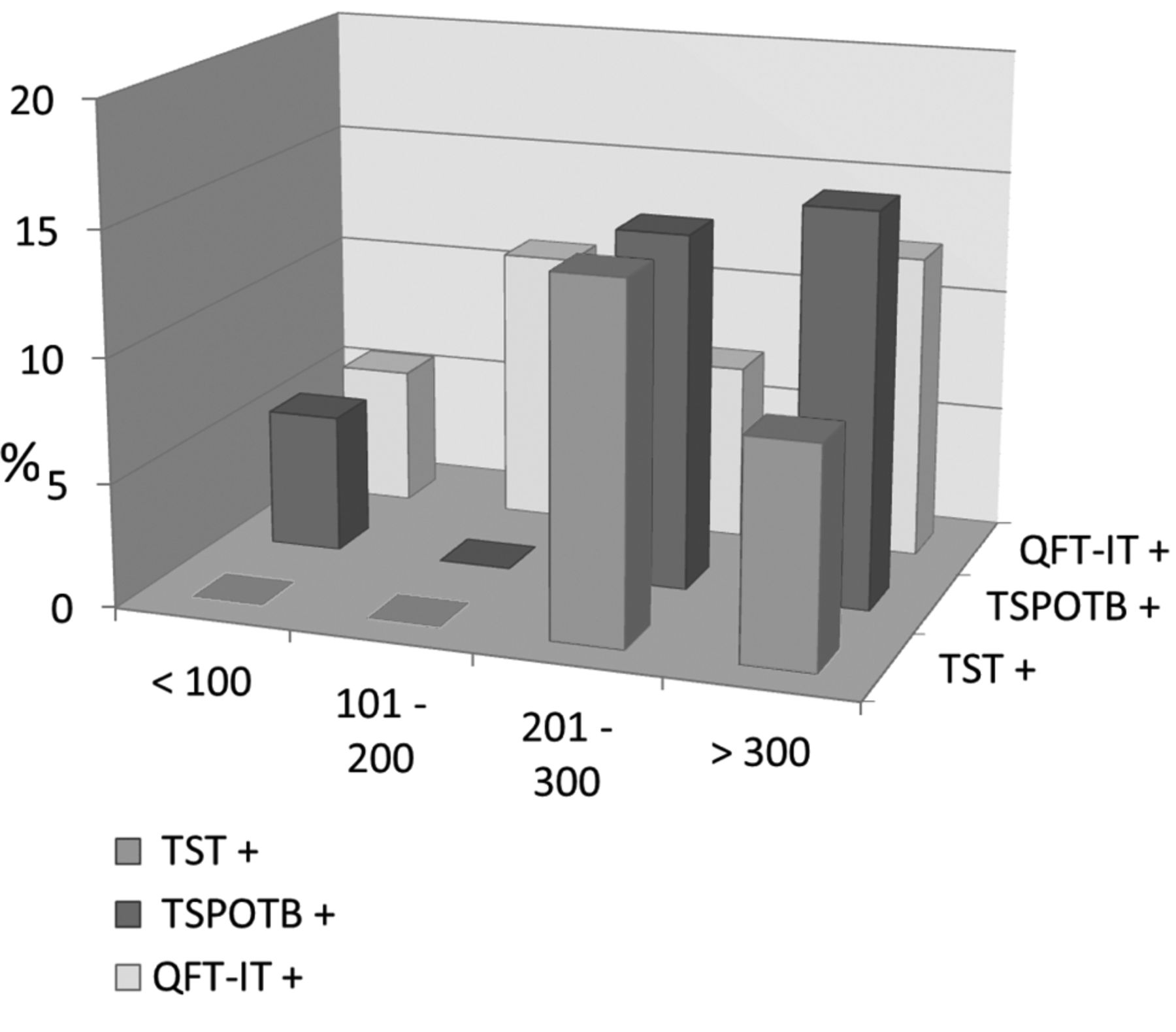
Percentage of positive tests in relation to the number of CD4.

TST was positive in 2 patients of group 3 (14.3%) and 5 patients of group 4 (8.8%). It was negative in 14 patients in group 1 (77.8%), 8 patients in group 2 (88.9%), 8 patients in group 3 (57.1%) and 44 patients in group 4 (77.2%). TST wasn't read in 4 patients of group 1 (22.2%), 1 patient of group 2 (11.1%), 4 patients of group 3 (28.6%) and 8 patients in group 4 (14%).

TSPOT-TB was positive in 1 patient in group 1 (5.5%), 2 patients in group 3 (14.3%) and 9 patients in group 4 (15.8%). The test showed a negative result in 15 patients in group 1 (83.3%), 7 patients in group 2 (77.8%), 11 patients in group 3 (78.6%) and 46 patients in group 4 (80.7%). It was indeterminate in 6 patients in group 1 (11.2%), 2 patients in group 2 (22.2%), 1 patient in group 3 (7.1%) and 4 patients in group 4 (3.5%).

Quantiferon-IT was positive in 1 patient in group 1 (5.6%), 1 patient in group 2 (11.1%), 1 patient in group 3 (7.1%) and 7 patients in group 4 (12.3%). The results were negative in 16 patients of group 1 (88.8%), 7 patients of group 2 (77.8%), 12 patients in group 3 (85.8%) and 49 patients of group 4 (85.9%). It was indeterminate in 1 patient of group 1 (5.6%), 1 patient of group 2 (11.1%), 1 patient of group 3 (7.1%) and 1 patient of group 4 (1.8%) (Figure 1).

Five patients showed a complete correspondence of TST, TSPOT-TB and QFT-IT.

Among patients with a negative TST, 4.1% showed a positive TSPOT-TB and 2.1% had a positive QFT-IT. 9.2% of all patients with a negative TST showed a positive result to IGRAs (Figure 2).

Figure 2.
  • Download figure
  • Open in new tab
  • Download powerpoint
Figure 2.

Percentage of positive IGRAs with negative TST.

Discordant results were observed in patients testing positive to IGRAs but negative to TST. Only 2 patients showed positive TST and negative IGRAs. Results of the study are summarized in Table II.

Discussion

TST, TSPOT-TB and Quantiferon-IT were positive in 7.1%, 12.2%, 10.2% cases respectively. Positive results of the tests seemed to associate with higher CD4 cell counts. Our study showed a poor concordance between TST and IFN-γ releasing assays, mainly in patients with a low CD4 cell count, as already reported by several articles in literature (13-15). The results of our study showed an increase in the number of patients that could undergo the preventive therapy for latent TB infection: among patients with negative TST, 4.1% had a positive TSPOT-TB, 2.1% had a positive QFT-IT; 9.2% patients had positive IGRAs, totally.

IGRAs should be able to identify TST negative subjects who could be treated with isoniazid. Interestingly, in our study, the blood tests provided low proportion of indeterminate results (13 cases for TSPOT-TB, 4 cases for QFT-IT). Most of these results seem to be related to a low CD4 cell count.

The undetectable results of IFN-γ release assays could be considered as “transient” results in patients with advanced immunosuppression.

Our results need to be confirmed by further investigations conducted in a larger number of cases, in order to establish the clinical impact and the effectiveness of dual testing approaches.

View this table:
  • View inline
  • View popup
  • Download powerpoint
Table II.

Comparison of TST, TSPOT-TB, QFT-IT in all 98 HIV-positive patients.

Footnotes

  • Funding

    The study was financed by CERIFARC/Regione Campania decree 176 of 03/07/2009 (BURC nr.45 of 20/07/2009) and selected among “Projects for improving of assistance, diagnosis and therapy of HIV infected patients: immunology, co-infection, information and prevention”.

  • Received July 17, 2014.
  • Revision received October 1, 2014.
  • Accepted October 7, 2014.
  • Copyright © 2015 International Institute of Anticancer Research (Dr. John G. Delinassios), All rights reserved

References

  1. ↵
    1. Diel R
    : Interferon gamma release assays for the diagnosis of latent Mycobacterium tubercolosis infection: a systematic review and meta analysis. Eur respire J 37: 88-99, 2011.
    OpenUrl
  2. ↵
    1. Menzies D
    : Meta analysis: new tests for the diagnosis of latent tubercolosis infection: areas of uncertainty and recommendations for research. Ann Intern Med 146: 340-354, 2007.
    OpenUrlCrossRefPubMed
  3. ↵
    1. American Thoracic Society,
    2. Centers for Disease Control and Prevention,
    3. Infectious Diseases Society of America
    : American Thoracic Society/Centers for Disease Control and prevention/Infectious Diseases Society of America: controlling tubercolosis in the United States. Am J Respir Crit Care Med 172: 1169-1127, 2005.
    OpenUrlCrossRefPubMed
  4. ↵
    1. Santin M
    : Interferon gamma release assays for the diagnosis of tubercolosis and tubercolosis infection in HIV infected adults: a systematic review and meta analysis. Plos one 7(3): e32482, 2012.
    OpenUrlCrossRefPubMed
    1. Giraldi E
    : Tubercolosis in HIV infected persons in the context of wide availability of highly active antiretroviral therapy. Eur Respir J 24: 11-17, 2004.
    OpenUrlAbstract/FREE Full Text
  5. ↵
    1. Ramos JM
    : Contribution of interferon gamma release assays testing to the diagnosis of latent tubercolosis infection in HIV infected patients: a comparison of Quantiferon TB Gold in Tube, TSPOT-TB and tuberculin skin test. BMC Infectious Diseases 12: 169, 2012.
    OpenUrlCrossRefPubMed
  6. ↵
    1. World Health Organization website
    . Available: http//www.who.int/tb/country/data/profiles/en/index.html. Accessed August 16, 2011.
  7. ↵
    1. Elzi DJ
    : Reducing tubercolosis incidence by tuberculin skin testing, preventive treatment, and antiretroviral therapy in an area of low tubercolosis transmission. Clin Infect Dis 44: 94-102, 2007.
    OpenUrlAbstract/FREE Full Text
  8. ↵
    1. Singh AR
    : Tubercolin test measurement-variability due to the time of reading. Chest 122: 1299-1301, 2002.
    OpenUrlCrossRefPubMed
  9. ↵
    1. Luetkemeyer AF
    : Comparison of an interferon gamma release assay with tuberculin skin testing in HIV infected individuals. Am J Respir Crit Care Med 175: 737-742, 2007.
    OpenUrlCrossRefPubMed
  10. ↵
    1. Stephan WR
    : Comparing Quantiferon tubercolosis gold, TSPOT tubercolosis and tuberculin skin test in HIV infected individuals from a low prevalence tubercolosis country. AIDS 22: 2471-2479, 2008.
    OpenUrlCrossRefPubMed
  11. ↵
    1. Palomino JC
    : Nonconventional and new methods in the diagnosis of tubercolosis: feasibility and applicability in the field. Eur Respir J 26: 339-350, 2005.
    OpenUrlAbstract/FREE Full Text
  12. ↵
    1. Talati NJ
    : Poor concordance between interferon gamma release assays and tuberculin skin tests in diagnosis of latent tubercolosis infection among HIV infected individuals. BMC Infectious Diseases 9: 15, 2009.
    OpenUrlCrossRefPubMed
  13. ↵
    1. Kang YA
    : Discrepancy between the tuberculin skin test and the whole blood interferon gamma assay for the diagnosis of latent tubercolosis infection in an intermediate tubercolosis-burden country. JAMA 293: 2756-2761, 2005.
    OpenUrlCrossRefPubMed
  14. ↵
    1. Cattamanchi A
    : Interferon gamma release assay for the diagnosis of latent tubercolosis infection in HIV individuals: a systematic review and meta analysis. J Acquir Immune Defic Syndr 56: 230-238, 2011.
    OpenUrlCrossRefPubMed
PreviousNext
Back to top

In this issue

In Vivo
Vol. 29, Issue 1
January-February 2015
  • Table of Contents
  • Table of Contents (PDF)
  • Index by author
  • Back Matter (PDF)
  • Ed Board (PDF)
  • Front Matter (PDF)
Print
Download PDF
Article Alerts
Sign In to Email Alerts with your Email Address
Email Article

Thank you for your interest in spreading the word on In Vivo.

NOTE: We only request your email address so that the person you are recommending the page to knows that you wanted them to see it, and that it is not junk mail. We do not capture any email address.

Enter multiple addresses on separate lines or separate them with commas.
Interferon Gamma Release Assays and Tubercolin Skin Test Performance in Different Settings of HIV Immunodeficiency
(Your Name) has sent you a message from In Vivo
(Your Name) thought you would like to see the In Vivo web site.
CAPTCHA
This question is for testing whether or not you are a human visitor and to prevent automated spam submissions.
2 + 2 =
Solve this simple math problem and enter the result. E.g. for 1+3, enter 4.
Citation Tools
Interferon Gamma Release Assays and Tubercolin Skin Test Performance in Different Settings of HIV Immunodeficiency
ROBERTO PARRELLA, VINCENZO ESPOSITO, MIRELLA ONOFRIO, GIOVANNI PARRELLA, ROSARIA VIGLIETTI, VINCENZO SANGIOVANNI, MIRIAM GARGIULO, FILOMENA DI MARTINO, ANNALISA DEL GIUDICE, GIULIA SANTORO, MARIANO BERNARDO, MARIA AURORA CARLEO, ANTONIO CHIRIANNI
In Vivo Jan 2015, 29 (1) 137-140;

Citation Manager Formats

  • BibTeX
  • Bookends
  • EasyBib
  • EndNote (tagged)
  • EndNote 8 (xml)
  • Medlars
  • Mendeley
  • Papers
  • RefWorks Tagged
  • Ref Manager
  • RIS
  • Zotero
Reprints and Permissions
Share
Interferon Gamma Release Assays and Tubercolin Skin Test Performance in Different Settings of HIV Immunodeficiency
ROBERTO PARRELLA, VINCENZO ESPOSITO, MIRELLA ONOFRIO, GIOVANNI PARRELLA, ROSARIA VIGLIETTI, VINCENZO SANGIOVANNI, MIRIAM GARGIULO, FILOMENA DI MARTINO, ANNALISA DEL GIUDICE, GIULIA SANTORO, MARIANO BERNARDO, MARIA AURORA CARLEO, ANTONIO CHIRIANNI
In Vivo Jan 2015, 29 (1) 137-140;
Reddit logo Twitter logo Facebook logo Mendeley logo
  • Tweet Widget
  • Facebook Like
  • Google Plus One

Jump to section

  • Article
    • Abstract
    • Patients and Methods
    • Results
    • Discussion
    • Footnotes
    • References
  • Figures & Data
  • Info & Metrics
  • PDF

Related Articles

  • No related articles found.
  • PubMed
  • Google Scholar

Cited By...

  • No citing articles found.
  • Google Scholar

More in this TOC Section

  • Efficacy of Percutaneous Direct Puncture Biopsy of Malignant Lung Tumors Contacting to the Pleura
  • Utility of Precision Oncology Using Cancer Genomic Profiling for Head and Neck Malignancies
  • Assessing the Risk of Stroke in the Elderly in the Context of Long-COVID, Followed Through the Lens of Family Medicine
Show more Clinical Studies

Similar Articles

Keywords

  • Latent tubercolosis infection
  • HIV infection
  • interferon gamma releasing assays
  • tuberculin skin test
In Vivo

© 2023 In Vivo

Powered by HighWire