Abstract
Background/Aim: The basic role of vascular endothelial growth factor (VEGF) in cancer is underscored by the approval of bevacizumab for first-line treatment of cancer patients. Recent anticancer therapeutics based on active tumor targeting by conjugating tumor-specific antibodies has become of great interest in oncology. Current progress in nanomedicine has exploited the possibility of designing tumor-targeted nanocarriers able to deliver specific molecule payloads in a selective manner to improve the efficacy and safety of cancer imaging and therapy. We herein aimed to determine the targeting ability of bevacizumab-conjugated quantum dots (QDs) in vitro and in vivo. Materials and Methods: We used QDs labeled with bevacizumab, in various in vitro experiments using cell lines derived from colorectal cancer (CRC) and breast cancer (BC). For a competition study of QD-bevacizumab complex and bevacizumab, the cells were pre-treated with bevacizumab (100 nmol/L) for 24 h before exposure to the QD-bevacizumab complex. The breast cancer cells (MDA-MB-231) were injected to 9 nude mice to make the xenograft tumor model. The QD-bevacizumab complex was injected into the tumor model and fluorescence measurements were performed at 1, 12, and 24 h post-injection. Results: Immunocytochemical data confirmed strong and specific binding of the QD-bevacizumab complex to the cell lines. The cells pre-treated with an excess of bevacizumab showed absence of QD binding. The in vivo fluorescence image disclosed that there was an increased signal of tumor after the injection of QDs. Ex vivo analysis showed 3.1±0.8%, 28.6±5.4% and 30.8±4.2% injected dose/g accumulated in the tumors at 1, 12 and 24 h respectively. Tumor uptake was significantly decreased in the animals pretreated with excess of bevacizumab (p=0.001). Conclusion: In conclusion, we could successfully detect the VEGF-expressing tumors using QDs-bevacizumab nanoprobes in vitro and in vivo, opening new perspectives for VEGF-targeted non-invasive imaging in clinical practice.
Cancer remains the leading cause of death worldwide (1). Despite intensive research in understanding cancer aetiopathogenesis, identification of cancer biomarkers, and improvements in surgery, chemotherapy and radiotherapy, patients survival rate from cancer has not significantly improved (1). Thus novel tools or agents for early cancer detection and diagnosis are urgently required.
It is well-accepted that tumor growth and metastatic dissemination basically depend on new blood vessel formation (angiogenesis). The vascular endothelial growth factor (VEGF)/VEGF receptor (VEGFR) signaling pathway is a key regulator of this process (2). In situ hybridization studies have shown high VEGF expression levels in the majority of human tumors (3, 4). The pivotal role of VEGF/VEGFR signaling pathway in cancer is underscored by the approval of bevacizumab (a humanized anti-VEGF antibody), for first-line treatment of cancer patients (5). Apart from treatment, as VEGF expression is localized in the tumor and VEGF is expressed during cancer development, this provides the opportunity for designing VEGF-targeted approaches for early cancer detection (6). Nowadays, anticancer therapeutics based on active tumor targeting by conjugating tumor-specific antibodies has become of great interest in oncology and nanomedicine, since this approach will increase therapeutic efficacy and will decrease systemic toxicity (7). Additionally, molecular imaging has emerged as a crucial tool in the field of cancer for in vivo monitoring of specific molecules and cellular processes, as well as targeted drug delivery (8-10).

Immunocytochemical studies of QDs-bevacizumab activity in cultured cell lines. A. DLD-1 cells as revealed by the presence of the QDs-bevacizumab complexes. B, MDA-MB-231 cells revealed by the presence of the QDs-bevacizumab complexes. Competition study of QDs-bevacizumab complexes and bevacizumab. After the addition of 100 nmol/L bevacizumab to DLD-1 (D) and MDA-MB-231 (C) cells. QDs-bevacizumab complex specific fluorescence was significantly reduced or absent.
Quantum dots (QDs) are semi-conductor nanocrystals with a quantum confinement property, which enables them to emit fluorescence from visible to infrared wavelengths on excitation (11). Recently due to their bright fluorescence, great photostability and their narrow and tunable emission spectrum, QDs have gained much interest for in vivo imaging applications (12).
The aim of the present study was to investigate whether the systemic delivery of bevacizumab conjugated to the surface of functionalized QDs led to target-specific ability in vitro, and accumulation in the tumor.
Materials and Methods
QDs-antibody conjugation. QDs which contain amine-derivatized, PEG-coated nanocrystals and the amine–thiol crosslinker (SMCC) was conjugated to bevacizumab with a Qdot605 Antibody Conjugation Kit (Invitrogen, Paisley, UK), according to the manufacturer's instructions. The molar ratio of bevacizumab fragments to the QDs at mixing is approximately 3:1. The final concentration of QD-bevacizumab complexes was determined by measuring the conjugate absorbance at 600 nm and using an extinction coefficient of 650,000 M-1 cm−1 at 600 nm.
Cell lines. The MDA-MB-231 breast cancer and the DLD-1 colorectal cancer cell lines were used for the in vitro experiments. Both cell lines were cells were cultured in Dulbecco's modified Eagle's medium supplemented with 5% fetal bovine serum. Conventional immunohistochemical techniques were used to determine the binding of ODs-bevacizumab complex to cells, using unconjugated QDs as a negative control. In these experiments QDs and QDs-bevacizumab (100 nmol/L) were incubated with cells for 1h at 37°C, washed, and photographed. For competition studies cells were pretreated with bevacizumab (100 nmol/L) for 24 h before exposure to the QDs-complex conjugate.
Tumor xenografts. All animal experiments were performed in compliance with the European legislation for animal welfare. Animal protocols have been approved by the Greek Authorities. Severe combined immunodeficiency (SCID) mice (average weight of 20 g) were obtained from the breeding facilities of the Institute of Biology NCSR “Demokritos” and were used for imaging studies. SCID mice were inoculated subcutaneously with 100 μl of cell suspension (approximately 107 MDA-MB-231 cells/animal) just above the left anterior leg, under sterile conditions. Tumors were allowed to grow for 3-5 weeks, or until well-palpable tumors developed. The animals were kept under aseptic conditions and had free access to food and water until the day of experimentation. All experiments were carried out in compliance with the relevant national laws relating to the conduct of animal experimentation.

Representative image of MDA-MB-231 human breast cancer-bearing mouse obtained at 12 h after intravenous injection of QDs-bevacizumab complexes. Arrows indicate the tumors.
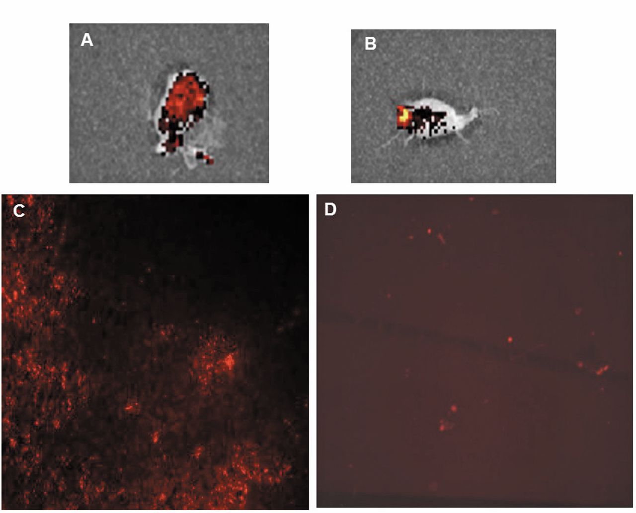
Tumor non-pre-treated (A) and pre-treated (B) with excess fold of bevacizumab. Tumor fluorescence in the pretreated group of mice is significantly reduced (p=0.001) (B). Representative histological image of tumors in mice treated with QDs-bevacizumab complexes (C) and in mice treated with QDs-bevacizumab complexes after pretreatment with excess bevacizumab (D).
In vivo optical tumor imaging. QDs-bevacizumab complexes were injected into the tail vein of mice at a concentration of 2 μmol/L and a volume of 100 μL. The animals were anesthetized with an i.p. injection of a ketamine and xylazine mixture at a dosage of 95 and 5 mg/kg respectively before the acquisition was started. In vivo fluorescence imaging was performed with an IVIS 200 small animal imaging system (Xenogen, Alameda, CA, USA). A DsRed filter (excitation wavelength 500-550 nm and emission wavelength 575-650 nm) was used for acquiring fluorescence imaging in vivo. The biodistribution of fluorescence intensity was monitored at 1, 12 and 24 h post-injection. In order to quantitatively estimate accumulation of the probe in tumors, animals were killed by decapitation. Tumors were excised and weighed. The fluorescence intensity was measured and normalized to photons per second with an ROI covering the entire tumor. The total fluorescence flux of each tumor was divided by its weight. The results were calculated as % injected dose/gram (% ID/g). For immunohistologic examination, tumors were fixed in 4% paraformaldehyde overnight and then transferred to ethanol before processing and paraffin embedding.
Statistical analysis. Data were expressed as mean±standard deviation. Means were compared using unpaired student's t-test. p-Values of less than 0.05 were considered statistically significant.
Results
In vitro study. QDs were conjugated to bevacizumab using the Qdot605 Antibody Conjugation Kit. Immunocytochemical data indicated strong and specific binding of the QDs-bavacizumab complex to both VEGF-expressing human breast cancer (MDA-MB-231) and colorectal cancer (DLD-1) cell lines (Figure 1A). QDs without antibody showed almost no binding to cells (Figure 1B). MDA-MB-231and DLD-1 cells pre-treated by excess bevacizumab also showed the absence of QDs-bavacizumab complex binding (Figure 1C).
In vivo imaging study with QDs-bevacizumab complex. After verifying that QDs-bevacizumab complexes have sensitive and specific binding ability to VEGF using the cell-binding assay described above, we proceeded to test it in living organisms. Figure 2 shows fluorescence images of mice bearing VEGF-positive MDA-MB-231 tumors at 12 h. In vivo fluorescence detection revealed distinct uptake in the VEGF expressing MDA-MB-231 tumors at 12 h after IV injection of QDs-bevacizumab complexes. The specific fluorescence signal was clearly visualized in the tumors area as compared with normal regions. The MDA-MB-231 tumor-bearing xenografts pre-treated with excess fold of bevacizumab showed a significant decrease in the accumulation of QDs-bevacizumab (p=0.001) (Figure 3). Quantitative analysis of the ex vivo tumor tissue fluorescence images post-injection with QDs-bevacizumab demonstrates the progressive accumulation of QDs-bevacizumab by the VEGF positive MDA-MB-231 tumors (Figure 4). Specifically, ex vivo analysis showed 3.1±0.8%, 28.6±5.4% and 30.8±4.2% injected dose/g accumulated in the tumors at 1, 12 and 24 h respectively. To further examine the specific binding of QDs-bevacizumab complexes in tumor tissues, tumors were harvested and paraffin-embedded. As shown in Figure 3C and D, sections from QDs-bevacizumab injected mice showed red fluorescence staining, whereas the tissues originating from animals pre-treated with bevacizumab before QDs-bevacizumab injections showed significantly reduced fluorescence signals.

Ex vivo study of QDs-bevacizumab in MDA-MB-231 human breast cancer-bearing mice at 1, 12 and 24 h. Data compare the percent injected dose per gram in the tumor after injection with QDs-bevacizumab complexes. Tumor uptake reached the peak at 12 h.
Discussion
The present study reports the development of QDs-bevacizumab conjugates for non-invasive tumor targeting and imaging of VEGF expression in human breast cancer xenografts in mice. Bevacizumab was chosen since, as it is mentioned above, it is a first-line treatment for several cancers and VEGF is a key mediator in tumor angiogenesis and cancer pathogenesis. Recently, nanotechnology has provided new perceptions for the development of cancer diagnosis by molecular imaging and targeted drug delivery platforms. Even if a lot of progress has been done, the poor penetration of drugs across the vascular barrier and into the tumor parenchyma remains a major problem for cancer therapy (13). In vivo imaging of QDs has been applied to many cases such as lymph node mapping, prostate cancer imaging and receptor-based specific tumor targeting (7, 14, 15).
QDs due to their optical and electrical properties have been intensively studied as a new class of nanoprobe for in vivo molecular imaging (12). QDs also have a modified surface with reactive functional groups for efficient conjugation of tumor-targeting ligands, antibodies and theurapeutic targets allowing for efficient and specific bioimaging (16). In the present study we used QDs-bevacizumab complexes for in vivo imaging studies. The major advantage of QDs for imaging in living organisms is that are photostable fluorophores in biological fluids (17). Furthermore, the therapeutic antibody component attached to the QDs, allows for differentiation between specific binding to specific molecular targets and also allows monitoring in a 12–24 h window as the antibody might stay bound for long time period (18). So, the QDs-bevacizumab can efficiently be used for non-invasive assess tumor, tumor angiogenesis and targeted drug delivery in vivo allowing several studies to be performed in experimental animals and humans. VEGF is strongly expressed in most of cancers (6, 14, 19), thus the specific localization of anti-VEGF-QDs in tumor indicate that can be used for improvement of imaging diagnostic accuracy and simultaneous targeted therapy (20). Concerning the QDs their potential toxicity is a problem for clinical applications, thus further studies concerning the optimal dose, toxicity and biodistribution are needed.
We showed the potential of specific in vitro and in vivo targeted-imaging regarding QDs-bevacizumab, which is among the potential nanoparticles for molecular imaging. This approach suggests that QDs conjugated with antibodies or therapeutics can be promising for cancer detection and targeted therapy.
Acknowledgements
This study was funded by Scholarship – Grant by the Experimental Research Center ELPEN Pharmaceuticals (E.R.C.E), and the Hellenic Society for Gastrointestinal Oncology (H.S.G.O.)
- Received August 5, 2014.
- Revision received October 2, 2014.
- Accepted October 9, 2014.
- Copyright © 2014 International Institute of Anticancer Research (Dr. John G. Delinassios), All rights reserved