
Main menu

  • Home
  • Current Issue
  • Archive
  • Info for
    • Authors
    • Advertisers
    • Editorial Board
  • Other Publications
    • Anticancer Research
    • Cancer Genomics & Proteomics
    • Cancer Diagnosis & Prognosis
  • More
    • IIAR
    • Conferences
  • About Us
    • General Policy
    • Contact
  • Other Publications
    • In Vivo
    • Anticancer Research
    • Cancer Genomics & Proteomics

User menu

  • Register
  • Subscribe
  • My alerts
  • Log in
  • My Cart

Search

  • Advanced search
In Vivo
  • Other Publications
    • In Vivo
    • Anticancer Research
    • Cancer Genomics & Proteomics
  • Register
  • Subscribe
  • My alerts
  • Log in
  • My Cart
In Vivo

Advanced Search

  • Home
  • Current Issue
  • Archive
  • Info for
    • Authors
    • Advertisers
    • Editorial Board
  • Other Publications
    • Anticancer Research
    • Cancer Genomics & Proteomics
    • Cancer Diagnosis & Prognosis
  • More
    • IIAR
    • Conferences
  • About Us
    • General Policy
    • Contact
  • Visit iiar on Facebook
  • Follow us on Linkedin
Research ArticleExperimental Studies

Anti-UV Activity of Kampo Medicines and Constituent Plant Extracts: Re-evaluation with Skin Keratinocyte System

TAKAO KATO, SHUNSUKE HINO, NORIO HORIE, TETSUO SHIMOYAMA, TADAYOSHI KANEKO, KAORU KUSAMA and HIROSHI SAKAGAMI
In Vivo July 2014, 28 (4) 571-578;
TAKAO KATO
1Department of Oral Surgery, Saitama Medical Center, Saitama Medical University, Kawagoe, Saitama, Japan
2Division of Pharmacology, Department of Diagnostic and Therapeutic Sciences, Saitama, Japan
  • Find this author on Google Scholar
  • Find this author on PubMed
  • Search for this author on this site
  • For correspondence: sakagami@dent.meikai.ac.jp takao@saitama-med.ac.jp
SHUNSUKE HINO
1Department of Oral Surgery, Saitama Medical Center, Saitama Medical University, Kawagoe, Saitama, Japan
  • Find this author on Google Scholar
  • Find this author on PubMed
  • Search for this author on this site
NORIO HORIE
1Department of Oral Surgery, Saitama Medical Center, Saitama Medical University, Kawagoe, Saitama, Japan
3Division of Pathology, Department of Diagnostic and Therapeutic Sciences, Saitama, Japan
  • Find this author on Google Scholar
  • Find this author on PubMed
  • Search for this author on this site
TETSUO SHIMOYAMA
1Department of Oral Surgery, Saitama Medical Center, Saitama Medical University, Kawagoe, Saitama, Japan
  • Find this author on Google Scholar
  • Find this author on PubMed
  • Search for this author on this site
TADAYOSHI KANEKO
3Division of Pathology, Department of Diagnostic and Therapeutic Sciences, Saitama, Japan
4Department of Oral and Maxillofacial Surgery, Nihon University School of Dentistry, Tokyo, Japan
  • Find this author on Google Scholar
  • Find this author on PubMed
  • Search for this author on this site
KAORU KUSAMA
3Division of Pathology, Department of Diagnostic and Therapeutic Sciences, Saitama, Japan
  • Find this author on Google Scholar
  • Find this author on PubMed
  • Search for this author on this site
HIROSHI SAKAGAMI
2Division of Pharmacology, Department of Diagnostic and Therapeutic Sciences, Saitama, Japan
  • Find this author on Google Scholar
  • Find this author on PubMed
  • Search for this author on this site
  • For correspondence: sakagami@dent.meikai.ac.jp takao@saitama-med.ac.jp
  • Article
  • Figures & Data
  • Info & Metrics
  • PDF
Loading

Abstract

Aim: In order to search for new biological activity of Kampo medicines and their constituent plant extracts, we investigated their ability to protect the cells from UV irradiation (referred to as ‘anti-UV activity’) using the human immortalised skin keratinocyte cell line HaCaT. Materials and Methods: Anti-UV activity was represented by the selectivity index (SI), defined as the ratio of the concentration that reduced the viable cell number by 50% to the concentration that increased the viability of UV-irradiated cells to 50%. Results: HaCaT cells were highly resistant to UV irradiation, approximately 20% of cells survived even when the exposure time was prolonged to 480 min. Sodium ascorbate, a popular antioxidant, used as positive control, had excellent anti-UV activity (SI=200). Among 10 Kampo medicines, Shosaikoto (SI=34) had the highest anti-UV activity, followed by Hangesyashinto (SI>28), Unseiin (SI>23) and Ninjinyoeito (SI=23), Saireito (SI>19), whereas another four Kampo medicines were much less active (SI<9.6). Among 25 constituent plant extracts, Scutellaria root had the highest anti-UV activity (SI=38), followed by Polyporus sclerotium (SI>26), Gardenia fruit (SI>23), Japanese Gentian (SI>20) and Saposhnikovia root (SI>20). Glycyrrhizin also had potent anti-UV activity (SI=36). The SI values determined with the present HaCaT system were generally one order higher than those obtained with previously reported HSC-2 human oral squamous cell carcinoma system, although there was good correlation between these two systems (R2=0.9118). Conclusion. The present study highlights the improved sensitivity of anti-UV activity detection with the HaCaT system, and suggests the possible application of Kampo medicines as a component of sunscreening cosmetics.

  • Glycyrrhizin
  • Kampo medicine
  • UV protection

Ultraviolet rays (UV) are invisible electromagnetic waves. Moderate doses of UV exert several favorable effects, such as sterilization and disinfection (1), induction of vitamin D synthesis (2), and stimulation of metabolism and skin resistance. However, an excessive dose of UV produces reactive oxygen species (ROS) which damage cellular DNA and proteins, leading to carcinogenesis (3). Guanine, the most susceptible DNA base, is oxidized to 7,8-dihydroxy-8-oxoguanine upon UV irradiation, and triggers the transversion of G:C to T:A, a cause of mutation (5). High doses of UV irradiation induced apoptotic cell death in human myelogenous leukemia cell lines, but induced other types of cell death in human T-cell leukemia, erythroleukemia, glioblastoma (6), oral squamous cell carcinoma (OSCC) cell lines and human normal oral cells (gingival fibroblasts, pulp cells, periodontal ligament fibroblast) (7). We recently established a method that can measure the activity of compound or extract in protecting cells from UV-induced injury (referred to as ‘anti-UV activity’), using human OSCC HSC-2 cells as target cells (7, 8). Using this method, we reported that alkaline extract of Sasa senanensis Rehder leaf (9-12), lignin–carbohydrate complex (13, 14), newly-synthesized water-soluble azulenes (15) and sodium ascorbate (vitamin C) (9-15) exhibited potent anti-UV activity. Their anti-UV activity was higher than that of hot water extracts of the leaves of green tea, oolong tea, black tea and barley tea, and their commercially available PET (polyethylene terephthalate) bottle drinks (13) and Kampo medicines and their constituent plant extracts (16). Alkaline extracts of green tea leaf, oolong tea leaf and orange flower exhibited significantly higher anti-UV activity than their corresponding hot water extract counterparts, possibly due to higher lignin–carbohydrate complex content of the alkaline extracts (17).

We previously reported that various Kampo medicines (18-20) and their constituents, such as glycyrrhizin (21) and flavones (22), inhibited cyclooxygenase-mediated prostaglandin E2 production by lipopolysaccharide-activated mouse macrophage-like RAW264.7 cells, suggesting its possible anti-inflammatory activity. We thought anti-UV activity should be evaluated using skin-derived cells for the screening of candidate products for skincare. We re-examined the possible anti-UV activity of 35 Kampo medicines and their constituent plant extracts using the human immortalised skin keratinocyte cell line HaCaT. We also investigated whether there is a good correlation between this newly-established HaCaT system and a previous HSC-2 cell-based system (16).

Materials and Methods

Materials. The following chemicals and reagents were obtained from the indicated companies: Glycyrrhizin, Wako Pure Chem. Ind. (Osaka, Japan); Dulbecco's modified Eagle medium (DMEM), Invitrogen Corp. (Carlsbad, CA, USA); fetal bovine serum (FBS), Gemini Bio-Products (Woodland, CA, USA); 3-[4,5-dimethylthiazol-2-yl]-2, 5-diphenyltetrazolium bromide (MTT) and dimethyl sulfoxide (DMSO), Sigma Chem. Ind. (St. Louis, MO, USA); Alisma rhizoma and Asiasarum root, Astragalus root, Atractylodes lancea rhizoma, Bupleurum root, Cimicifuga rhizoma, Cinnamon bark, Cnidium rhizoma, Coptis rhizoma, Gardenia fruit, ginger, ginseng, Glycyrrhiza, Japanese angelica root, Japanese Gentian, Jujube, Peony root, Phellodendron bark, Pinellia tuber, Platycodon root, Polyporus sclerotium, Poria sclerotium, Rehmannia root, Saposhnikovia root, Scutellaria root, Byakkokaninjinto, Hangesyashinto, Hotyuekkito, Juzentaihoto, Kikyoto, Ninjinyoeito, Rikkosan, Saireito, Shosaikoto and Unseiin were obtained from Tsumura Corp., Tokyo, Japan. Kampo medicines were supplied as dried powders, and dissolved in phosphate-buffered saline without calcium and magnesium [PBS (−)] prior to the experiments.

Assay for anti-UV activity. Spontaneously immortalized HaCaT keratinocytes, derived from adult human skin that remain highly related to their normal counterparts (CLS Cell Lines Service GmbH, Eppelheim, Germany) were inoculated at 1×10⁴ cells/0.1 ml in the inner 60 wells of a 96-microwell plate (Becton Dickinson Labware, Franklin Lakes, NJ, USA). The surrounding 36 exterior wells were filled with 0.1 ml of PBS(−) to minimize the evaporation of water from the culture medium. After 24 hours, the attached cells were replenished with PBS(−) containing different concentrations of samples. The cells were then placed at 20.5 cm from a UV lamp (wavelength=253.7 nm), taking off the lid, and exposed to UV irradiation (6 J/m2/min) for 90 seconds, unless otherwise stated. The media were immediately replaced with fresh DMEM plus 10% FBS and cells were cultured for a further 24 hours at 37°C in an incubator with 5% CO2 to determine the relative viable cell number by MTT method. In brief, the treated cells were incubated for another 4 hours in fresh culture medium containing 0.2 mg/ml MTT. Cells were then lysed with 0.1 ml of dimethyl sulfoxide (DMSO), and the absorbance at 540 nm of the cell lysate was determined using a microplate reader (Biochromatic Labsystem, Helsinki, Finland). From the dose–response curve, the 50% cytotoxic concentration against unirradiated cells and the concentration that increased the viability of UV-irradiated cells to 50% (EC50) were determined. The selectivity index (SI) was determined by the following equation: SI=CC50/EC50 (7, 8).

Figure 1.
  • Download figure
  • Open in new tab
  • Download powerpoint
Figure 1.

Effect of the time of exposure to UV irradiation on viable cell number. Near-confluent HaCaT cells were replenished with PBS(−) and then exposed to UV irradiation for the indicated periods. After incubation for 24 h in fresh culture medium, the viable cell number was determined as described in the Materials and Methods. Each value represents the mean±SD of three independent experiments.

Statistical analysis. Results are presented as the mean±standard deviation (SD) of triplicate assays.

Results

Optimum condition of UV irradiation. We first determined the minimum exposure time required to induce maximum cytotoxicity. When HaCaT cells were exposed to UV irradiation in PBS(−), the viable cell number determined 24 h after incubation in fresh culture medium declined to the lowest level at 90 sec, where approximately 25% of the cells are still viable, and this level was maintained up to 480 sec (Figure 1). Based on this finding, we used an exposure time of 90 sec for the subsequent experiments.

Anti-UV activity of Kampo medicines and their constituent plant extracts. Sodium ascorbate (vitamin C), a popular anti-oxidant used as positive control, showed excellent anti-UV activity (SI=200) (Figure 4, Table I). Kampo medicines and their constituent plant extracts protected HaCaT cells from the UV-induced cytotoxicity to various extents (Figures 2, 3 and 4). Among 10 Kampo medicines, Shosaikoto (SI=34) showed the highest anti-UV activity, followed by Hangesyashinto (SI>28), Unseiin (SI>23), Ninjinyoeito (SI=23) and Saireito (SI>19), whereas other four Kampo medicines were much less active (SI<9.6) (Figure 2, Table I).

Figure 2.
  • Download figure
  • Open in new tab
  • Download powerpoint
Figure 2.

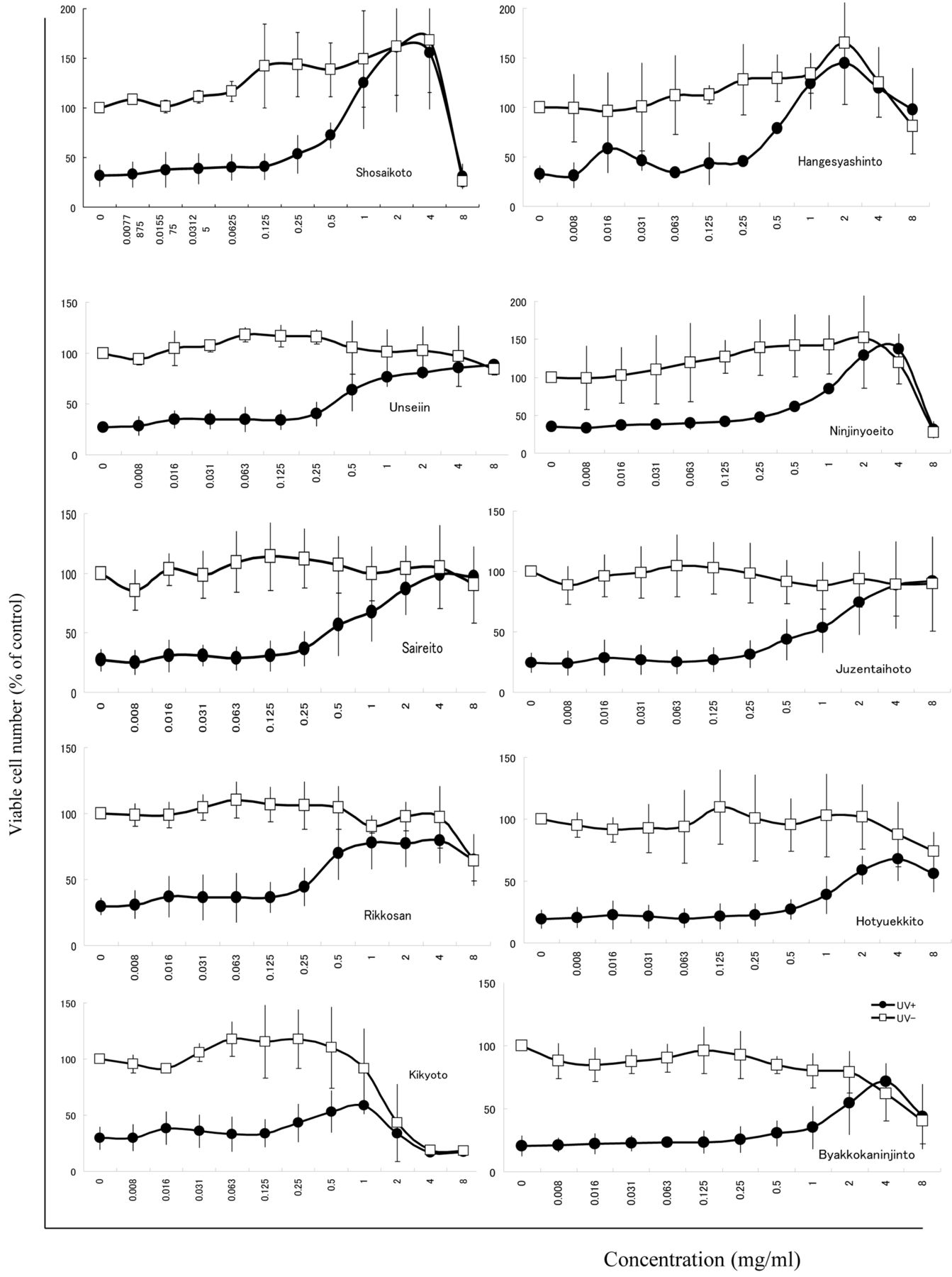
Anti-UV activity of 10 Kampo medicines. Near-confluent HaCaT cells were replenished with PBS(−) containing the indicated concentration of each Kampo medicine. The cells were then exposed to UV irradiation, and the viable cell number was determined after incubation for 24 h in fresh culture medium. Each value represents the mean±SD of three independent experiments.

Figure 3.
  • Download figure
  • Open in new tab
  • Download powerpoint
Figure 3.

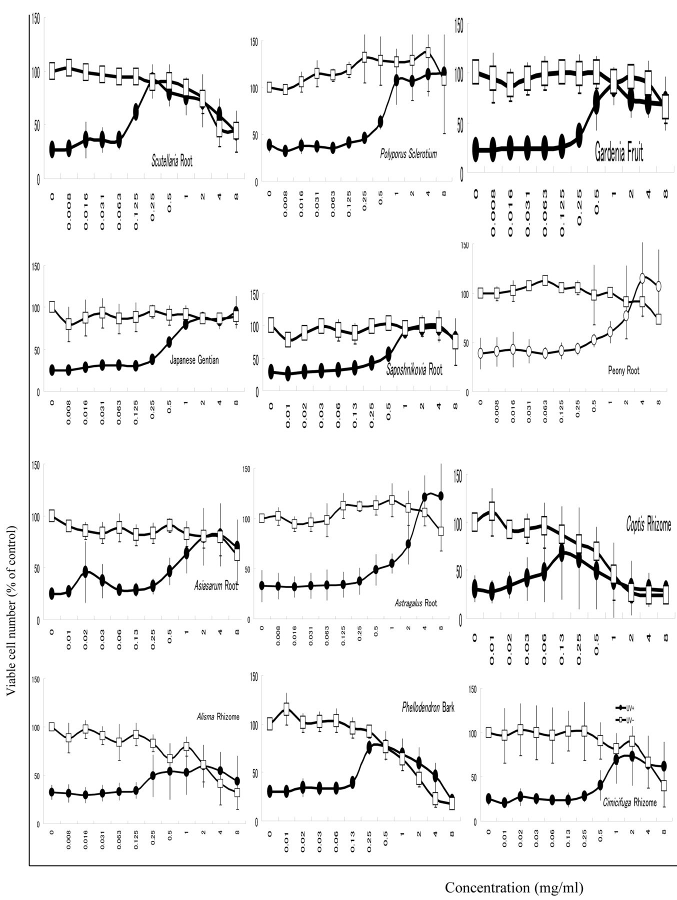
Anti-UV activity of 12 Kampo medicine constituent plant extracts. Near-confluent HaCaT cells were replenished with PBS(−) containing the indicated concentration of constituent plant extracts. The cells were then exposed to UV irradiation, and the viable cell number was determined after incubation for 24 h in fresh culture medium. Each value represents the mean±SD of three independent experiments.

Figure 4.
  • Download figure
  • Open in new tab
  • Download powerpoint
Figure 4.

Anti-UV activity of 13 Kampo medicine constituent plant extracts and vitamin C. Near-confluent HaCaT cells were replenished with PBS(−) containing the indicated concentration of each constituent plant extract glycrrizin, or sodium ascorbate (vitamin C). The cells were then exposed to UV irradiation, and the viable cell number was determined after incubation for 24 h in fresh culture medium. Each value represents the mean±SD of three independent experiments.

View this table:
  • View inline
  • View popup
  • Download powerpoint
Table I.

Anti-UV activity of Kampo medicines and constituent plant extracts against HSC-2 and HaCaT cells.

Among 25 plant extracts, Scutellaria root exhibited the highest anti-UV activity (SI=38), followed by Polyporus sclerotium (SI>26), Gardenia fruit (SI>23), Japanese Gentian (SI>20) and Saposhnikovia root (SI>20) (Figure 3 and 4). Glycyrrhizin also exhibited potent anti-UV activity (SI=36) (Figure 3, Table I).

Correlation of anti-UV activity between the present and previous systems. The SI values determined with the present HaCaT system were generally one order of magnitude higher than those determined by the previous HSC-2 system, although there was good correlation of these two systems (R2=0.9118) (Figure 5).

Discussion

The present study demonstrates that Kampo medicines and their constituent plant extracts exhibit some extent of anti-UV activity. The present HaCaT system may be suitable for the first detection of anti-UV activity, since the SI value (reflecting the anti-UV activity) determined by the present HaCaT system was one order higher than that predicted by previous method with HSC-2 cells.

Figure 5.
  • Download figure
  • Open in new tab
  • Download powerpoint
Figure 5.

Correlation of anti-UV activity [evaluated as selectivity index (SI) value] between the present HaCaT and previous HSC-2 systems. Each point was derived from the data of Table I.

The HaCaT cells were found to be much more resistant to UV irradiation (approximately 20-25% cells were viable upon exposure to 6 J/m2/min for 480 sec) compared to HSC-2 cells (fewer than 1% viable upon exposure for only 60 seconds) (7-15, 17), elevating the background level of the viability of UV-irradiated HaCaT cells. This suggests that HaCaT cells should possess a strong UV protection system. However, higher baseline viability (20-35% in Figures 2, 3 and 4) in the HaCaT system may make it difficult to accurately determine the EC50 values (see large deviation of the samples with lower SI values from the asymptotic line in Figure 5). Such deviating samples should be re-checked with the HSC-2 system for the accurate determination of the SI value.

In conclusion, the present study demonstrates the improved sensitivity of the HaCaT anti-UV activity detection system, and suggests the possible application of Kampo medicines as sunscreening components in cosmetics.

  • Received February 12, 2014.
  • Revision received April 1, 2014.
  • Accepted April 2, 2014.
  • Copyright © 2014 International Institute of Anticancer Research (Dr. John G. Delinassios), All rights reserved

References

  1. ↵
    1. Piluso LG,
    2. Moffatt-Smith C
    : Disinfection using ultraviolet radiation as an antimicrobial agent: A review and synthesis of mechanisms and concerns. PDA J Pharm Sci Technol 60: 1-16, 2006.
    OpenUrlAbstract/FREE Full Text
  2. ↵
    1. Barylsch MJ,
    2. Hofbauer GF,
    3. Dummer R
    : Vitamin D, ultraviolet exposure, and skin cancer in the elderly. Gerontology 56: 410-413, 2010.
    OpenUrlCrossRefPubMed
  3. ↵
    1. Ridley AJ,
    2. Whiteside JR,
    3. McMillan TJ,
    4. Allinson SL
    : Cellular and sub-cellular responses to UVA in relation to carcinogenesis. Int J Radiat Biol 55: 177-195, 2009.
    OpenUrl
    1. Cadet J,
    2. Berger M,
    3. Douki T,
    4. Morin B,
    5. Raoul S,
    6. Ravanat JL,
    7. Spinelli S
    : Effects of UV and visible radiation on DNA final base damage. Biol Chem 378: 1275-1286, 1997.
    OpenUrlPubMed
  4. ↵
    1. van Loon B,
    2. Markkanen E,
    3. Hübscher U
    : Oxygen as a friend and enemy: How to combat the mutational potential of 8-oxoguanine. DNA Repair 9: 604-616, 2010.
    OpenUrlCrossRefPubMed
  5. ↵
    1. Yanagisawa-Shiota F,
    2. Sakagami H,
    3. Kuribayashi N,
    4. Iida M,
    5. Sakagami T,
    6. Takeda M
    : Endonuclease activity and induction of DNA fragmentation in human myelogenous leukemic cell lines. Anticancer Res 15: 259-266, 1995.
    OpenUrlPubMed
  6. ↵
    1. Ueki J,
    2. Shimada A,
    3. Sakagami H,
    4. Wakabayashi H
    : Hormetic and UV-protective effects of azulene-related compounds. In Vivo 25: 41-48, 2011.
    OpenUrlAbstract/FREE Full Text
  7. ↵
    1. Kantoh K,
    2. Ono M,
    3. Nakamura Y,
    4. Nakamura Y,
    5. Hashimoto K,
    6. Sakagami H,
    7. Wakabayashi H
    : Hormetic and anti-radiation effects of tropolone-related compounds. In Vivo 24: 843-852, 2010.
    OpenUrlAbstract/FREE Full Text
  8. ↵
    1. Matsuta T,
    2. Sakagami H,
    3. Kitajima M,
    4. Oizumi H,
    5. Oizumi T
    : Anti-UV activity of alkaline extracts of the leaves of Sasa senanensis Rehder. In Vivo 25: 751-755, 2011.
    OpenUrlAbstract/FREE Full Text
    1. Sakagami H,
    2. Matsuta T,
    3. Satoh K,
    4. Ohtsuki S,
    5. Shimada C,
    6. Kanamoto T,
    7. Terakubo S,
    8. Nakashima H,
    9. Morita Y,
    10. Ohkubo A,
    11. Tsuda T,
    12. Sunaga K,
    13. Maki J,
    14. Sugiura T,
    15. Kitajima M,
    16. Oizumi H,
    17. Oizumi T
    : Biological activity of SE-10, a granulated powder of Sasa senanensis Rehder leaf extract. In Vivo 26: 411-418, 2012.
    OpenUrlAbstract/FREE Full Text
    1. Sakagami H,
    2. Amano S,
    3. Yasui T,
    4. Satoh K,
    5. Shioda S,
    6. Kanamoto T,
    7. Terakubo S,
    8. Nakashima H,
    9. Watanabe K,
    10. Sugiura T,
    11. Kitajima M,
    12. Oizumi H,
    13. Oizumi T
    : Biological interaction between Sasa senanensis Rehder leaf extract and toothpaste ingredients. In Vivo 27: 275-284, 2013.
    OpenUrlAbstract/FREE Full Text
  9. ↵
    1. Sakagami H
    1. Sakagami H,
    2. Matsuta T,
    3. Yasui T,
    4. Oguchi K,
    5. Kitajima M,
    6. Sugiura T,
    7. Oizumi T,
    8. Oizumi T
    : Chapter 8: Functional evaluation of Sasa Makino et Shibata leaf extract as group III OTC drug. In: Alternative Medicine. Sakagami H (ed.), pp. 171-200, 2012, InTech, Croatia.
  10. ↵
    1. Nanbu T,
    2. Matsuta T,
    3. Sakagami H,
    4. Shimada J,
    5. Maki J,
    6. Makino T
    : Anti-UV activity of Lentinus edodes mycelia extract (LEM). In Vivo 25(5): 733-740, 2011.
    OpenUrlAbstract/FREE Full Text
  11. ↵
    1. Nanbu T,
    2. Shimada J,
    3. Kobayashi M,
    4. Hirano K,
    5. Koh T,
    6. Machino M,
    7. Ohno H,
    8. Yamamoto M,
    9. Sakagami H
    : Anti-UV activity of lignin–carbohydrate complex and related compounds. In Vivo 27: 133-140, 2013.
    OpenUrlAbstract/FREE Full Text
  12. ↵
    1. Ueki J,
    2. Sakagami H,
    3. Wakabayashi H
    : Anti-UV activity of newly synthesized water-soluble azulenes. In Vivo 27: 119-126, 2013.
    OpenUrlAbstract/FREE Full Text
  13. ↵
    1. Kato T,
    2. Horie N,
    3. Matsuta T,
    4. Umemura N,
    5. Shimoyama T,
    6. Kaneko T,
    7. Kanamoto T,
    8. Terakubo S,
    9. Nakashima H,
    10. Kusama K,
    11. Sakagami H
    . Anti-UV/HIV activity of Kampo medicines and constituent plant extracts. In Vivo 26(6): 1007-1013, 2012.
    OpenUrlAbstract/FREE Full Text
  14. ↵
    1. Sakagami H,
    2. Ohkoshi E,
    3. Amano S,
    4. Satoh K,
    5. Kanamoto T,
    6. Terakubo S,
    7. Nakashima H,
    8. Sunaga K,
    9. Otsuki T,
    10. Ikeda H,
    11. Fukuda T
    : Efficient utilization of plant resources by alkaline extraction. Altern Integr Med 2, 2013 doi: 10.4172/2327-5162.1000133.
  15. ↵
    1. Horie N,
    2. Hashimoto K,
    3. Kato T,
    4. Shimoyama T,
    5. Kaneko T,
    6. Kusama K,
    7. Sakagami H
    : COX-2 as possible target for the inhibition of PGE2 production by Rikko-san in activated macrophage. In Vivo 22: 333-336, 2008.
    OpenUrlAbstract/FREE Full Text
    1. Kaneko T,
    2. Chiba H,
    3. Horie N,
    4. Kato T,
    5. Hashimoto K,
    6. Kusama K,
    7. Sakagami H
    : Effect of Sairei-to and its ingredients on the prostaglandin E2 production by mouse macrophage-like cells. In Vivo 22: 571-576, 2008.
    OpenUrlAbstract/FREE Full Text
  16. ↵
    1. Kaneko T,
    2. Chiba H,
    3. Horie N,
    4. Kato T,
    5. Kobayashi M,
    6. Hashimoto K,
    7. Kusama K,
    8. Sakagami H
    : Effect of Scutellariae radix ingredients on prostaglandin E2 production and COX-2 expression by LPS-activated macrophage. In Vivo 23: 577-582, 2009
    OpenUrlAbstract/FREE Full Text
  17. ↵
    1. Kato T,
    2. Horie N,
    3. Hashimoto K,
    4. Satoh K,
    5. Shimoyama T,
    6. Kaneko T,
    7. Kusama K,
    8. Sakagami H
    : Bimodal effect of glycyrrhizin on macrophage nitric oxide and prostaglandin E2 production. In Vivo 22: 583-586, 2008.
    OpenUrlAbstract/FREE Full Text
  18. ↵
    1. Kaneko T,
    2. Chiba H,
    3. Horie N,
    4. Kato T,
    5. Kobayashi M,
    6. Hashimoto K,
    7. Kusama K,
    8. Sakagami H
    : Inhibition of prostaglandin E2 production by flavone and its related compounds. In Vivo 24: 55-58, 2010.
    OpenUrlAbstract/FREE Full Text
PreviousNext
Back to top

In this issue

In Vivo: 28 (4)
In Vivo
Vol. 28, Issue 4
July-August 2014
  • Table of Contents
  • Table of Contents (PDF)
  • Index by author
  • Back Matter (PDF)
  • Ed Board (PDF)
  • Front Matter (PDF)
Print
Download PDF
Article Alerts
Sign In to Email Alerts with your Email Address
Email Article

Thank you for your interest in spreading the word on In Vivo.

NOTE: We only request your email address so that the person you are recommending the page to knows that you wanted them to see it, and that it is not junk mail. We do not capture any email address.

Enter multiple addresses on separate lines or separate them with commas.
Anti-UV Activity of Kampo Medicines and Constituent Plant Extracts: Re-evaluation with Skin Keratinocyte System
(Your Name) has sent you a message from In Vivo
(Your Name) thought you would like to see the In Vivo web site.
CAPTCHA
This question is for testing whether or not you are a human visitor and to prevent automated spam submissions.
15 + 1 =
Solve this simple math problem and enter the result. E.g. for 1+3, enter 4.
Citation Tools
Anti-UV Activity of Kampo Medicines and Constituent Plant Extracts: Re-evaluation with Skin Keratinocyte System
TAKAO KATO, SHUNSUKE HINO, NORIO HORIE, TETSUO SHIMOYAMA, TADAYOSHI KANEKO, KAORU KUSAMA, HIROSHI SAKAGAMI
In Vivo Jul 2014, 28 (4) 571-578;

Citation Manager Formats

  • BibTeX
  • Bookends
  • EasyBib
  • EndNote (tagged)
  • EndNote 8 (xml)
  • Medlars
  • Mendeley
  • Papers
  • RefWorks Tagged
  • Ref Manager
  • RIS
  • Zotero
Reprints and Permissions
Share
Anti-UV Activity of Kampo Medicines and Constituent Plant Extracts: Re-evaluation with Skin Keratinocyte System
TAKAO KATO, SHUNSUKE HINO, NORIO HORIE, TETSUO SHIMOYAMA, TADAYOSHI KANEKO, KAORU KUSAMA, HIROSHI SAKAGAMI
In Vivo Jul 2014, 28 (4) 571-578;
del.icio.us logo Digg logo Reddit logo Twitter logo CiteULike logo Facebook logo Google logo Mendeley logo
  • Tweet Widget
  • Facebook Like
  • Google Plus One

Jump to section

  • Article
    • Abstract
    • Materials and Methods
    • Results
    • Discussion
    • References
  • Figures & Data
  • Info & Metrics
  • PDF

Related Articles

  • No related articles found.
  • PubMed
  • Google Scholar

Cited By...

  • Prominent Anti-UV Activity and Possible Cosmetic Potential of Lignin-carbohydrate Complex
  • Google Scholar

More in this TOC Section

  • Systemic Administration of Lipopolysaccharide from Porphyromonas gingivalis Decreases Neprilysin Expression in the Mouse Hippocampus
  • Monitoring T-Cell Kinetics in the Early Recovery Period of Lung Transplantation Cases by Copy Number Levels of T-Cell Receptor Excision Circle
  • Successful Surgical Outcome of Feline Inductive Odontogenic Tumor in Three Cats
Show more Experimental Studies

Similar Articles

Keywords

  • glycyrrhizin
  • Kampo medicine
  • UV protection
In Vivo

© 2023 In Vivo

Powered by HighWire