


Abstract
Swine are becoming increasingly attractive as animal models for clinical research and the recently developed Microminipig (MMPig) has emerged as a possible experimental animal model. In this study, we demonstrated age-dependent changes in hematological parameters and coagulation activity in healthy MMPigs (58 male and 67 females, aged 0-34 months), and investigated white thrombus formation (WTF) using an in vitro microchip flow-chamber system (four males and four females, aged 22-23 months). There was no clear sex or age-dependent differences in any hematological parameters. While activated partial thromboplastin time (APTT) was shorter than prothrombin time (PT), with APTT:PT of 0.88:1, microchip flow-chamber system analysis showed that WTF time was shorter than that in humans, suggesting a possible thrombotic tendency in the MMPig. These results could be useful to life science researchers in the use of the MMPig as an experimental model animal for thrombus formation.
Swine have been used extensively in biomedical research, with a significant increase in recent decades, more than 60,000 pigs having been used in a year in the EU (1, 2). Because of their physiological and anatomical similarity to humans (3), swine are becoming increasingly attractive as animal models for clinical research. The microminipig (Brand: MMPig; registered with the Japanese Ministry of Agriculture, Forestry and Fisheries as a novel variety of swine; Fuji Micra Inc., Shizuoka, Japan) has emerged as a possible experimental animal model for non-clinical pharmacological/toxicological use (4-6). A female minipig, “Catherine” (the MMPig “Eve”), was the outcome from mating a pot-bellied pig and another type of minipig (4). The body weight (BW) of a young mature MMPig is <10 kg, enabling easy handling (3, 7-9). Except for coagulation activity, prothrombin time (PT) and activated partial thromboplastin time (APTT), the major hematological and biochemical parameters in the MMPig are similar to those found in Göttingen and Yucatan minipigs (7). The aim of the current study was to measure age-dependent changes in hematological parameters and coagulation activity, and to investigate white thrombus formation (WTF) in healthy MMPigs, using an automated microchip flow-chamber system.
Materials and Methods
Animals. All animals were maintained in the same animal unit at 24±3°C and relative humidity at 50±20%, with a 12 h light/dark cycle, and a maintenance space of 0.5-1.2 m2/animal. The amount of porcine diet (Marubeni Nisshin Feed Co.) provided was set according to age and body weight: 4-8%, 2-4%, and 1-3% of BW corresponding to 1 to 3 months, 4 to 6 months, and 7 months and older, respectively. The diet was composed of >13.0% crude protein, >2.0% crude fat, <8.0%crude fiber, <10.0% crude ash, >1.1% calcium, and >0.9% phosphorus. Tap water was available ad libitum. The animals used in this study were in good health and free of clinical signs of illness. They required no treatment or medication other than vaccination during the study. Data are presented as the mean±SD, and statistical analysis of differences was by F-test, and Student's t-test or Welch's t-test, at a significance level of p<0.05.
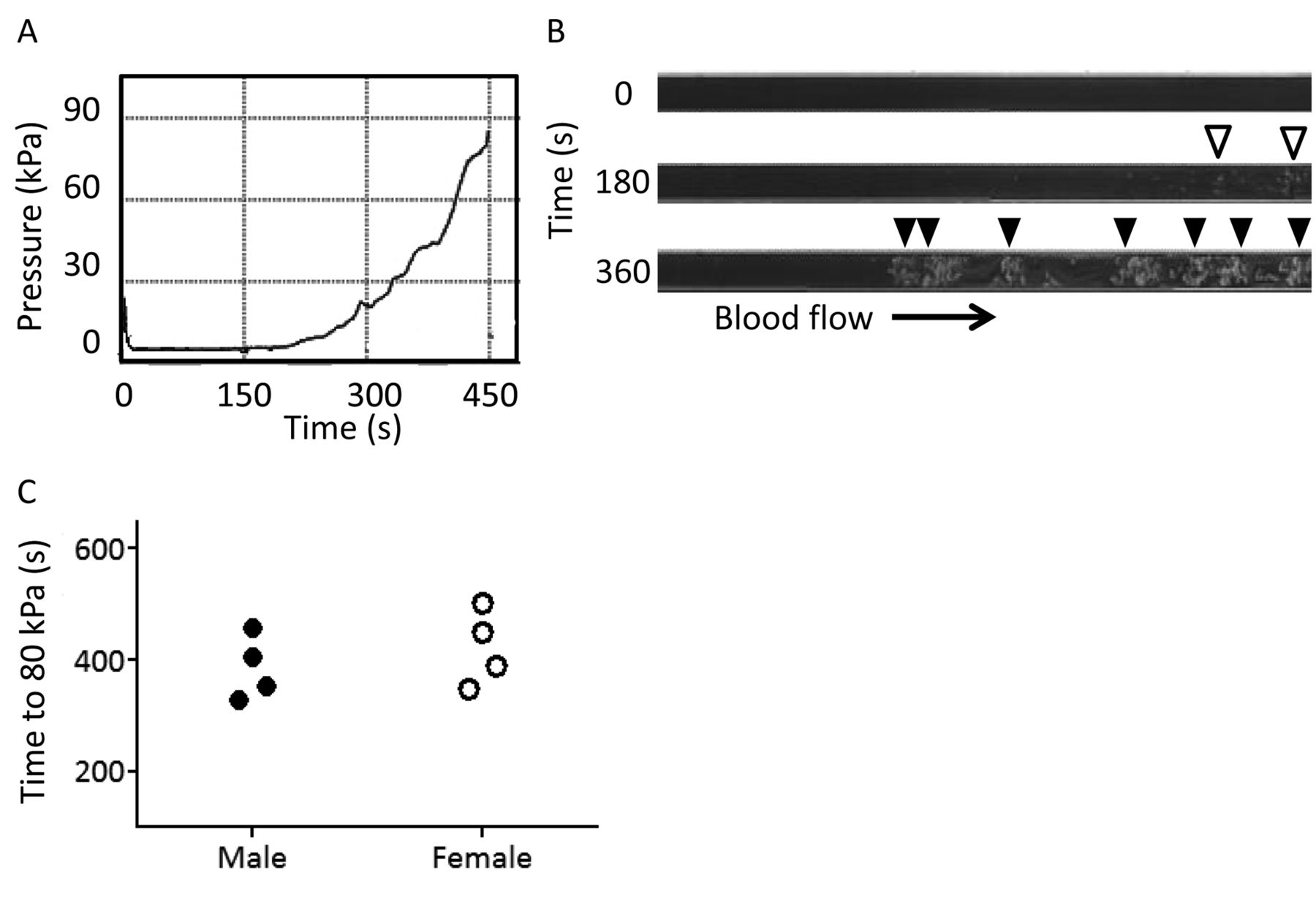
A: The flow-pressure waveform pattern during white thrombus formation (WTF). The black line represents the increasing pressure resulting from occlusion in the microchip flow-chamber by white thrombus formation. B: A typical image of white thrombus in the microchip flow-chamber. Open arrowheads indicate the initial small thrombus and closed arrowheads indicate mature WTF. C: The lag time for flow pressure to increase to 80 kPa in microminipigs measured by in vitro micro flow chamber system. p-Value is 0.4433 by the Student's t-test.
Blood collection. Blood samples were collected from the cranial vena cava of 125, fasted, conscious animals (58 males and 67 females) aged 0 to 34 months. For measurement of 23 hematological parameters, except PT and APTT, 1 ml of blood was collected with EDTA-2K as an anticoagulant and applied to an automatic analyzer (ADVIA 120, Siemens Healthcare Diagnostics Manufacturing Ltd., Munich, Bavaria, Germany). For measurement of PT and APTT, 1.5 ml of blood was collected with 150 μl of 3.8 w/v% sodium citrate solution as an anticoagulant. Plasma was obtained by centrifugation (4°C, 1,710 × g, for 15 min) and analyzed with an automatic analyzer (CA-7000, Sysmex Corporation, Kobe, Japan).
WTF assay. WTF assays were performed using an automated microchip flow-chamber system as previously stated (10). Blood (n=4 males and 4 females, aged 22 to 23 months, considered the most likely age for use in life science research) was collected into a tube containing 3.2% sodium citrate and mixed with 20 μl of 0.3 M CaCl2 containing 1.25 mg/ml of corn trypsin inhibitor immediately before application to the microchip. The mixture of blood and corn trypsin inhibitor was perfused over a microchip capillary coated with collagen and tissue thromboplastin at a flow rate of 10 μl/min. The WTF process is monitored by flow pressure changes in the capillary with a pressure sensor. As WTF spreads on the coated surface, the capillary is gradually occluded, increasing the flow pressure (Figure 1A and B). We calculated that the lag time for the flow pressure increase tothe 80 kPa (T80) from baseline, representing almost complete occlusion of the capillary by WTF.
Results
Hematological parameters and coagulation activity. Age-dependent hematological parameters and coagulation activity are listed in Tables I and II. Given statistical significance of p<0.01, hemoglobin values at age 3 to 5 months, APTT at age 9 to 12 months, and Red blood cell (RBC) at age 25 to 34 months were significantly lower and shorter respectively in females than those in males. Mean corpuscular hemoglobin concentration (MCHC) was higher in females aged 13 to 24 months. In the White blood cell (WBC) population, the basophil count was higher in females aged 13 to 24 months, corresponding to a higher basophil ratio and lower neutrophil ratio. No major parameter, including RBC and WBC, showed clear biological sex and/or age differences. APTT was shorter than PT (mean values) and the ratio of APTT to PT was approximately 0.88:1 in males, females, and both at the relevant age points.
Age-specific values in hematology in microminipigs.
WTF assay. The lag times for the flow pressure to increase by 80 kPa (T80) for male and female MMPigs was 386.3±50.4 min and 422.8±58.2 min, respectively (Figure 1C). There was no significant difference in T80 between the two groups (p=0.4433), indicating similar characteristics in thrombus formation in both males and females.
Discussion
Since the minipig is physiologically and anatomically similar to human, it is a suitable species for toxicological/pharmacological studies. However, despite continued efforts by breeders, minipigs are not yet widely used in life science research and one possible reason is the lack of reference values (11). We have reported reference values for hematological parameters in the newly-developed MMPig, the world's smallest minipig (7). In this study, we analyzed age-dependent changes in hematological and coagulation parameters for the MMPig to provide detailed information. There were no sex or age-dependent changes in hematological parameters during the experimental period. This indicates that there are no major differences in hematological parameters compared to those previously reported for the minipig (6).
Age-specific values in hematology in microminipigs.
However, the ratio of APTT to PT was different from that in other experimental animals. Although PT and APTT are commonly used as plasma-based assays for the assessment of coagulation activity in experimental animals, the exact time of each assay differs between species (12). In all species, except the rat (F344 strain), APTT is reported to be longer than PT and the ratio of APTT to PT is between 2:1 and 3:1. By contrast, in the MMPig, APTT is shorter than PT and the ratio is 0.88:1. Short APTT (11.2±1.0 s), as observed in the MMPig, is a unique characteristic when compared with previously reported results for minipigs; the APTT in the Gottingen minipig is in the range of 26 to 46 s and that for the Yucatan minipig an average of 15.46±1.15 s. APTT is longer than PT in both minipig species, consistent with other species (13, 14). Although it will be necessary to elucidate the biological mechanism of the shorter APTT observed in the MMPig, this hyper-coagulable response to the intrinsic pathway may suggest a thrombotic tendency in the MMPig.
APTT is assayed to evaluate the coagulation pathway, but does not fully reflect the interaction of coagulation factors or platelets in vivo. The newly-developed microchip-based flow chamber system (WTF assay) mimics in vivo blood flow and is influenced by both platelet activation and coagulation reactions over the collagen/tissue thromboplastin-coated surface (10). In this experiment, we used flow rates of 10 μl/min, corresponding to initial wall shear rates of 600× s−1, which simulates arterial blood flow in small to medium-sized arteries (15). T80 in the MMPig was 404.5±57.4 min compared with 558±90 in man, and the WTF assay indicated that white thrombus formation in the microminipig was markedly more rapid than that in human (10). This result further supports the conjecture that the MMPig has a thrombotic tendency, at least by in vitro thrombus formation assay.
Conclusion
In this study, we demonstrated age-dependent changes in hematological and coagulation parameters for MMPigs. All hematological parameters were within the normal range, with no major sex or age differences. APTT in the MMPig was shorter than PT and the ratio of APTT to PT was 0.88:1. We also investigated the thrombus formation activity and indicated a thrombotic tendency in the MMPig. These results could be useful to life science researchers in regard to the use of the MMPig as an experimental model animal for thrombus formation.
Acknowledgements
This work was partly supported by Health Labour Sciences Research Grant (no. 33361105) from the Ministry of Health, Labour and Welfare of Japan (to NM, HK, and AT), Adaptable and Seamless Technology transfer Program (A-Step No. AS2316907E) from the Ministry of Education Culture, Sports, Science and Technology of Japan (to AT and HK), Suzuken Memorial Foundation (to NM, HK and AT) and SENSHIN Medical Research Foundation (to NM, HK and AT). We are grateful to Mr. T. Motokado (SNBL, Ltd.), Dr. T. Nishimura, and Mr. N. Kaneko (Fuji Micra Inc.) for their advice and valuable technical assistance.
Footnotes
-
↵* These Authors contributed equally to this study.
-
Conflicts of Interest
TN and HS are employees of Fujimori Kogyo Co., Ltd. TI and IM hold endowed faculty positions in thrombosis research and have received funds from Medipolis Medical Research Institute, Shin Nippon Biomedical Laboratories, Asahi Kasei Pharma, and Asahi Kasei Medical.
- Received March 11, 2013.
- Revision received April 5, 2013.
- Accepted April 5, 2013.
- Copyright © 2013 International Institute of Anticancer Research (Dr. John G. Delinassios), All rights reserved
In this issue


Jump to section
Related Articles
Cited By...
- Development of a Microminipig Model of Atherosclerosis for the Evaluation of a HMGCR Inhibitor
- High Pathological Reproducibility of Diet-induced Atherosclerosis in Microminipigs via Cloning Technology
- NR6A1 Allelic Frequencies as an Index for both Miniaturizing and Increasing Pig Body Size
- Association Between HMGB1 and Thrombogenesis in a Hyperlipaemia-induced Microminipig Model of Atherosclerosis
- Diurnal Variation of Melatonin Concentration in the Cerebrospinal Fluid of Unanesthetized Microminipig
- Corpus luteum Regression Induced by Prostaglandin F2{alpha} in Microminipigs During the Normal Estrous Cycle
- The Microminipig as an Animal Model for Influenza A Virus Infection
- Profiles of Reproductive Hormone in the Microminipig During the Normal Estrous Cycle
- Comparison of the Genomic Sequence of the Microminipig, a Novel Breed of Swine, with the Genomic Database for Conventional Pig
- Investigation of Necessity of Sodium Cholate and Minimal Required Amount of Cholesterol for Dietary Induction of Atherosclerosis in Microminipigs
- Sex Differences of Serum Lipid Profile in Novel Microminipigs