


Abstract
Henoch-Shönlein purpura (HSP) is an acute, self-limited, systemic, small vessel vasculitis, that induces skin lesions, arthritis and abdominal pain. Palpable purpura is the most common manifestation in pediatric patients with HSP. We present an atypical case of HSP in a young patient and report successful treatment of the atypical skin lesions, while avoiding surgery.
- Henoch-Shönlein purpura (HSP)
- palpable purpura
- children
- bulla
- skin lesion
- hyaluronic acid-based dressings
Henoch-Shönlein purpura (HSP) is a rare non-thrombo-cytopenic IgA-mediated small vessel vasculitis of autoimmune hypersensitivity. The etiology is unknown and the typical first symptoms both in adults and children are skin lesions. Palpable purpura is the most common in pediatric patients with HSP, mainly on the lower extremities and the buttock (1). Skin necrosis rarely occurs in children (<5% of cases) and the probability of necrotic lesion increases with age (2).
We present a particular case of a 10-year-old male patient presented with ecchymotic lesions located in the left gluteal region and a very painful acute skin lesion that increased rapidly in size at the root of the right thigh. We report the association of the HSP with atopic dermatitis, autoimmune thyroiditis and ulcerative colitis. A successful combined treatment of dermal substitutes and advanced dressings, allowed us to achieve complete healing of the skin lesions, while avoiding surgery.
Case Report
A 10-year-old patient was admitted to the Pediatric Department with an ecchymotic skin lesion of the left buttock region. The patient underwent our plastic surgery consult. The mother told us that the initial lesions appeared as maculopapular erythematous rashes (varying in diameter from 1 to 15 millimeters), quickly met into palpable, purpuric, and ecchymotic injuries. Seven days from its onset, the skin lesion increased in volume, with very painful hemorrhagic blisters.
The patient was affected with atopic dermatitis and autoimmune thyroiditis. He had a history of hospitalization because of episodic bloody diarrhea. During his stay in the hospital, the patient complained of abdominal pain and underwent rectosigmoidoscopy which evidenced the presence of hyperemic areas, erosions and ulcers covered with fibrin in the rectum-sigma. The histopathological examination revealed the presence of an intraepithelial polymorphonuclear and lymphoplasmacellular infiltrate, with increase of basal plasma cells in the deep layer, increasing of plate cell trans-mucosal infiltration and widespread distortion of crypts and mucosal architecture, compatible with a diagnosis of ulcerative colitis. Mesalazine therapy was started. The patient reported pain in the joints of the lower limbs, especially in the knees, with difficulty in walking.
At first, our observation was that the skin lesion on the left side was covered with dark dry eschar, 5×4 cm in diameter, with erythematous surrounding halo, painful to digital pressure (Figure 1). A skin lesion biopsy was obtained. Histological examination revealed a thrombotic occlusion of the hypodermic artery with overhanging ischemic necrosis compatible with the clinical picture of vasculitis.
Eight days after the first observation, a new skin lesion appeared at the root of the right thigh. Similarly to the previous lesion, its color was bordeaux-purple, and was 5×3 cm in diameter, hot to the touch, painful and characterized by erythematous peri-wound skin. It increased rapidly in size reaching a diameter of 10 cm, extending in a few hours up to the right side, with the tendency to expand over the entire gluteal region (Figure 2A). Two days after its occurrence, we observed the presence of a dark dry eschar, 12×14 cm in diameter, with an erythematous surrounding halo, very painful on palpation (Figure 2B). The patient underwent a skin biopsy of the right lesion, with histological features similar to these of the left lesion biopsy, with a diagnosis of vasculitis.
Cultures of the skin ulcers were negative. Laboratory tests evidenced neutrophilic leukocytosis, c-ANCA- and ANA-positive and an increase of IgG anti-cardiolipin antibodies.
Palpable purpura together with abdominal pain and arthralgia led us to clinically diagnose this condition as HSP, although bullous and ulcerative skin lesions are rare in this disease. Immunosuppressive therapy with azathioprine at 75 mg/day and methyl prednisolone at 4 mg was started.
After two weeks from the appearance of first lesion, the patient underwent escharectomy of both wounds and wound bed debridement in order to obtain vital and well-perfused tissue ready for the subsequent application of Hyalomatrix PA® (Fidia Advanced Biopolymers, Italy). Hyalomatrix PA® was kept in place for 21 days. We performed a control inspection every 3 days and the monitoring through the external transparent layer showed the progression of dermal regeneration and the lack of wound infection.
Ten days after, the wound bed appeared covered by granulation tissue and the wound edges were re-epithelialized. After 21 days, a restoration of dermal component and partial re-epithelialization were apparent.
After one month of dressing, the lesion in the left buttock region had the appearance of a cavity and the wound bed was characterized by moderate exudate. Hyalogran® (Fidia Advanced Biopolymers) was applied for two weeks.
Finally, we used Jaloskin® (Anika Therapeutics, Inc., Bedford, MA, USA) in order to encourage a complete re-epithelialization.
Two months after initial treatment, we observed complete healing of the lesions with a good aesthetic result and resolution of abdominal pain and arthralgia (Figures 3 and 4). The patient underwent rectosigmoidoscopy which revealed a regression of ulcerative colitis.
Discussion
We present the case of a young patient who came to our attention with ecchymotic skin lesion of the left buttock region of uncertain diagnosis. This lesion rapidly increased in size and extended in depth in a short time, transforming into an extensive and painful ulcer with hemorrhagic blisters. The emergence of a new contralateral skin lesion that originated as a palpable purpura, and that turned within a few hours into hemorrhagic blisters and extensive skin necrosis led us to consider a systemic disease that could explain the onset of the lesions. The histological examination of the biopsy of the skin lesion was typical for vasculitis. The anamnesis and physical examination of the patient are essential for a correct diagnosis. Infact, palpable purpura, arthritis and abdominal pain are known as the ‘classic triad’ of HSP (3). More recent classifications, the 2006 European League Against Rheumatism (EULAR) and Pediatric Rheumatology Society (PReS) classification, include palpable purpura as a mandatory criterion, together with at least one of the following findings: diffuse abdominal pain, predominant IgA deposition (confirmed by skin biopsy), acute arthritis in any joint, and kidney involvement (as evidenced by the presence of blood and/or protein in the urine) (4). These diagnostic criteria confirmed our diagnosis.

Skin lesion of the left buttock region. Our first observation.
HSP, also known as anaphylactoid purpura, with an annual incidence of 13-20 per 100,000 children under 17 years old (5), is a systemic small vessel vasculitis. Purpura occurs in all cases, joint pains and arthritis in 80%, and abdominal pain in 62%. Forty percent of those afflicted have evidence of kidney involvement but only a quarter will have this to an extent sufficient to be noticeable without laboratory tests. To date, we have not noticed any kidney involvement in the current case. Lung and central nervous system involvement is generally rare (6).
There have been few reports of children with HSP exhibiting bullae as skin lesions and skin ulcers, as in this case; it is a rare phenomenon (7). Although vasculitis is usually limited to the upper layer of the dermis in HSP, it extended to the whole thickness of the dermis in a pediatric HSP patient with bullae (8). In this case, it is reasonable to assume that the thrombotic occlusion of the hypodermic artery may lead to damage in the corresponding skin region, leading to more severe skin manifestation, bullae or skin ulcers.
The purpura and skin lesions typically appear on the lower extremities and buttocks, as in this case, but may also be seen on the arms, face and trunk (9).
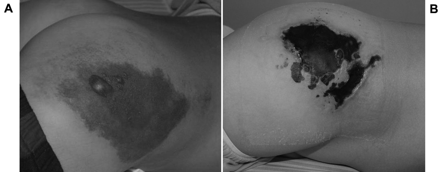
Skin lesion at the root of the right thigh at few hours after its occurrence (A) and skin two days after its occurrence (B).
We also evaluated the cause of abdominal pain. No abdominal masses or hepatosplenomegaly were observed. Abdominal ultrasound examination revealed colic wall thickening with the presence of reactive lymph nodes. Rectosigmoidoscopy with biopsy allowed us to make a diagnosis of ulcerative colitis, explaining the abdominal pain and bloody diarrhea.
Although the etiology of the vasculitis is unknown, it is postulated that certain triggers act as antigens that induce activation of the alternative complement pathway, while altered genetic markers determine susceptibility to HSP and predict disease severity (10). Furthermore, we report the association with other diseases such as atopic dermatitis and autoimmune thyroiditis in addition to ulcerative colitis. A state of hyperactivity of the immune system against such a common antigen may explain this association. But we have no evidence to support this theory. This case differs from those reported in literature, in that it had a rapid and dramatic onset, without any major prodromal symptoms and without known triggers or genetic markers.
Most patients with HSP require only supportive care. Analgesic and non steroidal anti-inflammatory drugs relieve joint and soft tissue discomfort. Corticosteroid therapy does not necessarily alter the likelihood of recurrence; however, if given early on in the course of the illness, corticosteroids appear to produce consistent benefits for several clinically-relevant HSP outcomes (11). In this case, we combined use of an immunosuppressant drug (azathioprine) with a corticosteroid drug (methyl prednisolone) due to the severity of the clinical symptoms. This type of therapy hinders the normal wound-healing process while providing good systemic benefits. Therefore, we considered the various treatment options for the two ulcers in this young patient.
Surgical management of these injuries consists of debridement and skin grafting of the areas involved. On occasion, flap repairs of these injuries have also been successful. In this case, invasive treatment should be avoided because of the patient's clinical condition. Furthermore a skin graft is contraindicated for a wound bed affected with vasculitis. Technical advances in tissue engineering have led to the development of biomaterials that are absolutely innovative and suited for wound managment. They are atraumatic, and do not require frequent changes. These therapeutic devices allow a conservative approach to be mainteined for to this type of injury. In this case we applied a dermal substitute, Hyalomatrix PA®, after escharectomy and debridement of the bed and margins of the wounds. Hyalomatrix PA® (12-15) is an acellular dermal substitute based on synthetic materials. It is composed of two layers: the internal layer is a three-dimensional dermis-like matrix consisting of fibers of a hyaluronic acid ester called Hyaff, while the external layer is a flexible and transparent elastomer film that operates as a semi-permeable barrier to external agents. The biological matrix acts by modifying the chemicol-physical characteristics of hyaluronic acid, a naturally occurring extracellular matrix playing an important role in the processes of tissue regeneration (16, 17). The transparent silicone membrane prevents liquid loss and allows for monitoring of the wound, without changing medication(18).
After 21 days of the application of Hyalomatrix PA®, a restoration of dermal component partial re-epithelialization were observed. Complete wound healing was achieved thanks to advanced dressings Hyalogran® and Jaloskin®.

Two months after initial treatment.
Hyalogran® is composed of adsorbing microgranules of sodium alginate and Hyaff.
The microgranules turn into a gel adapting to the lesion surface, entrapping the residues of devitalized tissue and exudate. At the same time, the presence of hyaluronic acid stimulates the processes of tissue repair and regeneration.
Jaloskin® is a hyaluronic acid-based dressing, consisting of a biomaterial made of esterified materials from hyaluronic acid that promotes cellular adhesion and three-dimensional structure. The dressing looks like a thin transparent film, which has to be adapted to the wound. Its characteristic of selective permeability to aqueous vapor allows for natural drainage of excess exudates, thus avoiding maceration of tissues. The selective permeability of Hyaff, on the other hand, keeps the wound moist, thus creating ideal conditions for rapid healing.
The transparency of the membrane permits constant visual monitoring of the underlying healing processes and it can be removed without causing microtraumas to the newly-formed tissues.
Conclusion
We describe an atypical case of HSP in a young patient. The peculiarity of the case comes from the type of skin lesions with ulcers and hemorrhagic bullae, which are rare in this disease in childhood. Skin biopsy allowed us to observe a thrombotic occlusion of the hypodermic artery that could explain this picture. Furthermore, we would like to emphasize the importance of the anamnesis and physical examination for a correct diagnosis because this atypical clinical picture in children with HSP may lead to a false diagnosis.

Two months after initial treatment.
We describe the association of HSP with atopic dermatitis, autoimmune thyroiditis and ulcerative colitis but we have no evidence to explain this association.
Anti-inflammatory and immunosuppressive therapy has allowed us to resolve the systemic symptoms with regression of arthralgia and ulcerative colitis resulting in the disappearance of abdominal pain. But this therapy hinders the physiological wound healing process. In addition the patient's clinical condition excluded surgical treatment, in particular a skin graft was contraindicated on a wound bed affected with vasculitis.
The combination of dermal substitutes and advanced dressings allowed us to maintain a conservative approach with overall excellent results for the quality of scars.
We hope that the present case will be helpful for a correct diagnosis in these atypical forms of HSP, while providing a useful treatment strategy for these types of skin lesions.
- Received September 30, 2012.
- Revision received November 5, 2012.
- Accepted November 6, 2012.
- Copyright © 2013 International Institute of Anticancer Research (Dr. John G. Delinassios), All rights reserved
References
In this issue


Jump to section
Related Articles
Cited By...
- No citing articles found.