


Abstract
Background: We have previously reported that azulene-related compounds can protect cells from UV-induced cytotoxicity. However, due to their high water insolubility, their anti-UV activity could not be accurately determined. In the present study, we newly-synthesized a total of nine derivatives with higher water solubility, and re-investigated their anti-UV activity. Materials and Methods: Cytotoxicity of these compounds against three human normal oral and three human oral cells squamous cell carcinoma cell lines (OSCCs) was evaluated by the 3-(4,5-dimethylthiazol-2-yl)-2,5-diphenyltetrazolium bromide (MTT) method. The concentration that reduced the viable cell number by 50% (CC50) and the concentration that increased the viability of UV-irradiated cells to 50% (EC50) were determined by the dose-response curves. Anti-UV activity (SI) was determined by the ratio of CC50 to EC50. The tumor specificity was determined by the ratio of the mean CC50 value for the normal cells to that for OSCC cells. Apoptosis induction was evaluated by DNA fragmentation and caspase activation. Results: All compounds except one (sodium 7-isopropyl-3-ethylazulene-1-sulfonate) were new compounds, and showed some tumor specificity (TS value=1.4 to 3.5), without induction of hormesis or apoptosis at lower and higher concentrations, respectively. Sodium 3-methylazulene-1-sulfonate showed the highest tumor specificity and potent anti-UV activity, approximately one half that of sodium ascorbate, the positive control. Conclusion: These data suggest the possible applicability of newly-synthesized water-soluble azulenes as skin care products protecting from UV irradiation.
Azulene (1-4), an isomer of naphthalene, has a dipole moment and a resonance energy with intermediate values between that of benzene and naphthalene, and is considerably more reactive, when compared with two arenes. Synthesis and chemical reactions of azulene derivatives have been investigated (5-7). They have been previously reported to show antibacterial (8), anti-ulcer (9), and relaxant activity (10), to inhibit thromboxane A2-induced vasoconstriction and thrombosis (11), and have been investigated for acute toxicity and local anesthetic activity (12), and chemotherapeutic activity against mucous membrane diseases (13, 14). However, the effects of azulene derivatives on cellular function have not yet been investigated in detail. We have reported tumor specificity (15-19), inhibitory action against nitric oxide production by activated macrophages (20-24), and certain anti-UV activity (15, 16, 25). However, due to high water insolubility, their actual anti-UV activity could not be determined. In the present study, we synthesized eight derivatives with much higher water solubility, and investigated their anti-UV activity together with sodium ascorbate as a positive control.
It has been reported that many toxic substances, environmental hormones, inorganic compounds, and even irradiation can modulate the growth of cultured cells in a bi-phasic fashion, stimulating or inhibiting the growth of cultured cells at lower and higher concentrations, respectively. This growth-stimulating effect at lower concentrations is known as hormesis (15). However, we recently found that three Chinese herbal extracts (26), sodium fluoride, and group I azulene compounds (16) had very week hormetic effects against various human normal and tumor cell lines. In order to confirm the generality of the occurrence of hormesis, we investigated whether the present eight compounds induce hormesis in human normal oral cells [gingival fibroblast (HGF), pulp cell (HPC) and periodontal ligament fibroblast (HPLF)] and three human oral squamous cell carcinoma (OSCC) cell lines (HSC-2, HSC-3, HSC-4).
Materials and Methods
Materials. The following chemicals and reagents were obtained from the indicated companies: Dulbecco's modified Eagle's medium (DMEM), from GIBCO BRL, Grand Island, NY, USA; fetal bovine serum (FBS) and 3-(4,5-dimethylthiazol-2-yl)-2,5-diphenyltetrazolium bromide (MTT) from Sigma-Aldrich Inc., St. Louis, MO, USA; dimethyl sulfoxide (DMSO) from Wako Pure Chem. Ind., Osaka, Japan; sodium ascorbate from Tokyo Chemical Industry Co., Ltd., Tokyo, Japan; and caspase-3 substrate, (DEVD-pNA) from MBVL, Nagoya, Japan.

Chemical structure of water-soluble azulenes: sodium 3-methylazulene-1-sulfonate [1], sodium 3-ethylazulene-1-sulfonate [2], sodium 3-propylazulene-1-sulfonate [3], sodium 7-isopropyl-3-methylazulene-1-sulfonate [4], sodium 7-isopropyl-3-ethylazulene-1-sulfonate [5], sodium 7-isopropyl-3-propylazulene-1-sulfonate [6], sodium 3-hexylazulene-1-sulfonate [7], sodium 7-isopropyl-3-heptylazulene-1-sulfonate [8] and disodium azulene-1,3-disulfonate [9].
Synthesis of test compounds. Sodium 3-methylazulene-1-sulfonate [1], sodium 3-ethylazulene-1-sulfonate [2], sodium 3-propylazulene-1-sulfonate [3], sodium 7-isopropyl-3-methylazulene-1-sulfonate [4], sodium 7-isopropyl-3-ethylazulene-1-sulfonate [5], sodium 7-isopropyl-3-propylazulene-1-sulfonate [6], sodium 3-hexylazulene-1-sulfonate [7], sodium 7-isopropyl-3-heptylazulene-1-sulfonate [8] and disodium azulene 1,3-disulfonate [9], were synthesized (Figure 1) according to previous references (11, 27-30). All compounds were dissolved in phosphate-buffered saline without calcium and magnesium [PBS(–)] at 80 mM and stored at −20°C before use. Cell culture. HGF, HPC and HPLF cells were established from first pre-molar tooth extracted from the lower jaw of a 12-year-old girl, as described previously (15). OSCC cell lines (HSC-2, HSC-3, HSC-4) were cultured as described previously (16, 17).
Assay for cytotoxic activity. Cells were inoculated at 3×10³ cells/0.1 ml in the inner 60 wells of a 96-microwell plate (Becton Dickinson Labware, NJ, USA). The surrounding 36 exterior wells were filled with 0.1 ml of PBS(–) to minimize the evaporation of water from the culture medium. After 48 h, the medium was removed by suction with aspirator, and replaced with 0.1 ml of fresh medium containing different concentrations of single-test compounds (0.31-20 mM). Cells were incubated for 48 h, and the relative viable cell number was then determined by the MTT method. In brief, treated cells were incubated for another 3 h in fresh culture medium containing 0.2 mg/ml MTT. Cells were then lysed with 0.1 ml of dimethyl sulfoxide (DMSO), and the absorbance of the cell lysate was determined at 540 nm, using a microplate reader (Biochromatic Labsystem, Helsinki, Finland).
Hormetic response of human oral normal and tumor cells after treatment with water-soluble azulenes: sodium 3-methylazulene-1-sulfonate [1], sodium 3-ethylazulene-1-sulfonate [2], sodium 3-propylazulene-1-sulfonate [3], sodium 7-isopropyl-3-methylazulene-1-sulfonate [4], sodium 7-isopropyl-3-ethylazulene-1-sulfonate [5], sodium 7-isopropyl-3-propylazulene-1-sulfonate [6], sodium 3-hexylazulene-1-sulfonate [7] or sodium 7-isopropyl-3-heptylazulene-1-sulfonate [8].
Assay for hormesis. The hormetic response was evaluated by the maximum response in each dose-response curve, as described previously (15).
Assay for DNA fragmentation. Cells (4×10⁴) were seeded on a 6-microwell plate and incubated for 48 h to allow for complete attachment. The medium was removed by suction with an aspirator, and replaced with 2 ml of fresh medium containing different concentrations of single-test compounds. Cells were then incubated for a further 6, 24 or 48 h. After washing twice with PBS(–), cells were collected by scraping with a rubber policeman on ice and spun-down in an eppendorf tube. Cells were lysed with 50 μl lysate buffer [50 mM Tris-HCl (pH 7.8), 10 mM EDTA, 0.5% (w/v) sodium N-lauroylsarcosinate]. The solution was incubated with 0.4 mg/ml RNase A and 0.8 mg/ml proteinase K for 2 h at 50°C and then mixed with 50 μl NaI solution [40 mM Tris-HCl (pH 8.0), 7.6 mM NaI, 20 mM EDTA-2Na], followed by 250 μl of ethanol. After centrifugation for 20 min at 20,000 ×g, the precipitate was washed with 1 ml of 70% ethanol and dissolved in TE buffer [10 mM Tris-HCl (pH 8.0), 1 mM EDTA-2Na]. Each sample (10-20 μl, equivalent to 2×10⁵ cells) was applied to 2% agarose gel electrophoresis in TBE buffer (89 mM Tris-HCl, 89 mM boric acid, 2 mM EDTA-2Na). After staining with ethidium bromide, the DNA was visualized by UV irradiation, and photographed as described previously (15). DNA from apoptotic HL-60 cells induced by UV irradiation (6 J/m2/min, 1 min) (15) were run in parallel as positive controls.
Assay for caspase-3 activation. The cells (8×10⁴) were seeded on a 6-microwell plate, and incubated for 48 h to allow for complete adherence. The medium was then removed by suction with an aspirator, and replaced with 2 ml of fresh medium containing different concentrations of single-test compounds. Cells were then incubated for a further 48 h. Cells were washed twice with PBS(–) and lysed with 100 μl of lysis solution [50 mM Tris-HCl, pH 7.5, 0.3% NP-40, 1 mM dithiothreitol (DTT)]. Cells were collected by scraping with a rubber policeman and transferred to an eppendorf tube. After resting the tube for 10 min on ice and centrifugation for 5 min at 10,000 ×g, the supernatant was collected. Lysate (50 μl, equivalent to 100 μg protein) was mixed with 50 μl lysis solution containing a substrate for caspase-3, DEVD-pNA. After incubation for 2, 4 or 24 h at 37°C, the absorbance of the liberated chromophore pNA was measured by microplate reader, at 405 nm as described previously (15).
Cytotoxicity of water-soluble azulenes towards human oral normal and tumor cells.
Assay for UV protection. The medium of cells attached to 96-microwell plates was replaced with PBS(–). Different concentrations of water-soluble azulenes were then added to the cell, all the plates were immediately placed at 21 cm from a UV lamp (wavelength 253.7 nm) and exposed to UV irradiation (6 J/m2/min) for different durations: 4 min for HGF cells, and 30 s for HSC-2 cells (15). The media were replaced with fresh DMEM plus 10% FBS and cells were then cultured at 37°C in an incubator with 5% CO2 until 48 h after the start of irradiation.
Statistical treatment. The difference between two groups was evaluated by the Student's t-test.
Results
Failure to induce hormesis. Eight water-soluble azulenes ([1]-[8]) did not induce any apparent hormetic stimulation of growth of three human oral normal cells (HGF, HPC, HPLF) and three OSCC cell lines (HSC-2, HSC-3, HSC-4). The maximum hormetic response was considerably low (<26.6%) (Table I), confirming our previous findings (15).
Tumor specificity. Compound [8] exhibited the highest cytotoxicity, followed by [7] > [6] > [5, 4, 3] > [2, 1]. On the other hand, the least cytotoxic compound [1] had the highest tumor specificity (TS >3.53), followed by [5] (TS=2.50) > [4] (TS=2.46) > [2] (TS=2.45) > [3] (TS=2.43) > [6] (TS=1.95) > [7] (TS=1.77) > [8] (TS=1.39) (Table II). There was negative correlation between the cytotoxicity and tumor specificity. Since compounds [1] and [5] exhibited the highest tumor specificity, the subsequent anti-UV activity experiments were carried out using these compounds.
Type of cell death induced by UV irradiation. Compounds [1] and [5] did not induce internucleosomal DNA fragmentation (such as that observed in UV-induced apoptotic HL-60 cells) nor did they produce large DNA fragments in HSC-2 and HSC-4 cells (Figure 2). With prolonged incubation time (2 → 4 → 24 hours), caspase-3 activity increased, however, the addition of 0-16 μM [1] and [5] did not significantly (p>0.05) change the caspase-3 activity for either of these cell lines (Figure 3). These data indicate that [1] and [5] did not induce apoptosis in either OCSS cell line.
Prominent anti-UV activity. We have previously reported that oral normal cells (HGF) were more resistant to UV irradiation as compared to OSCC (HSC-2) (15). Therefore, a longer exposure time (4 min) was necessary to induce apparent cytotoxicity in HGF cells, detected at 48 hours after the start of incubation. When water-soluble azulenes were added to PBS(–) during the exposure of cell to UV, UV-induced cytotoxicity was dramatically inhibited. Compound [1] had the highest anti-UV activity (SI=38.83, mean of two independent experiments in Table III, Figure 4A), followed by [4] (SI=28.98) > [2] (SI=28.74) > [9] (SI=26.15) > [5] (SI=16.16) > [3] (SI=12.63) > [6] (SI=4.38) > [7] (SI=2.61). The anti-UV activity of compound [1] for HGF cells was 40.49% of that of vitamin C (SI=95.88).
When HSC-2 cells were exposed to UV irradiation for a much shorter time (only 30 s), complete cell death was observed after 48 h. Addition of water-soluble azulenes in PBS(–) effectively protected the cells from UV-induced cytotoxicity. Compound [1] again exhibited the highest anti-UV activity (SI=68.50, mean of two independent experiments in Table III, Figure 4B), followed by [2] (SI=49.46) > [4] (SI=41.76)> [5] (SI=36.42) > [9] (SI=32.06) > [3] (SI=28.02) > [6] (SI=5.79)> [7] (SI=4.01). The anti-UV activity of compound [1] for HSC-2 cells was 53.01% of that of vitamin C (SI=129.21).
Discussion
We have synthesized a total of nine water-soluble azulenes. Among them, all compounds except [5] were new compounds. The present study demonstrated that water-soluble azulenes had generally higher anti-UV activity. Among them, compound [1] had the highest anti-UV activity for both HGF (SI=39) and HSC-2 cells (SI=69). The anti-UV activity of [1] was much higher than that of gallic acid (a component of tannin) (SI=5.4), epigallocatechin gallate (a major component of green tea) (SI=7.7), n-acetyl-l-cycteine (SI<1.0), catalase (SI<1.0), curcumin (SI<1.0), green tea extract (SI=3.4), black tea extract (SI<3.4), burley tea extract (SI<1.0) and Oolong tea extract (SI<1.0) (25), but amounts to approximately half that of vitamin C (SI=96 and 129, for HGF and HSC-2 cells respectively) (Table III).
Protective effect of water-soluble azulenes and sodium ascorbate (VC) on UV-induced cytotoxicity.
Compound [1] had the highest tumor specificity (TS >3.5) among nine water-soluble azulenes, without induction of hormesis (hermetic response=0%) at lower concentrations (Table I), or induction of apoptosis, i.e., internucleosomal DNA fragmentation (Figure 2) and caspase-3 activation (Figure 3), at higher concentrations. The tumor specificity of [1] was comparable with that of oligomeric hydrolyzable tannins (TS=4.4) and isoliquiritigenin (TS=4.0), but slightly higher than that of monomeric tannins (TS=1.5), quercetin (TS=2.2), curcumin (TS=1.7), kaempferol (TS=1.1) and sodium ascorbate (TS=2.5) (31).
In conclusion, the present study demonstrated, for the first time, that a new water-soluble azulene [1] has potent anti-UV activity with some tumor specificity, suggesting the possible applicability of this compound as a skin care product protecting from UV irradiation. There are at least three possibilities for the mechanism of anti-UV activity induction by [1]: (i) [1] may directly absorb the light of wavelength of 253.7 nm, (ii) [1] may scavenge any radicals produced from UV irradiation in PBS(–), or (iii) [1] may inhibit the signaling pathway of LOX-1 (32). Further study is underway in our laboratory to distinguish these possibilities.

Effect of water-soluble azulenzes on induction of DNA fragmentation. HSC-2 and HSC-4 cells were incubated for 6, 24 or 48 hours with the indicated concentrations of water-soluble azulene compounds, and lysed for the assay of DNA fragmentation by agarose gel electrophoresis. UV, DNA from UV-irradiated HL-60 cells.

Effect of water-soluble azulene compounds on induction of caspase-3 activity. HSC-2 and HSC-4 cells were incubated for 2 (indicated by black bars), 4 (white bars) or 24 (gray bars) hours with the indicated concentrations of water-soluble azulene compounds. The cells were lysed for determination of caspase-3 activity. As positive control, apoptotic HL-60 cells induced to apoptosis by UV irradiation were assayed for caspase-3 activity and this was defined as maximum activity (absorbance at 405 nm=0.18-0.33). Caspase-3 activity was determined by dividing the each value by that of maximum activity, and expressed as a percentage. Each value represents the mean±S.D. of triplicate assays. *p<0.05, **p<0.01 relative to the untreated cells.
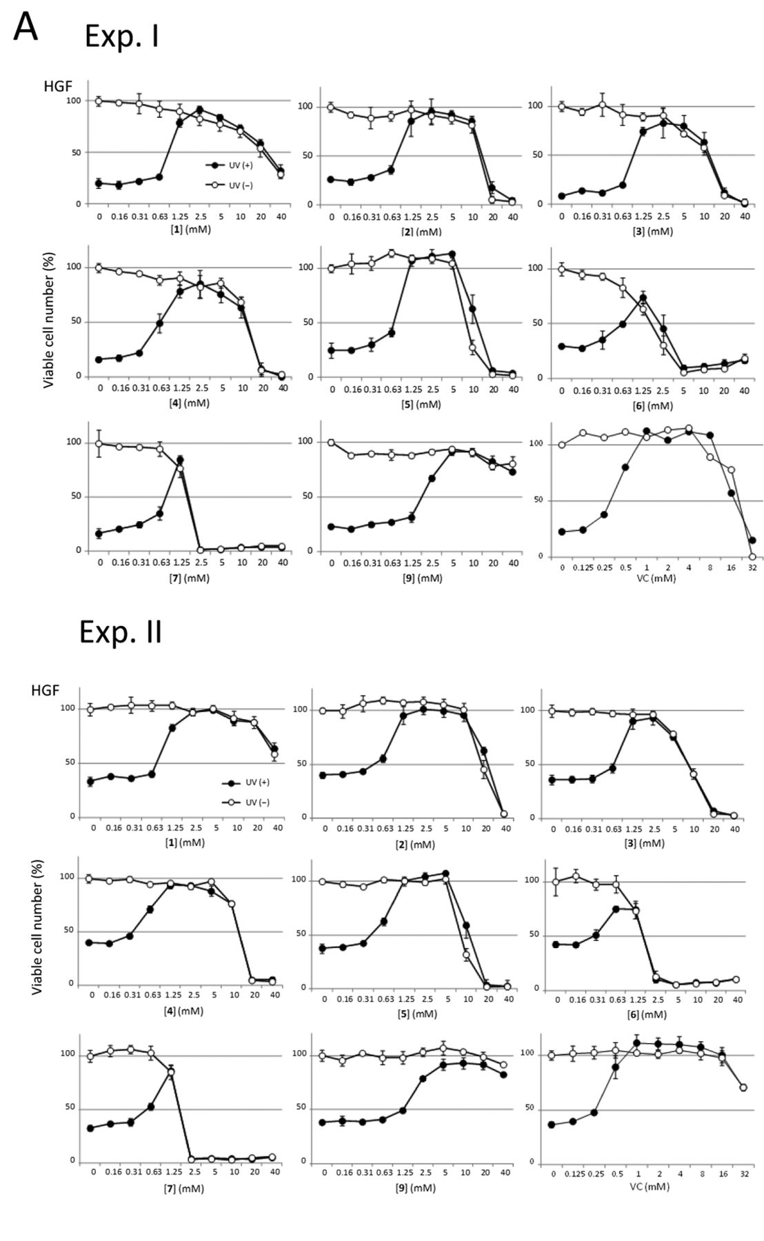

Effect of UV irradiation on the viability of cultured oral cells. HGF (18 PDL) (A) were exposed to UV irradiation (6 J/m2/min) for 4 minutes, and HSC-2 cells (B) were exposed to UV irradiation for only 30 seconds in PBS(–) containing the indicated concentrations of each azulene. After removing the sample, both cells were incubated for 48 hours in fresh azulene-free culture medium. The viable cell numbers were then determined by 3-(4,5-dimethylthiazol-2-yl)-2,5-diphenyltetrazolium bromide (MTT) method, and expressed as a percentage that of control cells not exposed to UV irradiation. Each value represents the mean±S.D. of triplicate assays in two independent experiments (Exp. I and Exp. II).
Acknowledgements
This study was supported in part by a Grant-in-Aid from the Ministry of Education, Science, Sports and Culture of Japan (Sakagami, No. 19592156).
- Received July 17, 2012.
- Revision received October 22, 2012.
- Accepted October 23, 2012.
- Copyright © 2013 International Institute of Anticancer Research (Dr. John G. Delinassios), All rights reserved
References
In this issue


Jump to section
Related Articles
Cited By...
- Prominent Anti-UV Activity and Possible Cosmetic Potential of Lignin-carbohydrate Complex
- Anti-UV Activity of Kampo Medicines and Constituent Plant Extracts: Re-evaluation with Skin Keratinocyte System
- Quantitative Structure-Activity Relationship Analysis of Cytotoxicity and Anti-UV Activity of 2-Aminotropones
- Cytotoxic Activity of Benzo[b]cyclohept[e][1,4]oxazines