


Article Figures & Data
Figures
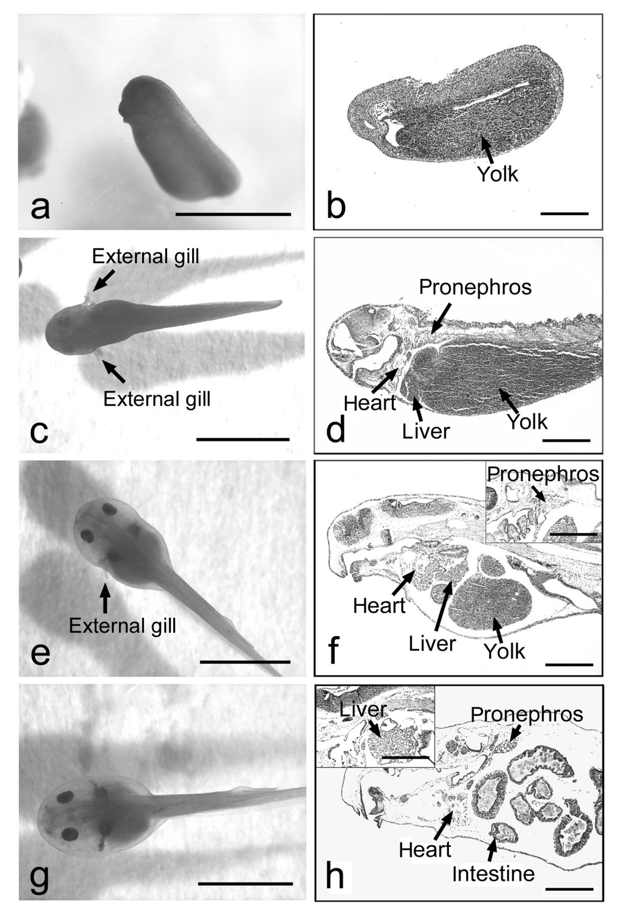
- Figure 1.
External appearances (a, c, e, g) and histological features (b, d, f, h) of the early embryos of Rana rugosa. Fertilized eggs were developed to the neurula (a, b) at two days after fertilization (DAF) and the embryos at stages 21 (c, d), 24 (e, f), and 25 (g, h) of Shumway (43) at 4, 6, and 8 DAF, respectively. At 2 DAF, the chemical treatments were terminated. Hatching occurred at 3 DAF. Two external gills can be seen at both sides of the embryo at stage 21 (c), and thereafter only one gill disappears in the embryo at stage 24 (e). A mass of cells containing yolk can be seen in the abdominal region of the neurula (b), and thereafter this diminished in the embryos during development (d, f, h). Organs such as the pronephros, heart, liver, and intestine, are differentiated in the embryos during development (d, f, h). Scale bar=2 mm.
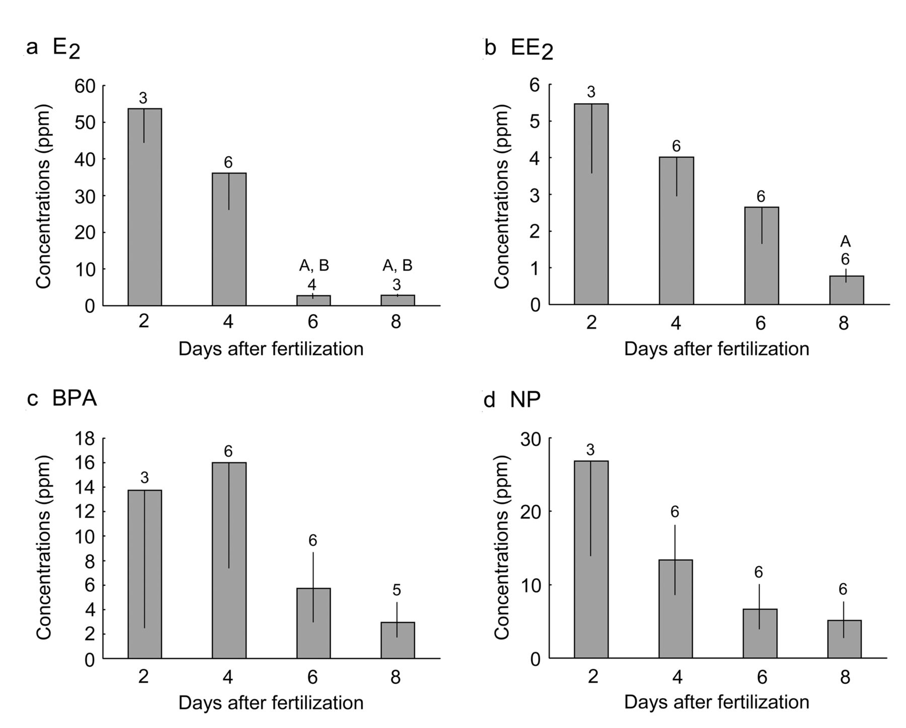
- Figure 2.
Concentrations of accumulated exogenous native estrogen and xenoestrogens in the embryo and jelly coat of Rana rugosa during development. The pre-cleaved embryos were exposed to 17β-estradiol (E2; a), 17α-ethynylestradiol (EE2; b), bisphenol A (BPA; c), and nonylphenol (NP; d) from 20 min after fertilization for two days. After two days, embryos were transferred to breeding water without estrogenic chemicals and reared for an additional six days. The breeding water was exchanged every day. The embryos were collected at 2, 4, 6, and 8 days after fertilization (DAF). Concentrations of the chemicals in the embryos were measured using a HPLC method. Numbers of samples examined are indicated above each column. Each column and the vertical line represent the mean±SE. Significance of differences: A, p<0.05 vs. 2 DAF; B, p<0.05 vs. 4 DAF.
Tables

In this issue


Jump to section
Related Articles
Cited By...
- No citing articles found.