


Abstract
Background/Aim: Use of zebrafish models may decrease the cost of screening new irradiation protectors and mitigators. Materials and Methods: Zebrafish (Danio rerio) models were tested for screening water-soluble radiation protectors and mitigators. Irradiation of embryos and monitoring survival, and measuring fibrosis of the caudal musculature of adults allowed for testing of acute and late effects, respectively. Results: Incubation of zebrafish embryos either before or after irradiation in ethyl pyruvate (1 mM) increased survival. Irradiation of adults to 15 to 75 Gy, delivered in single-fraction at 13 Gy/min, showed dose-dependent fibrosis at 30 days, quantitated as physiological decrease in swimming tail movement, and histopathological detection of collagen deposition in the dorsal musculature. Continuous administration of small-molecule radioprotector drugs in the water after irradiation reduced both acute and chronic injuries. Conclusion: The zebrafish is cost-effective for screening new radiation countermeasures.
A major complication of ionizing irradiation exposure is fibrosis (1, 19, 58). Patients completing a course of fractionated radiotherapy for treatment of head and neck, thoracic, and abdominal malignancies demonstrate dose-dependent appearance of late effects within 6-24 months after irradiation (59-60). While acute effects of radiation are well-documented for both total body and organ-specific exposure, a latent period following resolution of the acute effects has been difficult to relate to the onset of late effects (3, 8, 10, 19). In the C57BL/6J mouse model of radiation pulmonary fibrosis, elevation of cytokines associated with the acute effects (TGFβ, IL-1, TNFα) has been shown to resolve with resolution of acute effects at around two weeks after irradiation (3). A several-month latent period is associated with no detectable histopathological changes in the lungs, but is followed by onset of organizing alveolitis/fibrosis at around 100 to 120 days after irradiation, with concomitant elevation of TGFβ (3, 8). The molecular and cellular mechanisms associated with radiation pulmonary fibrosis, as well as fibrosis in other organs and organ systems have been difficult to elucidate (58-60).
The search for pharmacological agents to prevent or reduce radiation fibrosis has been the focus of an intense investigation. Small-molecule antioxidant inhibitors of cytokine elevation have been shown to reduce the severity of irradiation acute effects in both animal models and clinical trials (39-46). The application of pharmacological agents at the time of protection of the late effect, fibrosis, has met with incomplete success and there is need for continuous administration of anti-fibrotic agents (46, 51). The difficulty of measuring both uptake and efficacy of many test agents further complicates the evaluation of new drugs (57).
In the modern era of automated drug discovery, computational chemistry, and computer modeling of pharmacologically active small molecules, there is a need to rapidly assay potentially valuable target compounds, and then determine the structure and design of lead compounds (38, 42, 44). Lead compounds tested successfully in vitro, then require animal testing (38, 42, 44). While rodent models for acute irradiation effects have been the mainstay of clinical translational research in radiobiology, the costs of animals, animal care, and veterinary supervision services have challenged the apparent widespread use of mammalian/rodent models for clinical testing of lead compounds in many applications, including for the therapy of irradiation-induced side-effects (27).
The Zebrafish have been shown to be a valuable model for testing the effect of irradiation on teratogenetic effects (59), and potential effectiveness of radiation counter measures (59). The use of Zebrafish as a model for late irradiation effects has not been described.

Schematic of irradiation of zebrafish caudate. Six-month old zebrafish were anesthesized in 0.015% tricaine and placed in individual wells immersed in anesthetic. The fish were irradiated at 13.6 Gy per min. The fish were shielded to protect the internal organs, but not the tail.
In this current article, we present evidence for the efficacy of the zebrafish model in measuring radiation-induced long term effects and for screening small-molecule irradiation mitigators of late tissue fibrosis.
Materials and Methods
Zebrafish and aquatic maintenance facilities. The fish embryos and adults were maintained in conditions in accordance with the Institutional Animal Care and Use Committee of the University of Pittsburgh as previously described (59).
Irradiation procedure. Adult zebrafish were anesthetized by placing the fish in 0.015% tricaine in water. Once the fish were sedated, they were placed in individual wells in agarose, covered with water containing 0.015% tricaine and irradiated to the caudal portion of the fish to doses up to 75 Gy (Figure 1). The maintenance of anesthetic in the water allowed movement of the gill “operculum” to facilitate oxygenation of the fish while they were irradiated.
The holder device was placed on a series of Lucite blocks on the treatment table (patient assembly) of a Varian Linac 6 MV Varian CLINAC 2100C linear accelerator (Varian Medical Systems, Palo Alto, CA, USA). Dosimetry was carried out to deliver a 13 Gy/min dose rate of 6 MeV photons uniformly to the caudal region of the fish. Isodose curves for the radiation device and beam flatness were calculated to produce an error of <1% across the irradiated field.
The sedated fish were placed on a shallow petri dish containing agarose which had 10 slots to allow for irradiation of 10 fish at a time. The dish, filled with water containing 0.015% tricaine, was placed on a 15’’ stack of polystyrene on a raised LINAC table to bring the fish close to the radiation source. Source to prescription point, which was at the mid line of the fish, was kept at 55.7 cm to attain a dose rate of 13 Gy/Minute. Radiation field size of 40 cm × 7.5 cm was used to cover the entire petri dish. A 1.0-cm thick Superflab bolus material (Radiation Product Design, inc, USA) was placed above the entire irradiation area, so that the mid-line of the fish was at a depth of Dmax (1.5 cm for 6 MV beam). The area around the petri dish was filled with bolus material to maximize scatter condition at the prescription point.
A dose-response curve was created by irradiating fish in groups of 10 to doses of 15, 30, 45, 64, and 75 Gy at a dose rate of 1300 cGy per min. Fish were then allowed to recover and returned to aquarium tanks for observation. The maximum time from removal of fish to return to aquarium was approximately eight minutes. This interval has been shown to be consistent with other publications documenting the safety of Marcaine anesthesia for this interval. The fish were followed by development of fibrosis at which time they were sacrificed, fixed, sectioned, and stained with hematoxylin and Eosin (H&E) or Mallory's Trichrome Stain. All fish care protocols were approved by the Institutional Animal Care and Use Committee of the University of Pittsburgh. Fish care and veterinary care was provided by the Division of Laboratory Animal Research (DLAR) of the University of Pittsburgh.
Before irradiation, the anterior (cephalad) one half of the zebrafish was shielded by a 10½ value layer of lead block, so that the dose under the block was less than 10% of that in the irradiated posterior (caudal) section. This facilitated high dose irradiation of tail musculature and all associated structures.
To demonstrate that small molecules mitigate irradiation-induced fibrosis, zebrafish were irradiated to 32.5 Gy, as described above. The fish were then placed in water, containing 20 μM amifostine, 1 mM Tempol (Sigma Chemical Company, St. Louis, MO) or 20 μM EUK134 (Proteome Systems, Woburn, MA). The fish were followed for the development of fibrosis at which time they were sacrificed. In a separate experiment fish were irradiated to 30 Gy and then placed in water containing amifostine (Medimmune Prarma B.V., Nijmegen, Netherlands), tempol (Sigma/Aldrich Chemical Company, St. Louis, MO), or EUK134 (generously provided by Dr. Susan Doctrow, Proteome System, Inc., 6 Gill Street, Suite H, Woburn, MA 01801) as described above. The fish were sacrificed at day 60, fixed in paraformaldehyde, sectioned, and stained with either H&E or Mallory's Trichrome stain. The sections were examined microscopically and the percent of the caudate expressing fibrosis was determined.

Development of fibrosis in irradiated zebrafish. Zebrafish (6 months of age) were irradiated to doses ranging from 15 Gy (1500 cGy) to 75 Gy (7500 cGy) and followed for the development of fibrosis, observed as a curled stiff tail during swimming movements. This was observed at 2 months after irradiation (Figure 2A). An irradiation dose-response curve is shown in Figure 2B.
Observation and detection of survival and fibrosis. Embryo survival was calculated as published (59). Adult fish were returned to aquarium maintenance and observed daily for signs of radiation morbidity. Moribund or dying fish were removed from the aquarium. Fish surviving an additional ten days were observed for signs of fibrosis detected as a decrease in tail oscillation at swimming and alteration in the structure, shape, and function of tail, dorsal and caudal fins.
Histopathology. Sagittal sections of entire zebrafish were prepared for histopathological analysis, according to previously published methods.
Staining for fibrosis using the Mallory-Trichrome stain was carried out according to previous publications. A percent of cell musculature replaced by fibrosis was calculated for each individual.
Results
Radiation fibrosis dose response curve of adult Zebrafish. Fish receiving posterior/caudal half irradiation for doses between 15 and 75 Gy (Figure 1) were watched for development of fibrosis (Figure 2A). All fish irradiated at 45 Gy or higher developed fibrosis and were sacrificed by day 24. At 30 Gy, 25% of the fish did develop fibrosis as detected by abnormalities in swimming, stiff tail movement, and for changes in the shape and structure of tail and fin and survival (Figure 2B). Review of these results lead to the dose of 30 Gy being chosen for long-term observation as the maximum acutely tolerated dose, likely to result in irradiation late effect.

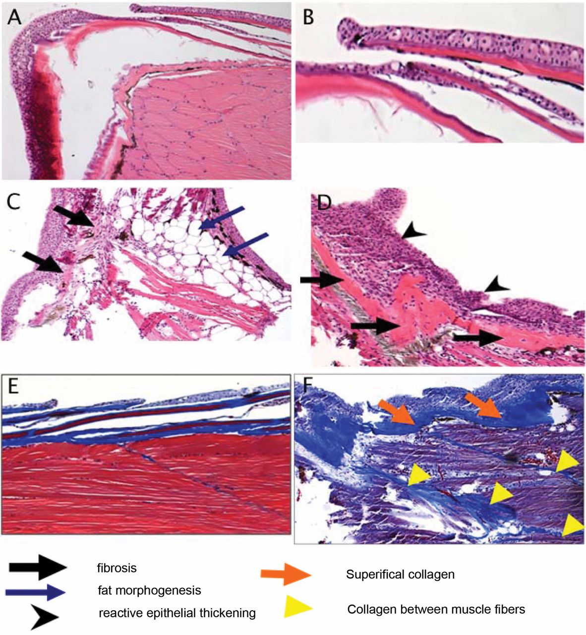
Histopathologic appearance of fibrotic changes in tails of irradiated zebrafish. Zebrafish were irradiated to 30 Gy and sacrificed 60 days later. The tails were removed from irradiated as well as nonirradiated control fish, fixed, sectioned and stained with H&E or Mallory's trichrome stain. Nonirradiated fish muscle and skin are shown in Figure 3A and 3B, respectively. Irradiated muscle and skin are shown in Figure 3C and 3D, respectively. Mallory's trichrome staining of collagen in nonirradiated and irradiated fish are shown in Figure 3E and 3F, respectively.
Pathological evidence of radiation-induced fibrosis. Sagittal sections of zebrafish were prepared for histopathological evaluation and Mallory trichrome staining, for evidence of fibrosis (Figure 3). Control non-irradiated muscle and skin is shown in Figure 3A and 3B, respectively. Irradiation of the caudal portion of the fish resulted in increased fibrosis as revealed by muscle atrophy, replacement of the musculature with both fatty/adipose cells, reactive skin with ongoing apoptotic cells, and abnormal layering and thickening of the skin (Figure 3C and 3D). Mallory-Trichrome staining also showed thickened areas of collagen, superficially (Figure 3E), as well as between muscle bundles (Figure 3F). These findings were similar to those seen in clinical radiation fibrosis (60-61).

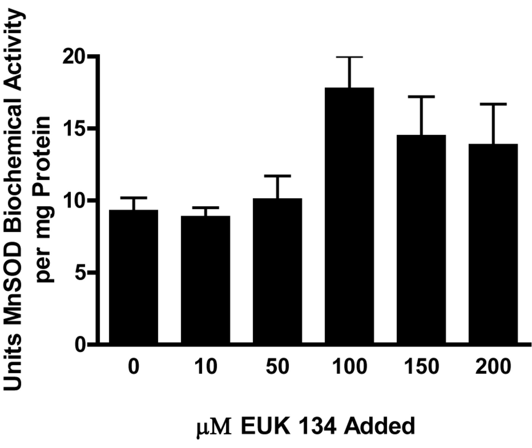
Increased SOD activity in zebrafish maintained in EUK139. Zebrafish were maintained free swimming in concentrations of EUK139 ranging from 0 to 200 μM for 1 hr, at which time the fish were sacrificed and then frozen in liquid nitrogen. The fish were thawed and the tail was removed, homogenized and the MnSOD biochemical activity determined. Fish maintained in 100 μM drug had a significantly increased MnSOD biochemical activity compared to control untreated fish (p=0.0178).
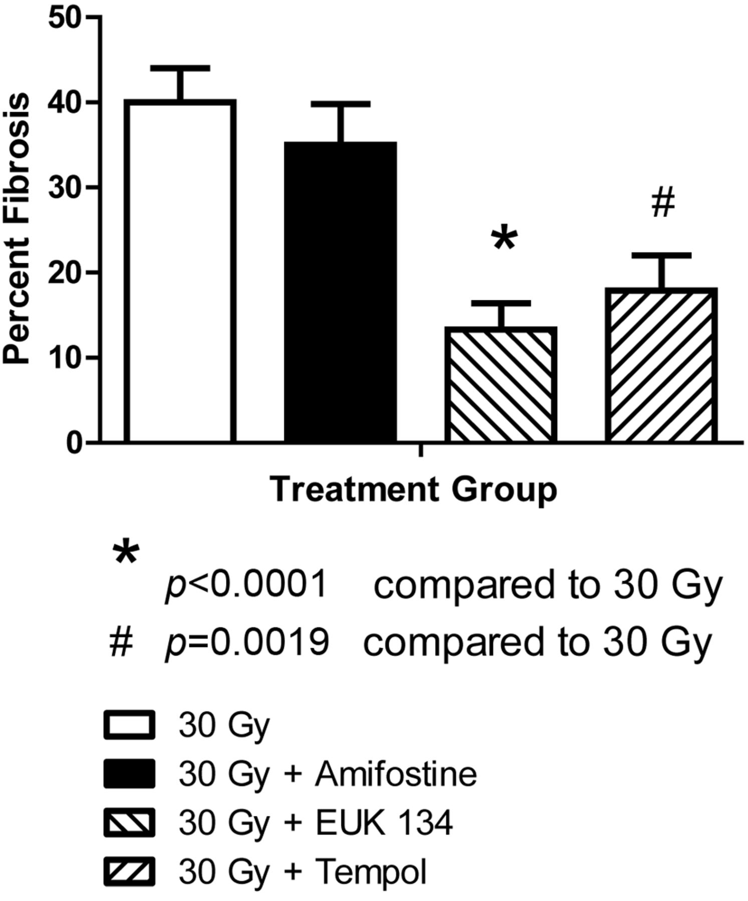
Effect of water-soluble small-molecule antioxidants on ameliorating irradiation fibrosis. We tested the effect of Amifostine, tempol and the SOD mimetic EUK 134 in preventing radiation fibrosis by determining if this radiation toxicity ameliorating drug altered on the magnitude of radiation fibrosis in fish receiving 32.5 Gy to the posterior body half. To determine if the drugs could be concentrated in the zebrafish tissue, zebrafish were incubated in the water containing EUK 134 at concentrations ranging from 0 to 200 μM for one hour at which time the SOD activity in the muscles of the tail was determined (Figure 4). Increased SOD activity was found in fish containing 100 μM EUK (p=0.0178). To demonstrate that the small molecule antioxidants could mitigate irradiation damage, zebrafish were irradiated to 32.5 and placed in water containing amifostine, tempol, or EUK134. The fish were followed for the development of fibrosis at which time they were sacrificed. All control fish irradiated at 35 Gy were dead by 25 days (Figure 5). Fish incubated in amifostine, tempol, or EUK134 had increased survival compared to control-irradiated fish (p<0.0001, =0.0016 or =0.0001, respectively, Figure 5). Not only did EUK134 and tempol increase survival, fish irradiated to 30 Gy and incubated in tempol or EUK134 also demonstrated less fibrosis compared to control-irradiated fish at 60 days after irradiation (p<0.0001 or =0.0019, respectively, Figs. 6 and 7). Although amifostine increased survival after 32.5 Gy, it did not decrease fibrosis at 60 days after 30 Gy (Figures 6 and 7).

EUK139 or tempol added after irradiation improves survival of irradiated zebrafish. Fish were irradiated to 30 Gy, placed in water containing amifostine (20 μM), tempol (1 mM), EUK139 (50 μM) or vehicle control and followed for survival. Zebrafish treated with EUK139, Amifostine or Tempol after irradiation showed an increased survival compared to control fish. Drugs added pre-irradiation were toxic.
Discussion
One potential advantage of the zebrafish model for testing new small-molecule agents as potential clinical translational use in the prevention of radiation fibrosis is the clear economic advantage. The zebrafish model is cos-effective relative to rodent models of radiation fibrosis. The standard model for radiation pulmonary fibrosis is the C57BL/6J mouse (1, 3, 19). Since the onset of fibrosis is 100 to 120 days after irradiation, maintenance of significantly large numbers of animals (particularly 12 per group) for 120 days to 200 days, 5 to 9 thousand dollars. In contrast, the maintenance in significant numbers of zebrafish in multiple doses of a drug would be significantly less expensive.

EUK139 mitigates irradiation induced fibrosis. Zebrafish were irradiated to 30 Gy to the tails as described in Methods, and then placed in water containing amifostine (20 μM), tempol (1 mM), or EUK139 (50 μM). Two months later surviving fish were sacrificed, fixed, and tails stained with Mallory's trichrome stain and examined microscopically for fibrosis. Panel 6A shows the control fish, and Panel 6B is 60 days after 30 Gy. Fish placed in water containing 50 μM EUK139, after 30 Gy, are shown in Panel 6C. Panels 6D and 6E show fish maintained in tempol or amifostine, respectively, after 30 Gy. The percent of tissue showing fibrosis was determined in fish maintained in EUK139 or tempol compared to control-irradiated fish and showed significantly decreased fibrosis compared to 30-Gy, control-irradiated fish (p<0.0001 or p=0.0019, respectively, Figure 7).

EUK134 or tempol prevents fibrosis in groups of 20 Zebrafish irradiated to 30 Gy to the caudal body measured at 60 days. Results are presented as the percent fibrosis±SEM from each group of fish described in the legend to Figure 6.
Water-soluble agents can be monitored in a water quite effectively and this is as an advantage over taking blood or plasma samples from mice. The ability to sacrifice small numbers of fish at serial time points after administration of a drug and quantitate drug deposition in tissue relative to concentration of the water also greatly facilitates cost-effective analyses of drug uptake relative to effect. A potential disadvantage of the zebrafish system is the requirement for water solubility of small molecule agents or in the case of drug screening small molecules for potential clinical use (33, 38, 42, 44). The zebrafish model facilitates rapid translation to the clinic of agents likely to be delivered by oral and topical application which means rapid delivery to the circulation would be desirable for systemic administration. In contrast, for those agents used for organ specific radioprotection, such as Manganese Superoxide Dismutase-Plasmid Liposome protection of the lung (1), esophagus (2, 8), or oral cavity/oropharynx (16), bladder (12), a lipid-soluble and organ-specific application might be desirable (57). However, even in these situations, organ-specific radioprotection systemic tests of agents in animal models are desirable to rule-out potential systemic toxicities of a locally delivered agent. The present studies provide support for the use of the zebrafish model as a test agent for new radiation counter measures against both acute and late effects of irradiation including the prevention of late fibrosis.
Acknowledgements
Supported by NIH Grant of the NIAID U19 A168021.
- Received October 4, 2012.
- Accepted October 16, 2012.
- Copyright © 2012 International Institute of Anticancer Research (Dr. John G. Delinassios), All rights reserved


