
Main menu

  • Home
  • Current Issue
  • Archive
  • Info for
    • Authors
    • Advertisers
    • Editorial Board
  • Other Publications
    • Anticancer Research
    • Cancer Genomics & Proteomics
    • Cancer Diagnosis & Prognosis
  • More
    • IIAR
    • Conferences
  • About Us
    • General Policy
    • Contact
  • Other Publications
    • In Vivo
    • Anticancer Research
    • Cancer Genomics & Proteomics

User menu

  • Register
  • Subscribe
  • My alerts
  • Log in
  • My Cart

Search

  • Advanced search
In Vivo
  • Other Publications
    • In Vivo
    • Anticancer Research
    • Cancer Genomics & Proteomics
  • Register
  • Subscribe
  • My alerts
  • Log in
  • My Cart
In Vivo

Advanced Search

  • Home
  • Current Issue
  • Archive
  • Info for
    • Authors
    • Advertisers
    • Editorial Board
  • Other Publications
    • Anticancer Research
    • Cancer Genomics & Proteomics
    • Cancer Diagnosis & Prognosis
  • More
    • IIAR
    • Conferences
  • About Us
    • General Policy
    • Contact
  • Visit iiar on Facebook
  • Follow us on Linkedin
Case ReportClinical Studies

A Case of Hydroxyurea-induced Leg Ulcer after Definitive Treatment Suspension in a Patient Affected by Thrombocythemia: Effectiveness of a New Collagenase

PAOLO FIORAMONTI, PASQUALE FINO, PAOLA PARISI, NICOLO SCUDERI and MARIA G. ONESTI
In Vivo November 2012, 26 (6) 1053-1056;
PAOLO FIORAMONTI
Department of Plastic, Reconstructive and Aesthetic Surgery, Policlinico Umberto I, Sapienza, University of Rome, Rome, Italy
  • Find this author on Google Scholar
  • Find this author on PubMed
  • Search for this author on this site
PASQUALE FINO
Department of Plastic, Reconstructive and Aesthetic Surgery, Policlinico Umberto I, Sapienza, University of Rome, Rome, Italy
  • Find this author on Google Scholar
  • Find this author on PubMed
  • Search for this author on this site
  • For correspondence: pasquale.fino@gmail.com pasquale.fino@uniroma1.it
PAOLA PARISI
Department of Plastic, Reconstructive and Aesthetic Surgery, Policlinico Umberto I, Sapienza, University of Rome, Rome, Italy
  • Find this author on Google Scholar
  • Find this author on PubMed
  • Search for this author on this site
NICOLO SCUDERI
Department of Plastic, Reconstructive and Aesthetic Surgery, Policlinico Umberto I, Sapienza, University of Rome, Rome, Italy
  • Find this author on Google Scholar
  • Find this author on PubMed
  • Search for this author on this site
MARIA G. ONESTI
Department of Plastic, Reconstructive and Aesthetic Surgery, Policlinico Umberto I, Sapienza, University of Rome, Rome, Italy
  • Find this author on Google Scholar
  • Find this author on PubMed
  • Search for this author on this site
  • Article
  • Figures & Data
  • Info & Metrics
  • PDF
Loading

Abstract

Hydroxyurea (HU) is an antitumor agent effective in the treatment of myeloproliferative disorders. It is usually well-tolerated and has low toxicity but its use is associated with several adverse cutaneous effects. Among them, leg ulcers have been noted in association with long-term administration. Poor response to traditional local and systemic therapy is a typical feature of HU-induced leg ulcers, and discontinuation of the drug is often required to achieve complete wound healing. We present a case of circumferential HU-induced leg ulcer in a patient affected by essential thrombocythemia, in which the lesion occured after the definitive suspension of the treatment. We propose a conservative management with a new collagenase (Bionect Start®) for skin lesions in this type of patient, for whom a surgical treatment could be more complicated, due to the underlying disease.

  • Hydroxyurea
  • thrombocythemia
  • Bionect Start®
  • circumferential wound
  • leg ulcers

Hydroxyurea (HU) is an antitumor agent, a hydroxylated derivative of urea, that has been recognized since 1960 as being effective against cancer (1). It is a cytostatic agent and through inhibition of the enzyme ribonucleotide reductase, it promotes the inhibition of cellular DNA synthesis and cell death in the S phase of the cell cycle (2).

Chronic myeloid leukaemia and other myeloproliferative disorders (3, 4), such as essential thrombocythemia (5) and polycythemia vera (PV) (6) are the most common indications for HU therapy.

The cutaneous side-effects of hydroxyurea include xerosis, hyperpigmentation, nail discoloration, scaling, lichen planus-like lesions, poikiloderma and atrophy of the skin and subcutaneous tissues (7-9). Another rare and incompletely-characterized complication, described in patients with myeloproliferative-receiving high-dose long-term HU treatment, is the occurrence of painful leg ulcers (10). Leg ulcers induced by HU have common features; most of them are localized to the malleolar region, and are refractory to meticulous wound care. Poor response to traditional local and systemic therapy is a typical feature of HU-induced leg ulcers, and discontinuation of the drug is often required to achieve complete wound healing (6, 8, 11). Suspension of the drug usually improves the skin ulcer; although, in some cases, the ulcer remains and additional therapies, such as skin grafting, are needed (12).

We present a case of circumferential leg ulcer in a patient affected by essential thrombocythemia, previously treated with HU therapy, in which the lesion occurred after the definitive suspension of the treatment, probably triggered by a traumatic event. We also propose conservative management with a new collagenase (Bionect Start®) for skin lesions in this type of patient, for whom a surgical treatment could be more complicated, due to the underlying disease.

Case Report

A 74-year-old female, affected by essential thrombocythemia, presented to the Department of Plastic and Reconstructive Surgery of Policlinico Umberto I of Rome with a circumferential wound of the right leg.

The patient referred that she had been treated with HU, receiving 1 g daily of HU, for 7 years and that the treatment had been suspended for six months. During the treatment, she did not develop any cutaneous or soft tissue complications.

She reported that the lesion appeared one month after a leg trauma, caused by the hit of a soccer ball and that she had undergone other outpatient care with poor results. The skin lesion progressed to become a deeper, severely painful and hemorrhagic ulcer. The complete blood cell count revealed a hemoglobin level of 9.2 g/dl, and a platelet count of 788×103/μ/l.

At local clinical examination, the lesion was found to extend from the knee to the ankle (25×30 cm), involving the cutaneous and subcutaneous tissues. The lesion had regular margins and definite limits. The bed of the ulcer was highly fibrinous and appeared to be contaminated (Figure 1).

Before starting any treatment, a bacterial examination was performed. The treatment consisted of conservative management and antibiotic therapy, administered due to Staphyloccocus aureus and Pseudomonas aeruginosa wound contaminations.

The wound care consisted of saline solution washes, disinfection with Amukine and Betadine, application of Bionect Start® (Fidia Farmaceutici SpA, Medical Devices, Abano Terme, Italy) and occlusive dressing. Zinc oxide paste was also applied to the skin surrounding the wound. No advanced dressings that could vary healing time was used. The treatment was performed daily by the patient personally and every three days at our outpatient clinic, to examine for changes and progress.

We observed a progressive and rapid improvement of the condition, with resolution of the contamination and reduction of secretion, exudates and pain or burning during dressing. (Figure 2).

The complete re-epithelialization of the extensive loss of substance was achieved after three months, obtained with antibiotic therapy and dressing-alone without surgery (Figure 3).

Discussion

HU is an antitumor agent, which is usually well-tolerated and has low toxicity (1). Adverse cutaneous effects associated with HU have been occasionally described in the literature (8, 13). In addition, leg ulcers have been noted in association with long-term HU administration. HU-induced skin ulcers were first reported in 1985 by Stahl and Silber (10). Approximately nine percent of patients receiving HU medication develop this complication (7, 14). HU effects on actively proliferating epithelial cells have been less described (14). The pathogenesis of HU-induced ulceration may be multifactorial. The main assumptions are: interruption of the microcirculation due to leukocytoclastic vasculitis or arterial microthrombi related to platelet dysregulation (13, 15), cumulative toxicity in the basal layer of the epidermis through inhibition of DNA synthesis (8); and repeated mechanical injury in areas subject to trauma (16). Literature data reveal that most of the patients with HU-induced leg ulcer had been treated with more than 1 g of HU per day for at least one year (8).

Poor response to traditional local and systemic therapy is a typical feature of HU-induced leg ulcers, and discontinuation of the drug is often required to achieve complete wound healing (6, 8).

In the case described here, a new ulcer had developed even after 5 months from cessation of HU administration. Probably in this case, a traumatic event triggered the onset of the first event and since the direct cytotoxic effect of HU (8) may continue even after the withdrawal of the drug, the repair of the first injury may have been inhibited. Recurrence of HU-related leg injuries after discontinuation of the therapy has already been described (17), but our case is particularly interesting because the recurrence occurred many years after discontinuation of HU therapy.

Frequently, management of resistant HU-related ulcers requires surgical therapy such as skin graft. We propose a conservative management for this type of patient, for whom a surgical treatment could be more complicated, due to the underlying disease.

We proceeded with disinfection and use of Bionect Start®. Bionect Start® is a topical cream containing hyaluronic acid, bacterially-fermented sodium hyalunorate (0.2% w/w) salt and bacterial collagenase obtained from non-pathogenic Vibrio alginolyticus (>2.0 nkat/g) (18). The use of collagenase is based on its lysis of fibrin and necrotic tissue. The topical administration of collagenase increases the effect of macrophagic collagenase, which is responsible for wound debridement by splitting and breaking down proteins which hold dead and devitalised material on the wound (19). Bionect Start® provides an optimal moist enviroment and wound preparation which facilitates the natural healing process (20, 21). This drugs also contains hyaluronic acid (HA) (21), which above all generates a microenvironment stimulating the secretion of growth factors and proliferation and migration of fibroblasts, endothelial cells, keratinocytes and angiogenesis (19, 21) and has a positive effect on the inflammatory response (22-24). Moreover HA is also capable of regulating the water balance, acting on osmotic pressure and flow resistance and selectively regulating the diffusion of plasma and matrix proteins (25).

With conservative management and the use of Bionect Start®, we obtained complete resolution and re-epithelialization of the circumferential lesion in three months.

Conclusion

We suggest to HU-treated patients, that it is important to be aware of the possibility that this type of complication may occur, even years after HU treatment suspension, as did in our case. We advise such patients to avoid conditions that may lead to traumatic events which could create discomfort and reduce their quality of life.

We also propose conservative management of such lesions in patients with essential thrombocythemia because the underlying pathology could complicate major surgical treatment. Bionect Start® appears to be useful for this indication and to permit rapid wound healing of the lesion with no morbidity for the patient.

Figure 1.
  • Download figure
  • Open in new tab
  • Download powerpoint
Figure 1.

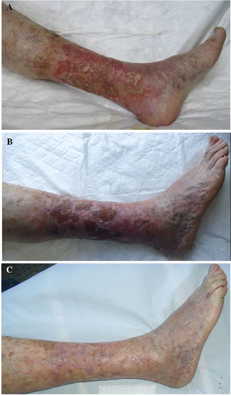
A: At presentation, the lesion extended from the knee to the ankle (25×30 cm), involving the cutaneous and subcutaneous tissues; the bed of the ulcer was highly fibrinous and contaminated. B: Progressive and rapid improvement of the condition occurred, with resolution of the contamination and reduction of secretion, exudates and pain or burning during dressing after one month and a half. C: Complete re-epithelizlization of the extensive loss of substance was apparent after 3 months of therapy.

Acknowledgements

Special thanks to Dr. Franco Bartolomei for his help in preparing this manuscript.

Footnotes

  • Conflicts of Interest

    The Authors have no financial interest to declare in relation to the content of this manuscript.

  • Received June 21, 2012.
  • Revision received September 13, 2012.
  • Accepted September 20, 2012.
  • Copyright © 2012 International Institute of Anticancer Research (Dr. John G. Delinassios), All rights reserved

References

  1. ↵
    1. Boyd AS,
    2. Neldner KH
    : Hydroxyurea therapy. J Am Acad Dermatol 25: 518-524, 1991.
    OpenUrlPubMed
  2. ↵
    1. Yarbro JW
    : Mechanism of action of hydroxyurea. Semin Oncol 19: 1-10, 1992.
    OpenUrlPubMed
  3. ↵
    1. Goldman JM
    : Therapeutic strategies for chronic myeloid leukemia in the chronic (stable) phase. Semin Hematol 40: 10-17, 2003.
    OpenUrlPubMed
  4. ↵
    1. Rice L,
    2. Baker KR
    : Current management of the myeloproliferative disorders: A case-based review. Arch Pathol Lab Med 130: 1151-1156, 2006.
    OpenUrlPubMed
  5. ↵
    1. Demirçay Z,
    2. Cömert A,
    3. Adigüzel C
    : Leg ulcers and hydroxyurea: Report of three cases with essential thrombocythemia. Int J Dermatol 41: 872-874, 2002.
    OpenUrlPubMed
  6. ↵
    1. Bader U,
    2. Banyai M,
    3. Böni R,
    4. Burg G,
    5. Hafner J
    : Leg ulcers in patients with myeloproliferative disorders: Disease- or treatment-related? Dermatology 200: 45-48, 2000.
    OpenUrlPubMed
  7. ↵
    1. Najean Y,
    2. Rain JD
    : Treatment of polycythemia vera: The useof hydroxyurea and pipobroman in 292 patients under the age of 65 years. Blood 90: 3370-3377, 1997.
    OpenUrlAbstract/FREE Full Text
  8. ↵
    1. Weinlich G,
    2. Schuler G,
    3. Greil R,
    4. Kofler H,
    5. Fritsch P
    : Leg ulcers associated with long-term hydroxyurea therapy. J Am Acad Dermatol 39: 372-374, 1998.
    OpenUrlCrossRefPubMed
  9. ↵
    1. Daoud MS,
    2. Gibson LE,
    3. Pittelkow MR
    : Hydroxyurea dermopathy: A unique lichenoid eruption complicating long-term therapy with hydroxyurea. J Am Acad Dermatol 36: 178-182, 1997.
    OpenUrlCrossRefPubMed
  10. ↵
    1. Stahl RL,
    2. Silber R
    : Vasculitic leg ulcers in chronic myelogenousleukemia. Am J Hematol 78: 869-872, 1985.
    OpenUrl
  11. ↵
    1. Stagno F,
    2. Guglielmo P,
    3. Consoli U,
    4. Fiumara P,
    5. Russo M,
    6. Giustolisi R
    : Successful healing of hydroxyurea-related leg ulcers with topical granulocyte-macrophage colony-stimulating factor. Blood 94: 1479-1480, 1999.
    OpenUrlFREE Full Text
  12. ↵
    1. Kato N,
    2. Kimura K,
    3. Yasukawa K,
    4. Yoshida K
    : Hydroxyurea-related leg ulcers in a patient with chronic myelogenous leukemia: A case report and review of the literature. J Dermatol 26: 56-62, 1999.
    OpenUrlPubMed
  13. ↵
    1. Sirieix ME,
    2. Debure C,
    3. Baudot N,
    4. Dubertret L,
    5. Roux ME,
    6. Morel P,
    7. Frances C,
    8. Loubeyres S,
    9. Beylot C,
    10. Lambert D,
    11. Humbert P,
    12. Gauthier O,
    13. Dandurand M,
    14. Guillot B,
    15. Vaillant L,
    16. Lorette G,
    17. Bonnetblanc JM,
    18. Lok C,
    19. Denoeux JP
    : Leg ulcers and hydroxyurea: 41 cases. Arch Dermatol 135: 818-820, 1999.
    OpenUrlCrossRefPubMed
  14. ↵
    1. Montefusco E,
    2. Alimena G,
    3. Gastaldi R,
    4. Carlesimo OA,
    5. Valesini G,
    6. Mandelli F
    : Unusual dermatologic toxicity of long-term therapy with hydroxyurea in chronic myelogenous leukaemia. Tumori 72: 317-321, 1986.
    OpenUrlPubMed
  15. ↵
    1. Chaine B,
    2. Neonato MG,
    3. Girot R,
    4. Aractingi S
    : Cutaneous adverse reactions to hydroxyurea in patients with sickle cell disease. Arch Dermatol 137: 467-470, 2001.
    OpenUrlPubMed
  16. ↵
    1. Saravu K,
    2. Velappan P,
    3. Lakshmi N,
    4. Shastry BA,
    5. Thomas J
    : Hydroxyurea induced perimalleolar ulcers. J Korean Med Sci 21: 177-179, 2006.
    OpenUrlPubMed
  17. ↵
    1. Kikuchi K,
    2. Arita K,
    3. Tateishi Y,
    4. Onozawa M,
    5. Akiyama M,
    6. Shimizu H
    : Recurrence of hydroxyurea-induced leg ulcer after discontinuation of treatment. Acta Derm Venereol 91(3): 373-374, 2011.
    OpenUrlPubMed
  18. ↵
    1. Ponzio I,
    2. Pierangeli M,
    3. Marchesini A,
    4. Carboni A,
    5. Talevi D,
    6. Grassetti L,
    7. Petrucci E,
    8. Bertani A,
    9. Scalise A
    : Gel piastrinico, valutazione del ruolo nel wound bed preparation e sua analisi clinico-qualitativa. Acta Vulnologica 6: 25-30, 2008.
    OpenUrl
  19. ↵
    1. Mekkes JR,
    2. Zeegelaar JE,
    3. Westerhof W
    : Quantitative and objective evaluation of wound debriding properties of collagenase and fibrinolysin/“des”oxyribonuclease in a necrotic ulcer animal model. Arch Dermatol Res 290(3): 152-157, 1998.
    OpenUrlPubMed
  20. ↵
    1. Ramundo J,
    2. Gray M
    : Collagenase for enzymatic debridement: A systematic review. J Wound Ostomy Continence Nurs 36(6 Suppl): 4-11, 2009.
    OpenUrl
  21. ↵
    1. Claeys A,
    2. Gaudy-Marqueste C,
    3. Pauly V,
    4. Pelletier F,
    5. Truchetet F,
    6. Boye T,
    7. Aubin F,
    8. Schmutz JL,
    9. Grob JJ,
    10. Richard MA
    : Management of pain associated with debridement of leg ulcers: A randomized, multicentre, pilot study comparing nitrous oxide-oxygen mixture inhalation and lidocaïne-prilocaïne cream. J Eur Acad Dermatol Venereol 25(2): 138-144, 2010.
    OpenUrlPubMed
  22. ↵
    1. Anderson I
    : Debridement methods in wound care. Nurs Stand 20(24): 65-66, 68, 70, 2006.
    OpenUrlPubMed
    1. Hollander D,
    2. Schmandra T,
    3. Windolf J
    : Using an esterified hyaluronan fleece to promote healing in difficult-to-treat wounds. J Wound Care 9(10): 463-466, 2000.
    OpenUrlPubMed
  23. ↵
    1. Wisniewski HG,
    2. Hua JC,
    3. Poppers DM,
    4. Naime D,
    5. Vilcek J,
    6. Cronstein BN
    : TNF/IL-1-inducible protein TSG-6 potentiates plasmin inhibition by inter-alpha-inhibitor and exerts a strong anti-inflammatory effect “in vivo”. J Immunol 156(4): 1609-1615, 1996.
    OpenUrlAbstract
  24. ↵
    1. Kangesu T,
    2. Manek S,
    3. Terenghi G,
    4. Gu XH,
    5. Navsaria HA,
    6. Polak JM,
    7. Green CJ,
    8. Leigh IM
    : Nerve and blood vessel growth in response to grafted dermis and cultured keratinocytes. Plast Reconstr Surg 101(4): 1029-1038, 1998.
    OpenUrlPubMed
PreviousNext
Back to top

In this issue

In Vivo: 26 (6)
In Vivo
Vol. 26, Issue 6
November-December 2012
  • Table of Contents
  • Table of Contents (PDF)
  • Index by author
  • Back Matter (PDF)
  • Ed Board (PDF)
  • Front Matter (PDF)
Print
Download PDF
Article Alerts
Sign In to Email Alerts with your Email Address
Email Article

Thank you for your interest in spreading the word on In Vivo.

NOTE: We only request your email address so that the person you are recommending the page to knows that you wanted them to see it, and that it is not junk mail. We do not capture any email address.

Enter multiple addresses on separate lines or separate them with commas.
A Case of Hydroxyurea-induced Leg Ulcer after Definitive Treatment Suspension in a Patient Affected by Thrombocythemia: Effectiveness of a New Collagenase
(Your Name) has sent you a message from In Vivo
(Your Name) thought you would like to see the In Vivo web site.
CAPTCHA
This question is for testing whether or not you are a human visitor and to prevent automated spam submissions.
2 + 13 =
Solve this simple math problem and enter the result. E.g. for 1+3, enter 4.
Citation Tools
A Case of Hydroxyurea-induced Leg Ulcer after Definitive Treatment Suspension in a Patient Affected by Thrombocythemia: Effectiveness of a New Collagenase
PAOLO FIORAMONTI, PASQUALE FINO, PAOLA PARISI, NICOLO SCUDERI, MARIA G. ONESTI
In Vivo Nov 2012, 26 (6) 1053-1056;

Citation Manager Formats

  • BibTeX
  • Bookends
  • EasyBib
  • EndNote (tagged)
  • EndNote 8 (xml)
  • Medlars
  • Mendeley
  • Papers
  • RefWorks Tagged
  • Ref Manager
  • RIS
  • Zotero
Reprints and Permissions
Share
A Case of Hydroxyurea-induced Leg Ulcer after Definitive Treatment Suspension in a Patient Affected by Thrombocythemia: Effectiveness of a New Collagenase
PAOLO FIORAMONTI, PASQUALE FINO, PAOLA PARISI, NICOLO SCUDERI, MARIA G. ONESTI
In Vivo Nov 2012, 26 (6) 1053-1056;
del.icio.us logo Digg logo Reddit logo Twitter logo CiteULike logo Facebook logo Google logo Mendeley logo
  • Tweet Widget
  • Facebook Like
  • Google Plus One

Jump to section

  • Article
    • Abstract
    • Case Report
    • Discussion
    • Conclusion
    • Acknowledgements
    • Footnotes
    • References
  • Figures & Data
  • Info & Metrics
  • PDF

Related Articles

  • No related articles found.
  • PubMed
  • Google Scholar

Cited By...

  • No citing articles found.
  • Google Scholar

More in this TOC Section

  • Evaluation of TET Family Gene Expression and 5-Hydroxymethylcytosine as Potential Epigenetic Markers in Non-small Cell Lung Cancer
  • Automated Non-coplanar Volumetric Modulated Arc Therapy Planning for Maxillary Sinus Carcinoma
  • The Influence of the Rapid Increase in the Number of Adverse Event Reports for COVID-19 Vaccine on the Disproportionality Analysis Using JADER
Show more Clinical Studies

Similar Articles

Keywords

  • hydroxyurea
  • thrombocythemia
  • Bionect Start®
  • circumferential wound
  • leg ulcers
In Vivo

© 2023 In Vivo

Powered by HighWire