


Article Figures & Data
Figures
- Figure 1.
A: In vivo migration of tumor cells. Tumor cell chemotaxis and metastatic homing of CXC chemokine receptor 4 (CXCR4)-positive tumor cells is mediated by stromal-cell derived factor-1alpha (SDF-1α) expression in destination organs e.g. bone marrow, liver and lung. B: In vitro chemotaxis: Boyden chamber principle. A cell suspension is placed in the upper chamber, while the medium containing the chemoattractant is placed in the lower chamber. Migratory cells that are attracted by the specific chemoattractant pass through the filter membrane.
- Figure 2.
Metastatic potential and CXCR4 expression of OE19 cells. Representative images of hematoxylin eosin (H.E.) and CXC chemokine receptor 4 (CXCR4) immunostaining of primary tumor (PT), liver, lung and lymph node (LN) tissue from an animal with orthotopically implanted esophageal tumor (magnification ×100). CXCR4 expression is observed in all tumor-bearing tissues.
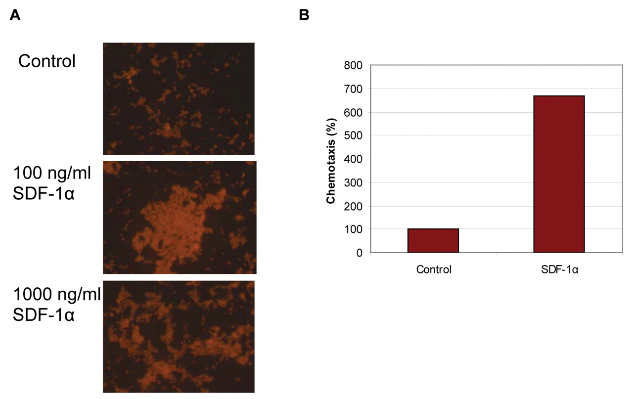
- Figure 3.
Tumor cell migration in Boyden Chamber. A: Microscopic evaluation of the Boyden chamber-based migration assay shows a dose-dependent effect of stromal-cell derived factor-1alpha (SDF-1α)-stimulated chemotaxis on OE19 cells. B: A relevant migratory effect of SDF-1α-mediated migration on CXCR4-positive cells is observed at 250 ng/ml compared to unstimulated cells.
- Figure 4.
Cell migration in vivo. A: Compartment-mimicking principle of an in vivo model of chemotaxis. Tumor cells are injected into the flank of the animal (I, compartment or origin). Metastases are monitored in the abdominal and thoracic tissues (II, destination compartments: lung, liver, peritoneum, retroperitoneum) after intraperitoneal stimulation. B: Metastatic spread to liver, lung, peritoneum and retroperitoneal tissue is analyzed by real-time polymerase chain reaction (PCR), according to the level of human polymerase (RNA) II polypeptide 2 (POL2). Means of cycle threshhold (ct) values were used for graphic depiction of results. There is a relevant effect of stromal-cell derived factor-1alpha (SDF-1α) stimulation (100 and 500 ng) on all organs compared to the sham-treated group (DPBS). Detection of human, and thus metastatic, cells was even greater in lung and peritoneum in the group treated with 500 ng SDF-1α, compared to the positive tumor control, indicating a strong metastatic effect.


In this issue


Jump to section
Related Articles
Cited By...
- No citing articles found.