


Article Figures & Data
Figures
- Figure 1.
Cytotoxicity of sorafenib on Huh7/NF-κB-luc2 cells occurs in a dose-dependent manner. Cells were treated with 0-15 μM sorafenib for 6 and 24 h, respectively. Cell viability was determined by the 3-(4,5-Dimethylthiazol-2-yl)-2,5-diphenyltetrazolium bromide (MTT) assay. Data are presented as means±SD (n=3). **p<0.01 as compared with that of dimethyl sulfoxide (DMSO)-treated control. The experiments were repeated three times.
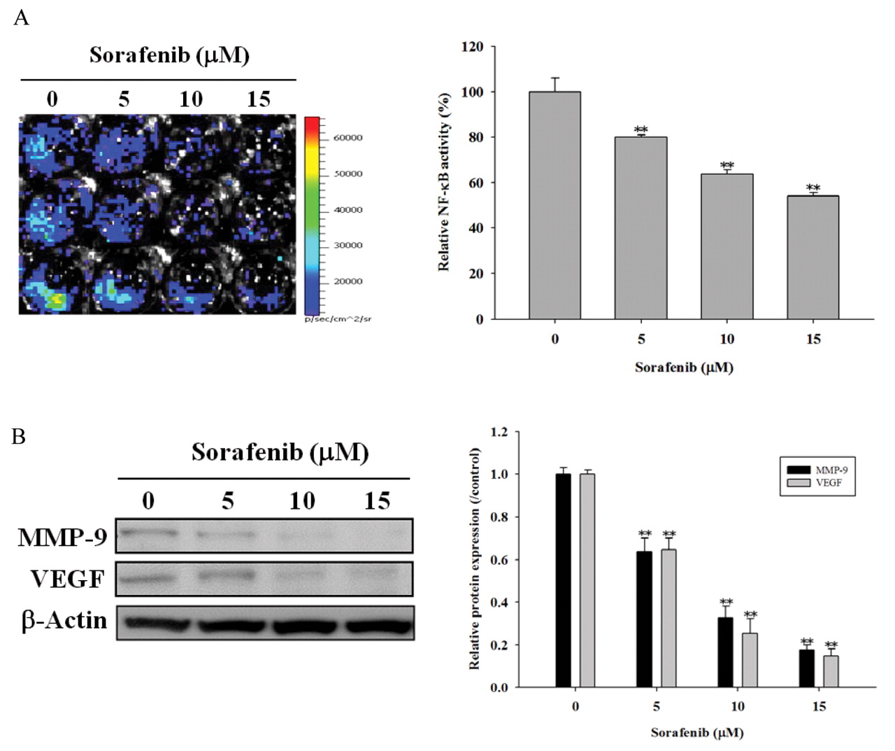
- Figure 2.
Intrinsic nuclear factor kappaB (NF-κB) activity, matrix metalloproteinase-9 (MMP-9) and vascular endothelial growth factor (VEGF) expressions are inhibited by sorafenib in a dose-dependent manner in Huh7/NF-κB-luc2 cells. Cells were incubated with different concentration of sorafenib for 6 hours. A: Left: Bioluminescent imaging (BLI) of the relative NF-κB activity versus the concentration of sorafenib; right: quantification of the relative NF-κB activity. B: Top: Western blotting of MMP-9 and VEGF expressions using β-actin as the internal control; bottom: quantification of the western blotting. The ratios of MMP-9/β-actin and VEGF/β-actin in sorafenib-treated groups are compared with those of the dimethyl sulfoxide (DMSO)-treated control. Data are presented as means±SD (n=3). **p<0.01 as compared with that of DMSO-treated control. The experiments were repeated three times.
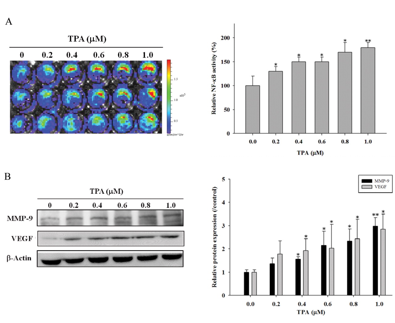
- Figure 3.
Intrinsic nuclear factor kappaB (NF-κB) activity, matrix metalloproteinase-9 (MMP-9) and vascular endothelial growth factor (VEGF) expressions are increased by 12-O-tetradecanoylphorbol-13-acetate (TPA) in a dose-dependent manner in Huh7/NF-κB-luc2 cells. Cells were incubated with various concentrations of TPA for 6 hours. A: Top: Bioluminescent imaging (BLI) of the relative NF-κB activity versus the concentration of TPA; bottom: quantification of the relative NF-κB activity. B: Top: Western blotting of MMP-9 and VEGF expressions using β-actin as the internal control; bottom: quantification of the western blotting. Data are presented as means±SD (n=3). *p<0.05, **p<0.01 as compared with that of dimethyl sulfoxide (DMSO)-treated control. The experiments were repeated three times.
- Figure 4.
12-O-Tetradecanoylphorbol-13-acetate (TPA)-induced nuclear factor kappaB (NF-κB) activity, matrix metalloproteinase-9 (MMP-9) and vascular endothelial growth factor (VEGF) overexpressions are inhibited by sorafenib in Huh7/NF-κB-luc2 cells. Cells were treated with dimethyl sulfoxide (DMSO) and sorafenib 30 min prior to the addition of TPA, respectively, and cultured for another 6 hours. A: Top: Bioluminescent imaging (BLI) of the relative NF-κB activity; bottom: quantification of the relative NF-κB activity. B: Top: Western blotting of MMP-9 and VEGF expressions using β-actin as the internal control; bottom: quantification of the western blotting. Data are presented as means±SD (n=3). **p<0.01 as compared with that of DMSO-treated control; #p<0.05, ##p<0.01 as compared with that of TPA-treated group. The experiments were repeated three times.
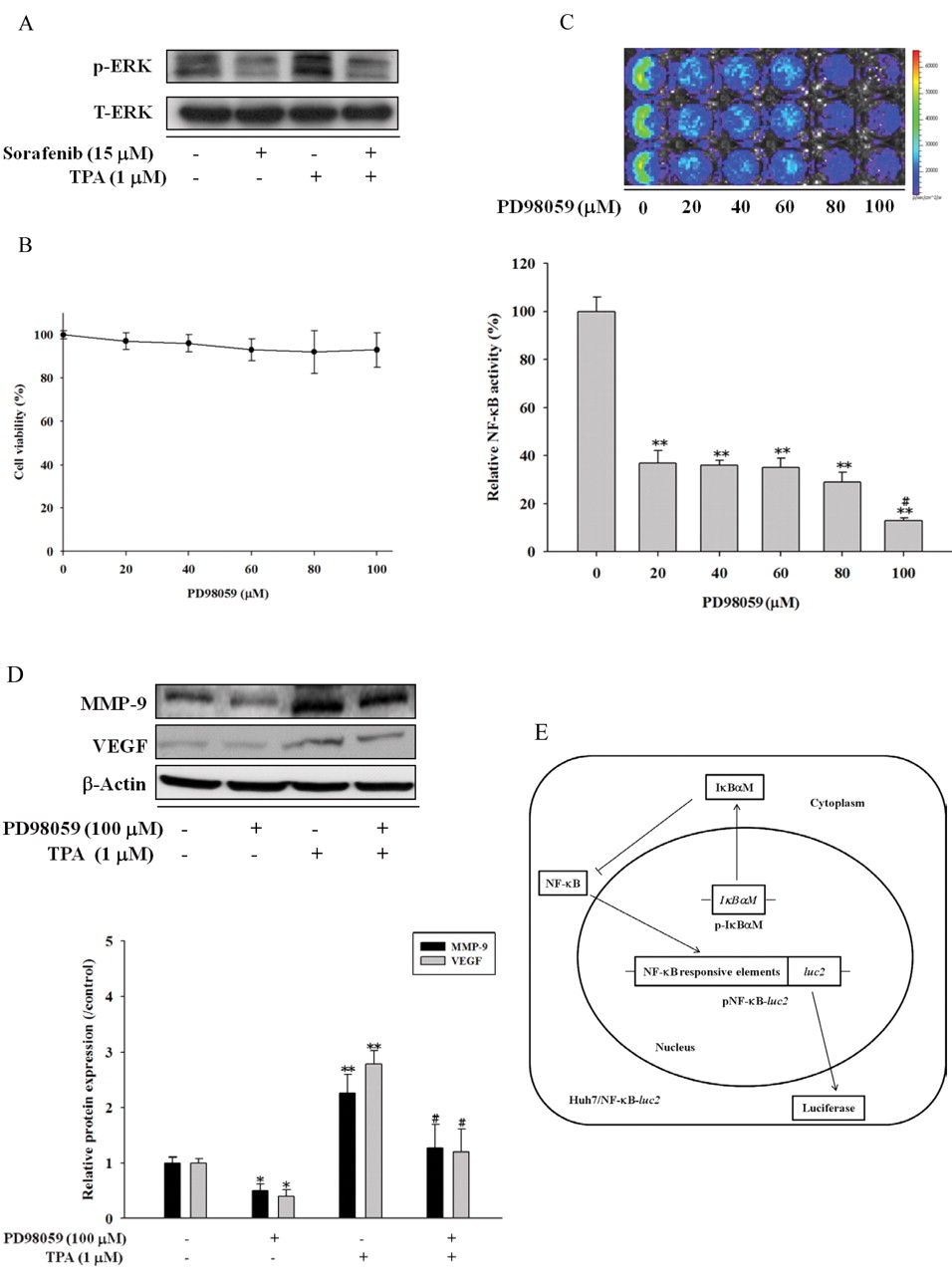
- Figure 5.
12-O-Tetradecanoylphorbol-13-acetate (TPA)-induced nuclear factor kappaB (NF-κB) activity, matrix metalloproteinase-9 (MMP-9) and vascular endothelial growth factor (VEGF) expressions suppressed by sorafenib in Huh7/NF-κB-luc2 cells via the suppression of extracellular signal-regulated kinase (ERK) signal pathway. Cells were treated with dimethyl sulfoxide (DMSO), sorafenib, and PD98059 30 minutes prior to the addition of TPA, respectively, and incubated for another 6 hours. A: TPA-induced ERK phosphorylation is inhibited by sorafenib. B: No cytotoxicity was found in cells treated with various concentrations of PD98059 for 6 hours. C: Intrinsic NF-κB activity is inhibited by the treatment with PD98059 for 6 hours. Top: Bioluminescent imaging (BLI) of the relative NF-κB activity; bottom: quantification of BLI. D: Intrinsic and TPA-induced MMP-9 and VEGF expressions are suppressed by PD98059. Top: Western blotting; bottom: quantification of the western blotting. E: Inhibitor of kappaB-α mutant vector (p-IκBαM), a super repressor of NF-κB, bound to NF-κB with high affinity, preventing its phosphorylation by inhibitor of kappaB kinase (IKK). As a consequence, NF-κB is sequestered in the cytoplasm. F: Intrinsic and TPA-induced NF-κB activities are decreased in cells treated with sorafenib, PD98059, and transfection with p-IκBαM, respectively. Top: BLI of the relative NF-κB activity; bottom: quantification of BLI. G: Intrinsic and TPA-induced NF-κB/DNA binding activities are blocked in cells treated with sorafenib, PD98059, and transfection with p-IκBαM, respectively. Top: NF-κB/DNA binding activity as determined by EMSA; bottom: quantification of EMSA. Data are presented as means±SD (n=3). *p<0.05, **p<0.01 as compared with that of DMSO-treated control; #p<0.05, ##p<0.01 as compared with that of TPA-treated only group. The experiments were repeated three times.
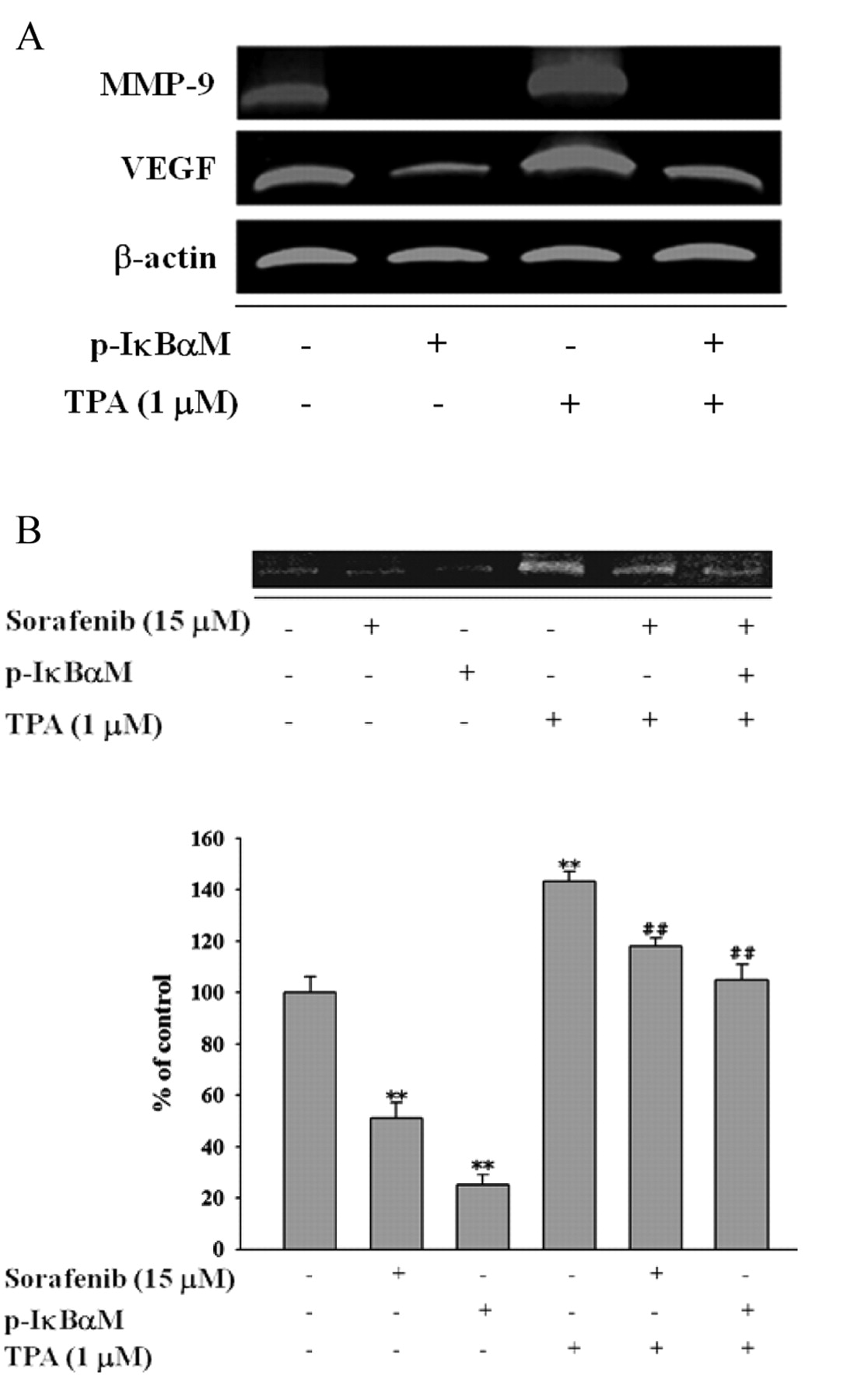
- Figure 6.
Blockade of nuclear factor kappaB (NF-κB) activity results in the decrease of 12-O-tetradecanoylphorbol-13-acetate (TPA)-induced matrix metalloproteinase-9 (MMP-9) and vascular endothelial growth factor (VEGF) expressions, and the secretion of MMP-9 into the cultured medium of Huh7/NF-κB-luc2 cells. Cells were treated with sorafenib and inhibitor of kappaB-α mutant vector (p-IκBαM) 30 minutes prior to the addition of TPA, and incubated for another 6 hours. A: The mRNA expressions of MMP-9, VEGF, and β-actin were assayed with RT-PCR. B: Top: the MMP-9 protein released into the cultured medium was analyzed with gelatin zymography; bottom: quantification of the top panel. Data are presented as means±SD (n=3). **p<0.01 as compared with that of dimethyl sulfoxide (DMSO)-treated control; ##p<0.01 as compared with that of TPA-treated only group. The experiments were repeated three times. C: The suggested mechanism for TPA-induced MMP-9 and VEGF expressions inhibited by sorafenib through extracellular signal-regulated kinase (ERK)/NF-κB pathway.


In this issue


Jump to section
Related Articles
Cited By...
- Lenvatinib Suppresses Protein Kinase B Signaling and Induces Apoptosis in Osteosarcoma Cells
- Sorafenib Induces Apoptosis and Inhibits NF-{kappa}B-mediated Anti-apoptotic and Metastatic Potential in Osteosarcoma Cells
- Astragaloside IV Induces Apoptosis, G1-Phase Arrest and Inhibits Anti-apoptotic Signaling in Hepatocellular Carcinoma
- Magnolol Induces Apoptosis and Inhibits ERK-modulated Metastatic Potential in Hepatocellular Carcinoma Cells
- Fluoxetine Inhibits DNA Repair and NF-ĸB-modulated Metastatic Potential in Non-small Cell Lung Cancer
- Amentoflavone Inhibits ERK-modulated Tumor Progression in Hepatocellular Carcinoma In Vitro
- Hyperforin Suppresses Tumor Growth and NF-{kappa}B-mediated Anti-apoptotic and Invasive Potential of Non-small Cell Lung Cancer
- Amentoflavone Enhances the Therapeutic Efficacy of Sorafenib by Inhibiting Anti-apoptotic Potential and Potentiating Apoptosis in Hepatocellular Carcinoma In Vivo
- Amentoflavone Induces Apoptosis and Inhibits NF-ĸB-modulated Anti-apoptotic Signaling in Glioblastoma Cells
- Regorafenib Induces Apoptosis and Inhibits Metastatic Potential of Human Bladder Carcinoma Cells
- Amentoflavone Induces Anti-angiogenic and Anti-metastatic Effects Through Suppression of NF-{kappa}B Activation in MCF-7 cells
- Synergistic Effect of Sorafenib with Ionizing Radiation on Human Oral Cancer Cells
- Simultaneous Imaging of Temporal Changes of NF-{kappa}B Activity and Viable Tumor Cells in Huh7/NF-{kappa}B-tk-luc2/rfp Tumor-bearing Mice