


Abstract
Aim: Comparative study of the growth inhibition by different types of fluoride compounds used in dentistry has been limited. We investigated the effects of sodium fluoride (NaF), diammine silver fluoride [Ag(NH3)2F] and 5-fluorouracil (5-FU) on the growth of eleven human normal and tumor cells in total. Materials and Methods: Viable cell number was determined by 3-(4,5-dimethylthiazol-2-yl)-2,5-diphenyltetrazolium bromide (MTT) assay. Apoptosis induction was evaluated by caspase-3 activation and DNA fragmentation. Fluoride was determined using a fluoride-specific electrode. Results: All compounds had little or no growth stimulating effect (hormesis) on all cells. Ag(NH3)2F exhibited the highest cytotoxicity towards both normal and tumor cells. 5-FU had the selective cytostatic activity towards oral squamous cell carcinoma cell lines, whereas NaF was selectively cytotoxic towards glioblastoma cell lines. None of the compounds induced internucleosomal DNA fragmentation and only 5-FU induced slight activation of caspase-3 in an oral squamous cell carcinoma cell line (HSC-2). Cytotoxicity of fluoride compounds was not reduced by superoxide dismutase and catalase, reducing the possibility of the involvement of reactive oxygen species in the mechanism of action. Approximately 0.01-0.09% initially added NaF was recovered from the cells, whereas the cellular uptake of Ag(NH3)2F and 5-FU was below the detection limit. Conclusion: Cytotoxicity of fluoride compounds may not be directly linked to their tumor specificity nor to their apoptosis-inducing activity.
Sodium fluoride (NaF) (1), diammine silver fluoride [Ag(NH3)2F] (2) and 5-fluorouracil (5-FU) (3) are frequently used for the prevention of caries, the endodontic treatment and chemotherapy of oral cancer, respectively. We have previously reported that the introduction of fluoride into the backbone structures of vitamin D derivatives (4), acyloins (5) and acetylazulenes (6, 7) significantly enhanced the cytotoxicity compared with that of the parent compounds. However, the relative cytotoxicity of NaF, Ag(NH3)2F and 5-FU toward both normal and tumor cells from oral tissues has not been yet investigated. We investigated here the relative cytotoxicity of these fluoride compounds against a total of eleven human cultured cells: normal oral cells [gingival fibroblast (HGF), pulp cells (HPC), periodontal ligament fibroblast (HPLF)], oral squamous cell carcinoma (OSCC) cell lines (HSC-2, HSC-3, HSC-4, NA, Ca9-22), glioblastoma cell lines (T98G, U87MG) and promyelocytic leukemia cells (HL-60). Since there are at least three types of cell death, apoptosis (type I programmed cell death characterized by blebbing, chromatin condensation, internucleosomal DNA fragmentation and the loss of cell surface microvilli), autophagy (type II programmed cell death characterized by the formation of autophagosomes and autophagolysosomes engulfing the broken organelles) and necrosis (characterized by swelling of cells and organella) (8, 9), we also investigated whether these compounds induce apoptosis in HSC-2 cells, which are the most sensitive to the test compounds among eight non-leukemic tumor cell lines, using apoptotic HL-60 cells as positive control.
Materials and Methods
Materials. The following materials were obtained from the indicated companies: Dulbecco's modified Eagle's medium (DMEM: GIBCO BRL, Grand Island, NY, USA); fetal bovine serum (FBS: JRH Bioscience, Lenexa, KS, USA); NaF and dimethyl sulfoxide (DMSO: Wako Pure Chem. Ind., Ltd., Osaka, Japan); Ag(NH3)2F (Bee Brand Medico Dental Co., Osaka, Japan); 5-FU (Kyowa, Tokyo, Japan); sodium ascorbate (Tokyo Chemical Industry Co., Ltd., Tokyo, Japan); 3-(4,5-dimethylthiazol-2-yl)-2, 5-diphenyltetrazolium bromide (MTT: Sigma-Aldrich, St. Louis, MO, USA); Culture plastic dishes and plates (6- and 96-well: Becton Dickinson, Franklin Lakes, NJ, USA).
Cell culture. Normal oral cells prepared from the periodontal tissues (HGF, HPC, HPLF) (9, 10), OSCC cell lines (HSC-2, HSC-3, HSC-4, NA, Ca9-22) (provided by Professor Nagumo, Showa University) and glioblastoma cell lines (T98G, U87MG) (provided by Dr. Iida, Showa University) were cultured at 37°C in DMEM supplemented with 10% heat-inactivated FBS in a humidified atmospher. These cells were detached by 5% CO2 trypsin in phosphate-buffered saline without magnesium and calcium [PBS(−)], and were used for subculture or inoculation for experiments.
HL-60 cells (provided by Professor Nakaya, Showa University) were cultured in suspension in RPMI-1640 medium supplemented with 10% heat-inactivated FBS.
Determination of viable cell number. Cells were inoculated at 3×10⁴/ml in 96-microwell plates and incubated for 48 hours to allow for complete attachment. The cells were replenished with fresh culture medium and were incubated for 48 hours with various concentrations of test compounds [NaF (0-32 mM), Ag(NH3)2F (0-320 μM) and 5-FU (0-1000 μM)]. The viable cell number was determined using the MTT colorimetric method. In brief, cells were incubated for 4 hours with 0.2 mg/ml MTT in the culture medium. After removal of the medium, the cells were lysed with 0.1 ml of DMSO. The absorbance at 540 nm of the lysate was determined using a microplate reader (Multiskan, Biochromatic, Labsystem, Osaka, Japan) (10). The concentration that reduced the viable cell number by 50% (CC50) was determined from the dose‘response curve. Maximum hormetic response (%) was determined by the following equation: maximum hormetic response=[(maximum viable cell number in treated cells – viable cell number in untreated cells)/(viable cell number in untreated cells)] ×100 (11, 12).
Determination of tumor specificity value. The tumor specificity (TS) was derived from the following equation:
TS (OSCC vs. normal oral cells)=(CC50[HGF] + CC50[HPC] + CC50[HPLF])/(CC50[HSC-2] + CC50[HSC-3] + CC50[HSC-4] + CC50[NA] + CC50[Ca9-22] × (5/3).
TS (glioblastoma vs. normal oral cells)=(CC50[HGF] + CC50[HPC] + CC50[HPLF]/(CC50[T98G] + CC50[U87MG]) × (2/3).
Cellular uptake. HL-60 cells in suspension (3×10⁷ cells/30 ml) or adherent HSC-2 cells (5-7.5×10⁶ cells/10 ml) were incubated for 0-2.5 hours with NaF (2.5-20 mM) or Ag(NH3)2F (0.026-0.2 mM). HL-60 cells were pelleted down with low-speed centrifugation, and were washed four times with 1 ml of cold PBS(−) to sufficiently dilute out extracellular fluoride. The cells were then suspended in 5 ml of chilled water and frozen at −40°C. HSC-2 cells were washed four times with 5 ml of cold PBS(−), scraped with a rubber policeman in 5 ml of chilled water, and were then frozen. The frozen cells were subsequently thawed, disrupted by a bath-type sonicator (UC-0515, 150 W, 25 kHz, UENO-SEISAKUSHO, Tokyo, Japan), and the concentration of ionic fluoride in the cell lysate was determined with a fluoride-specific electrode (Orion 9609BNWP; Thermo Fisher Scientific Inc., Beverly, MA, USA) connected to an ion analyzer (model 290A; Orion Research Inc., Boston, MA, USA). A calibration curve was produced with three different standard solutions of fluoride (0.1, 1, 10 ppm).
Role of ROS. Cells were inoculated at (3×10⁴/ml) in 96-microwell plates and incubated for 48 hours at 37°C to allow for complete attachment. The cells were replenished with fresh culture medium and incubated for 48 hours with the indicated concentrations of sodium ascorbate (0-32 mM), NaF (0-20 mM), Ag(NH3)2F (0-400 μM) and 5-FU (0-1000 μM) in the presence or absence of catalase (3000 units/ml) and SOD (300 units/ml), in 96-well culture plates. After the incubation, 0.2 mg/ml MTT was added in the fresh culture medium and were incubated for four hours, then the medium was removed. The cells were lysed with 0.1 ml of DMSO. The absorbance of the lysate was determined at 540 nm using a microplate reader (Multiskan, Biochromatic, Labsystem, Osaka, Japan) (10).
Assay for DNA fragmentation. Cells were lysed with 50 μl lysate buffer [50 mM Tris-HCl (pH 7.8), 10 mM EDTA, 0.5%(w/v) sodium N-lauroyl-sarcosinate]. The lysate was incubated with 0.5 mg/ml RNase A and 0.5 mg/ml proteinase K for 1-2 hours at 50°C, and then mixed with 50 μl NaI solution [7.6 M NaI, 20 mM EDTA-2Na, 40 mM Tris-HCl, pH 8.0], and 250 μl of ethanol. After centrifugation for 20 min at 15,000 ×g, the precipitate was washed with 1 ml of 70% ethanol, dissolved in TE buffer (10 mM Tris-HCl, 1 mM EDTA; pH 3-5), and then applied to 2% agarose gel electrophoresis in TBE buffer (89 mM Tris-HCl, 89 mM boric acid, 2 mM EDTA; pH 8.0). A DNA molecular marker (Takara, Shiga, Japan) and DNA from HL-60 cells, induced to apoptosis by ultraviolet (UV) irradiation (6 J/m2/min, 1 min), were used for calibration. The DNA fragmentation pattern was examined in photographs taken under UV illumination (12).
Assay for caspase activation. Treated and control cells were washed with cold PBS(−) and lysed in lysis solution. After standing for 10 min on ice and centrifugation for 10 min at 10,000 ×g, the supernatant was collected. The lysate (50 μl, equivalent to 100 μg protein) was mixed with 50 μl of reaction buffer (MBL, Nagoya, Japan), containing substrates for caspase-3 [(DEVD)-pNA (p-nitroanilide)]. After incubation for 3 hours at 37°C, the absorbance at 405 nm of the liberated chromophore pNA was measured by microplate reader (13).
Statistical analysis. Statistical analysis for multiple comparison between groups was performed using one-way or two-way ANOVA, followed by Bonferroni multiple t-test as post hoc test (SPSS II: IBM, New York, USA). And the Student's t-test was carried out to analyze the statistical difference between two groups. Differences were considered significant at p<0.05. Results are presented as mean±standard deviation (SD).
Results
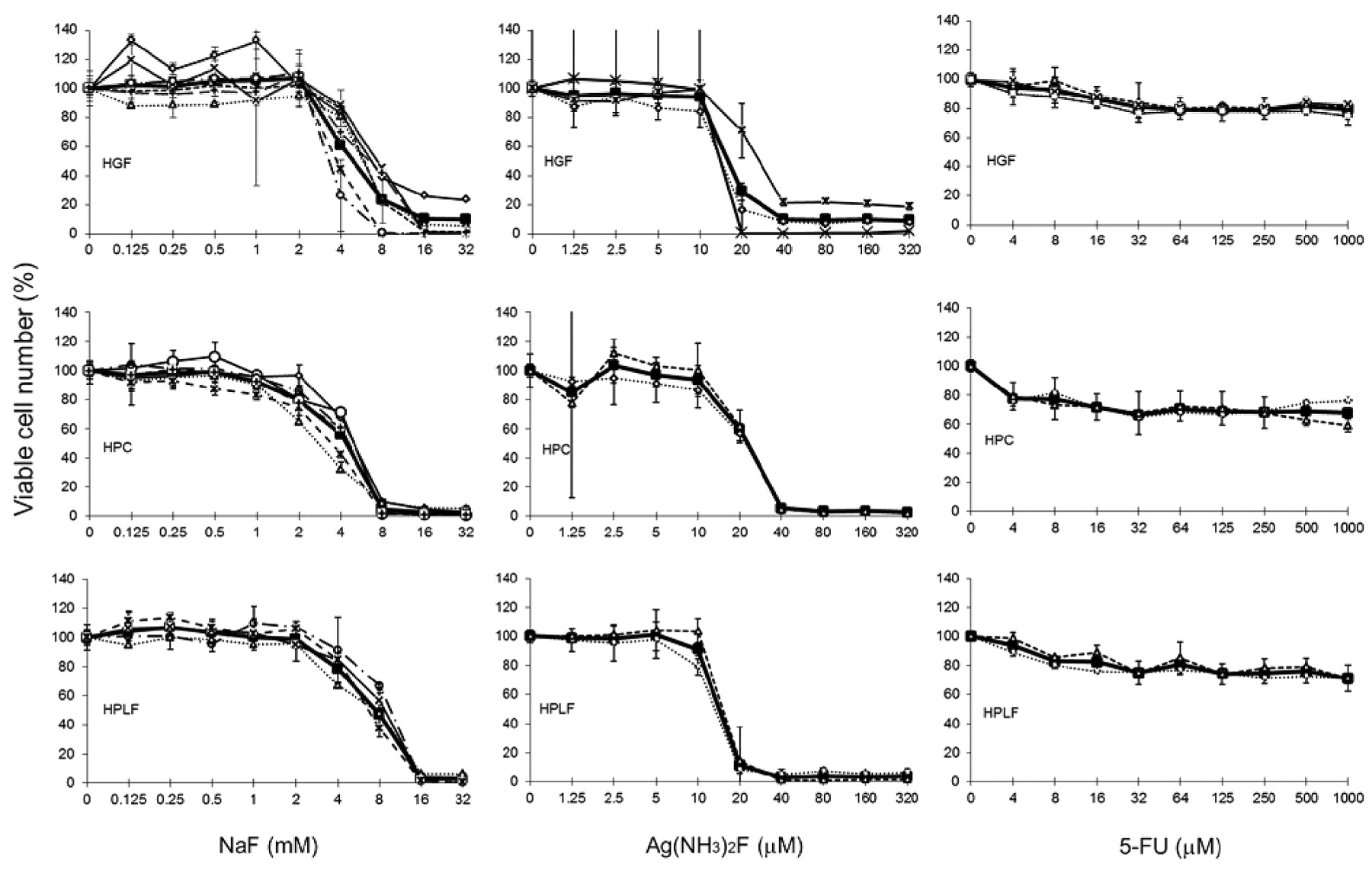
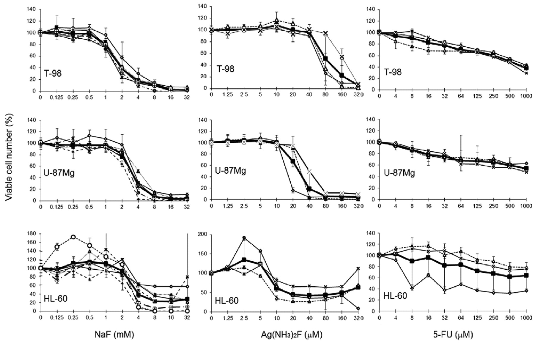
Cytotoxicity. NaF did not have any apparent growth-stimulatory effect (hormesis) (hormetic response <10%) at lower concentrations (0.125-1 mM), but it reduced the viable cell number at higher concentrations, essentially killing all cells at 32 mM (Figures 1, 2 and 3). NaF exhibited higher cytotoxicity against OSCC (HSC-2, HSC-3, HSC-4, NA, Ca9-22) (CC50=2.360-3.740 mM) and glioblastoma cell lines (T98G, U87MG) (CC50=1.733-3.350 mM), as compared to normal oral cells (HGF, HPC, HPLF) (CC50=4.367-8.350 mM), yielding a TS of 2.3 and 2.4, respectively (Table I). Promyelocytic leukemia cells HL-60 were rather resistant (CC50=4.914 mM).

Effect of fluoride compounds on the growth of normal human oral cells. Near confluent cells were incubated for 48 hours with the indicated concentrations of NaF, Ag(NH3)2F or 5-fluorouracil (5-FU), and the viable cell number was determined by the MTT method. Each value represents the mean from 6 (NaF), and 3 Ag(NH3)2F and 5-FU experiments. The mean value of all experiments is shown by the thick line.
Ag(NH3)2F also exhibited very little or no hormesis in most of the cells, except for HL-60 cells, where approximately a 30% maximum hormetic response was observed. Ag(NH3)2F was two orders more cytotoxic, as compared with NaF, killing all cells at 40 μM (Figures 1, 2 and 3). HL-60 cells were the most sensitive to Ag(NH3)2F (CC50=13 μM). Normal oral cells (CC50=20-23 μM) were more sensitive than OSCC (CC50=24-32 μM) and glioblastoma cell lines (CC50=41-105 μM), yielding lower TS values (0.76 and 0.3, respectively) (Table I).
5-FU did not exhibit hormesis, but did dose-dependently inhibit growth (cytostatic effect) without killing all cells tested (Figures 1, 2 and 3). The effect of 5-FU was more prominent on OSCC cell lines (CC50=42-128 μM) as compared with normal oral cells (CC50=>338->640 μM), being somewhat tumor-specific (TS>3.8). The effect of 5-FU on glioblastoma cell lines was much smaller (TS=1.1), due to the highly resistant nature of these cell lines (CC50>356->566 μM) (Table I).
Cytotoxicity of three fluoride compounds.

Effect of fluoride compounds on the growth of five (OSCC) cell lines. Near confluent cells were incubated for 48 hours with the indicated concentrations of NaF, Ag(NH3)2F or 5-FU, and the viable cell number was determined by the MTT method. Each value represents the mean from 6 (NaF), and 3 (Ag(NH3)2F and 5-FU) experiments. The mean value of all experiments is shown by the thick line.
Cellular uptake. We first confirmed that precipitation of NaF, by complex formation with calcium was not observed (data not shown). Only 0.0078-0.03% of the initially added NaF was recovered from HL-60 cells (Experiments I, II and III in Table II). The uptake or cell binding of NaF was increased in an unsaturated manner (Experiment I in Table II). The amount of NaF recovered from the cells was the highest at 30 min after addition, and thereafter declined (Experiments II and III in Table II), suggesting that the normal cellular function was already disrupted at 30 min when a very high concentration of NaF (10 mM) was used.
Relatively higher amounts of NaF (approximately 0.022-0.091% of that initially added) were recovered from HSC-2 cells (Experiments IV and V in Table II).
Amounts of Ag(NH3)2F and 5-FU (monitored by fluoride in the molecule) recovered from the cells were below the detection limit (<0.1 ppm) (data not shown).

Effect of fluoride compounds on the growth of human glioblastoma and promyelocytic leukemia cell lines. Near confluent cells were incubated for 48 hours with the indicated concentrations of NaF, Ag(NH3)2F or 5-FU, and the viable cell number was determined by the MTT method. Each value represents the mean from 6 (NaF), 3 (Ag(NH3)2F and 5-FU) experiments. The mean value of all experiments is shown by the thick line.
Role of ROS. Addition of SOD and catalase significantly reduced the cytotoxicity of sodium ascorbate, against HSC-2 cells (concentration of sodium ascorbat: F(9,11)=186.4, p<0.001; dose of Ag(NH3)2F: F(9,1)=219.04, p<0.001; Two way ANOVA) inhibited by co-application of SOD (300 units/ml) p<0.001 and CAT ((3000 units/ml): p<0.05). Differences between groups were measured (p<0.05; Student's t-test), confirming our previous findings (14), but did not reduce the cytotoxicity of NaF and Ag(NH3)2F, or the cytostatic action of 5-FU (Figure 4). This result reduced the likelihood of the involvement of ROS in cell death induction by fluoride compounds.
Type of cell death. Treatment of HSC-2 cells with either NaF (2-32 mM), Ag(NH3)2F (10-160 μM) or 5-FU (20-320 μM) did not induce internucleosomal DNA fragmentation (Figure 5).
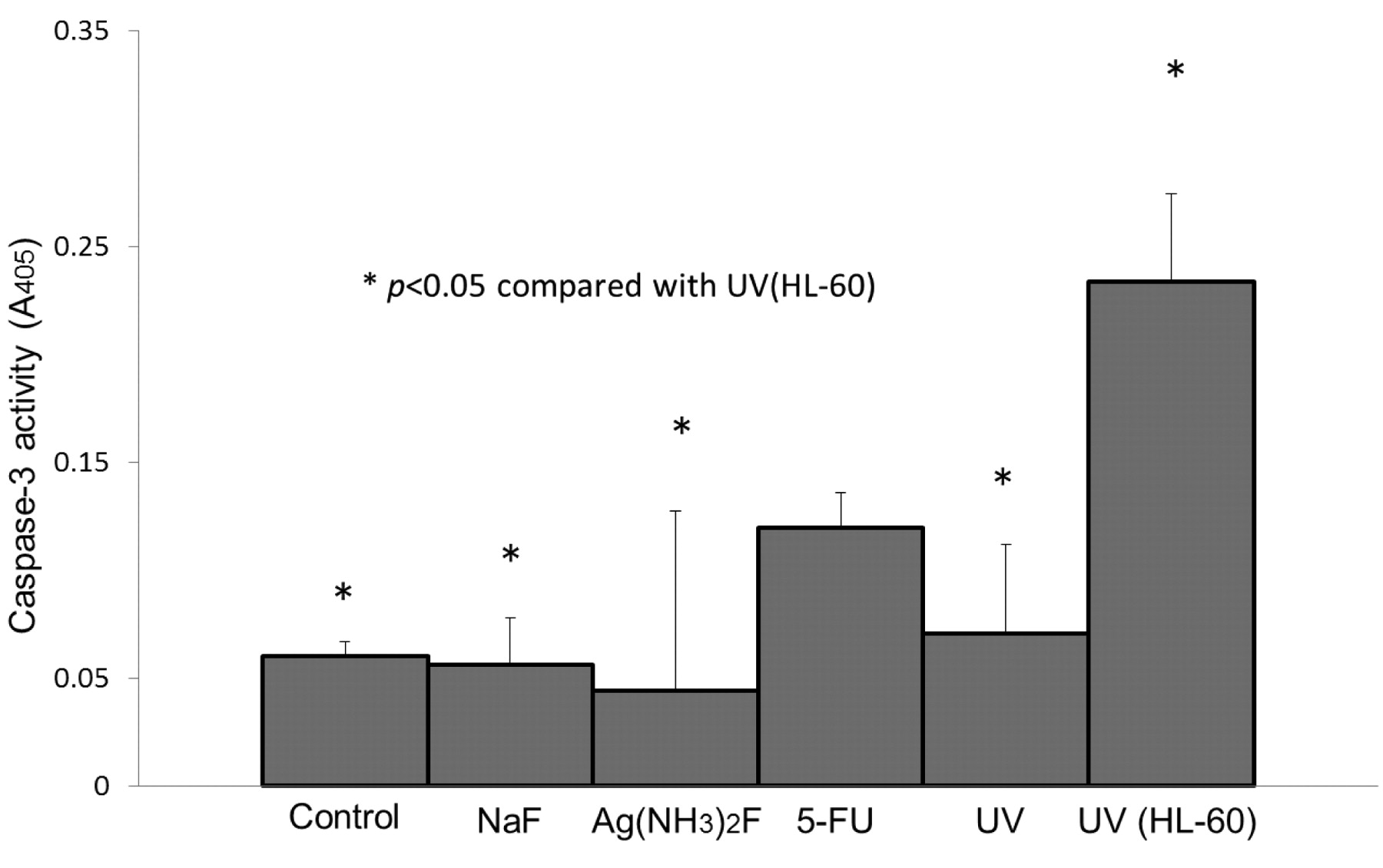
We first confirmed that apoptotisis of HL-60 cells, induced by UV irradiation (positive control) exhibited the highest caspase-3 activity (Figure 6).
Although they were significant (F (5,12)=9.402, p<0.01: One way ANOVA; Bonferroni multiple t-test: p<0.01). Only 5-FU treatment resulted in no significant difference from the positive control, whereas treatment with NaF, Ag(NH3)2F and UV irradiation induced significantly (p<0.05) lower caspase-3 activity in HSC-2 cells.
Fluoride recovery from cells after incubation with NaF during culture.

Role of ROS in induction of cytotoxicity by fluoride compounds. Near confluent HSC-2 cells were incubated for 48 hours with the indicated concentrations of sodium ascorbate (A), NaF (B), Ag(NH3)2F (C), or 5-FU (D) in the presence (●) or absence (○) of catalase (3000 units/ml) and SOD (300 units/ml), and then assayed for the viable cell number by the MTT method. Each value was obtained from three independent experiments, which came out in triplicates per experiment. *p<0.05 (Student's t-test) as compared to cells without SOD or catalase. Reproducible results were obtained in another experiment.
Discussion
The present study demonstrated that three fluoride compounds, NaF, Ag(NH3)2F and 5-FU, did not induce any apparent hormetic response in most of the cells, except HL-60 cells, confirming our previous finding for NaF in normal oral cells (15). This may be an advantage that there is a little likelihood of inducing adverse effects, such as gingival hyperplasia, reported to be induced after treatment with antiepileptic drugs and calcium channel antagonists (16). However, how much damage these compounds cause in oral tissues needs to be further monitored. We observed that NaF and Ag(NH3)2F induced some extent of hormetic response (10-30%) in HL-60 cells, known to be easily committed to apoptosis by many inducers. None of the fluoride compounds induced internucleosomal DNA fragmentation, a biochemical hallmark of apoptosis, nor hormesis. These circumstantial evidences of two cases suggest a possibility of the link between hormesis and apoptosis.
We found that these three compounds affect cellular growth differently: NaF and Ag(NH3)2F were cytotoxic, while 5-FU was cytostatic. Moreover, Ag(NH3)2F exhibited higher cytotoxicity towards both normal cells and tumor cells, being poorly tumor-specific (TS=0.3-0.76). On the other hand, NaF and 5-FU were slightly more specific towards OSCC (TS=2.4) and glioblastoma cells (TS>3.8), respectively. We also found that NaF, as well as other fluoride compounds, did not induce apoptosis markers, in agreement with our recent finding that NaF induced apoptosis in HSC-2 cells only at selected concentrations (~3 mM) (13). These data suggest that the cytotoxicity of fluoride compounds may not be directly related to their tumor specificity, nor to their apoptosis-inducing activity. The different effects of these compounds may be derived from their different sites of action. NaF has been reported to inhibit the glycolytic enzyme, enolase (17), and also interfere with calcium (18). Ag(NH3)2F is used as a root canal irrigant because of its antimicrobial effect and the deposition of silver compounds (19). 5-FU is known to induce antitumor activity by inhibiting the synthesis of precursors of DNA, as well as by inhibiting the function of RNA (20). It remains to be investigated whether prolonged incubation with 5-FU induces much higher apoptosis.

Induction of DNA fragmentation by fluoride compounds. Near confluent HSC-2 cells were incubated for six hours with the indicated concentrations of NaF (A), Ag(NH3)2F (B), or 5-FU (C), respectively, or exposed to UV for 1 min followed by 4 hours incubation (indicated by UV), and DNA was prepared for the detection of DNA fragmentation by agarose gel electrophoresis. UV(HL), DNA from HL-60 cells induced to apoptosis by UV irradiation (6 J/m2/min, 1 min) followed by 3 hours incubation in regular culture medium. M, DNA marker. Reproducible results were obtained in another experiment.

Activation of caspase-3 by fluoride compounds. Near confluent HSC-2 cells were incubated for 4 hours without (control) or with 6 mM NaF, 60 μM Ag(NH3)2F, 80 μM 5-FU, irradiated with UV (HSC-2) and UV (HL-60) (positive control), and the cell lysate was prepared for the assay of caspase-3. *p<0.05 (Bonferroni t-test, vs. UV(HL-60). Each value was obtained from three independent experiments, which came out in triplicates per experiment.
At present, no information is available regarding the point of action of NaF, whether it acts intracellularly, or extracellularly. Our preliminary study of the intracellular uptake of fluoride demonstrated that the amounts of fluoride in the cells increased with increasing amounts of NaF added to the cells, in an unsaturated manner. There was a possibility that we might only have measured the NaF and the calcium complex that may have precipitated with the cells by centrifugation. This possibility, however, was eliminated by our finding that essentially no fluoride was detected in the pellet in the absence of the cells (data not shown). Therefore, it was demonstrated that NaF was bound to or incorporated into the cells. Further study will be necessary to distinguish which of these occurs.
Time-kinetics experiments demonstrated that the amounts of NaF recovered from the cells peaked at 30 min, and declined thereafter. This indicates that a very high concentration of NaF (10 mM) may instantly disturb the normal cellular metabolism. The experiment with SOD and the catalase suggests that these fluoride compounds themselves, but not the secondary metabolite such as ROS, attacks the cells. Further study using metabolomics is needed to identify the intracellular target molecule of each fluoride compound.
In conclusion, the present study suggests that the cytotoxicity of fluoride compounds may not be directly linked to their tumor specificity nor to their apoptosis-inducing activity.
- Received January 25, 2012.
- Revision received February 24, 2012.
- Accepted February 27, 2012.
- Copyright © 2012 International Institute of Anticancer Research (Dr. John G. Delinassios), All rights reserved


