


Abstract
Background: We previously reported that contact with copper (Cu) induced immediate cell death via an oxidation-involved mechanism, and the Cu-induced oxidation and cell death were effectively alleviated under hypoxic conditions. In order to explore alternative strategies for the protection from the Cu-induced cytotoxicity, we investigated whether the inclusion of gold (Au) in the Cu plate, as alloy,has a protective effect. Materials and Methods: Human gingival fibroblast (HGF) cells, established from periodontal tissues, were inoculated on Au/Cu alloy of different Au ratios. After incubation at 37°C for different times under normoxic conditions, cellular viability and amino acid consumption were determined. Changes in the elemental composition of the alloy and in the culture medium were chemically analyzed by X-ray photoelectron spectroscopy and by inductively coupled plasma-optical emission spectrometry. Results: Contact with the Cu plate induced cytotoxicity and cystine oxidation in time-dependent manners. Inclusion of Au at more than 10% in the alloy, completely abrogated the cytotoxicity and reduced the oxidation of Cu and the elution of Cu from the alloy. Conclusion: Inclusion of Au as a component of alloy reduces the cytotoxicity of the Cu plate, possibly by reducing its oxidation.
Dental alloys have been reported to induce allergic reactions in the oral cavity, although infrequently (1). This may be caused by the stimulated release of metal ions from the alloys in the acidic environments of the oral cavity, possibly produced by inflammation, bacterial infection, and the intake of soft drinks and coffee (2, 3). Metal ions may be incorporated into cells, possibly via metal transporter-mediated endocytosis (4, 5). Although numerous studies have shown cytotoxic activity and tissue-damaging activity of metal extracts (6-8), the study of cytotoxicity induced by direct contact with metals has been limited. We recently constructed an assay system to investigate the interaction of cells with metals (9). Direct contact of cells with a copper (Cu) plate was found to induce rapid non-apoptotic cell death, characterized by a smear pattern of DNA fragmentation, little or no caspase activation, loss of the membrane barrier, cytoplasmic damage prior to nuclear damage and vacuolization, without loss of cell surface microvilli in the human promyelocytic leukemic cell line HL-60 (9). On the other hand, other metals such as gold (Au), silver and palladium were essentially inactive (9).
It is very important to explore methods by which they cytotoxicity of Cu can be reduced. We have proposed two strategies for this purpose: the first is to investigate the protective effects of factors affecting the oral environment (such as saliva, tea polyphenol and hypoxia), and the second is to manufacture the least cytotoxic alloy e.g. using different atomic ratios of Au to Cu. We previously tested the efficacy of the first strategy, and reported that (i) Cu-induced oxidation (evaluated by consumption of easily oxidizable cysteine, methionine and histidine) was most effectively reduced under hypoxic conditions, as compared with that observed in the presence of epigallocatechin gallate, or saliva; and (ii) Cu-induced cytotoxicity was also significantly reduced under hypoxic conditions (10).
In the present study, we tested the efficacy of the second strategy, by investigating the effect of contact of an Au/Cu alloy on the viability of human gingival fibroblast (HGF), on cystine consumption by the cells, and on changes in the elemental composition of the alloy, using X-ray photoelectron spectroscopy (XPS) and inductively coupled plasma-optical emission spectrometry (ICP-OES).
Materials and Methods
Materials. The following metals, chemicals and reagents were obtained from the indicated companies: Cu plate (99.99%, 20×20×0.5 mm), Au/Cu alloy (containing 3, 5, 10, 25 and 75% of Au) (20×20×0.5 mm) (Tokuriki Honten, Co, Japan); Dulbecco's modified Eagle's medium (DMEM; GIBCO BRL, Grand Island, NY, USA); fetal bovine serum (FBS, Sigma Chem. Co., St. Louis, MO, USA); trichloroacetic acid (TCA, Wako Pure Chem. Co, Tokyo, Japan).
Polishing of metal plate surface. The metal plates were polished using alumina slurry in water (micropolish, Buehler, Waukegan Road Lake Bluff, IL, USA) down to 0.05 μm particle size. After polishing, their surfaces were examined using scanning electron microscopy (JSM-6360LV, JEOL, Tokyo, Japan) to confirm the consistency of surface smoothness.
Cell culture. HGF cells were prepared from periodontal tissues, according to the guideline of the Meikai University Ethic Committeee (no. A0808), after obtaining the informed consent from the patients, and were cultured in DMEM supplemented with 10% heat-inactivated FBS under a humidified normoxic 5% CO2 atmosphere (Ikemoto Rika Kogyo, Tokyo). HGF cells were harvested by detaching them from the culture plate with 0.25% trypsin-0.025% EDTA-Na in phosphate-buffered saline without magnesium and calcium [PBS (−)] and subcultured at a 1:4 split ratio once a week, with one medium change in between. Since HGF cells have a limited lifespan, ceasing proliferation at a 20 population doubling level (PDL) (11), the cells at 9-12 PDL were used in the present study.
Cytotoxicity of direct contact with metal plates. HGF cells were trypsinized and resuspended at a cell density of 2×10⁶/ml in fresh medium. Five hundred microliters of HGF cells (2×10⁶/ml) in DMEM with 10% FBS were inoculated onto the Cu or Au/Cu alloy plates (in a 3.5 cm dish), and incubated for 30 min at 37°C. Cells were recovered from the metal plates by gentle pipetting, or with trypsinization when necessary. The viability of the cells was determined by cell counting with a hemocytometer after 0.15% trypan blue dye staining.
Determination of the changes in extracellular free amino acids. Five hundred microliters of HGF cell suspension were inoculated onto the metal plates which were then incubated for 30 min at 37°C. Culture supernatant (medium fraction), obtained after centrifugation (1500 ×g for 3 min), was mixed with an equal volume of 10% TCA, and was left on ice for 30 min. After centrifugation for 5 min at 10000 ×g, the deproteinized supernatant was collected and stored at −40°C. The supernatants (20 μl) were analyzed with a JEOL LC-300 amino acid analyzer, and amino acids were detected by using the ninhydrin reaction (9).
The consumption of each amino acid during incubation was calculated using the following equation: Consumption (%)={([AA]before – [AA]after)/[AA]before} ×100, where [AA]before and [AA]after represent the concentration of each amino acid before and after incubation for the indicated times, respectively.
Surface analysis of Au/Cu alloys plates. Before and after immersion in culture medium without the cells, the surface of Au/Cu alloy plates was analyzed using X-ray photoelectron spectroscopy (XPS). The XPS analysis was performed using a photoelectron spectroscope (Axis-Ultra, Kratos, Kyoto, Japan). The X-ray resource was monochronized AlKα (filament voltage: 15 kV, emission current: 10 mA), the area of measurement was 200×600 μm, and take-off angle of photoelectrons was 90°.
Quantification of eluted elements. The elemental composition of the alloy and the concentration of Cu eluted into the culture medium was determined by ICP-OES (Vista-MPX, SII, Japan), as described previously (12).
Statistical analysis. Statistical differences among the samples were evaluated using ANOVA, followed by Scheffè's multiple comparison test or Student's t-test, at α=0.05.
Results
Au inhibited Cu-induced cytotoxicity. When HGF cells contacted a pure Cu plate, the viability declined time-dependently (p<0.05) (Figure 1A). However, contact with a plate of Au/Cu alloy that contained Au at more than 25%, did not significantly change the viability (p>0.05), but the viability was significantly higher than the one for the Cu plate at each time point (p<0.05) (Figure 1A).
Next, we investigated the effect of reducing the ratio of Au to Cu in the alloy, on the Cu-induced cytotoxicity, in order to determine the critical Au ratio (Figure 1B). Contact with alloy that contained Au at 3, 5, or 10% resulted in significantly higher viability than that with a plate of pure Cu (2.2%) (p<0.05). Contact with alloy that contained Au at 10% resulted in 99.3% viability, not significantly different from that with the pure Au plate (p>0.05). On the other hand, contact with alloy that contained Au at 3 or 5% resulted in significantly lower viability than that with Au at 10% (p<0.05). There was no significant difference in cell viability between plates with 3% and 5% Au (p>0.05) (Figure 1B).
Au inhibited Cu-induced oxidation. When HGF cells were inoculated onto a Cu plate, the consumption of cystine increased with the duration of contact with the Cu plate (Figure 2A), confirming that cystine is a sensitive marker for Cu-induced oxidation. When Au was included at 25, 50, 70 or 100%, the consumption of cystine was not significant (Figure 2A).
Next, the effect of reducing the ratio of Au to Cu in the alloy on Cu-induced cystine consumption was investigated in order to determine the critical Au ratio (Figure 2B). The inclusion of 10% Au also completely abrogated the cystine consumption, whereas the inclusion of only 3 or 5% Au significantly (p<0.05), but not completely, inhibited cystine consumption (Figure 2B).

Effect of contact with alloy plates on cell viability. Cells were inoculated onto alloy plates. After incubation for 0, 30, 90 or 180 min (A) or 90 min (B), cell viability (%) was determined. Each value represents the mean±S.D. from three independent experiments. *p<0.05 compared vs. pure Cu plate (0% Au). **p<0.05 compared to 3 and 10% Au. ***p<0.05 compared to 5 and 10% Au.
Change in the surface constituents of Au/Cu alloy. We next investigated possible changes in the surface constituents of Au/Cu alloy by XPS, before (Figure 3A-C) and after (Figure 3D-F) incubation in culture medium. The survey spectra of Au/Cu alloys after immersion in culture medium shows larger oxygen, carbon and nitrogen peaks than that before immersion of alloys, as can be seen in Figures 3A and D, suggesting the adsorption or binding of amino acids or serum proteins in the culture medium. It should be noted that the adsorption or binding of these components was reduced with increasing Au content.
Alloy elements were detected in all alloy samples, and therefore the chemical state of alloy component elements was investigated. Analysis of Cu2p region of the sample before the immersion into culture medium shows that the satellite peak of Cu2+ (indicated by black circle) was detected (Figure 3B). After immersion in culture medium, the satellite peak of Cu2+ was diminished to an undetectable level with increasing Au, whereas the Cu2+ peak was rather augmented without Au (Figure 3E). Analysis of Cu Auger region of the Cu plate before immersion in culture medium (Figure 3C), shows that Cu was detected as prominent peaks of Cu0 and Cu+. The peak height of Cu+ was higher than that of Cu0, suggesting the occurrence of slight oxidation. After immersion in culture medium, the ratio of Cu0 was increased with the high Au content (Figure 3F). These results suggest that oxidation of Cu in Au/Cu alloy is retarded dose-dependently with the presense of Au content in the alloy.

Effect of contact with alloy plates on cystine consumption. Cells were inoculated on to alloy plates. The consumption of cystine during 30, 90, or 180 min (A) or 90 min (B) was then determined. Each value represents the mean±S.D. from three independent experiments. *p<0.05 compared vs. pure Cu plate (0% Au).
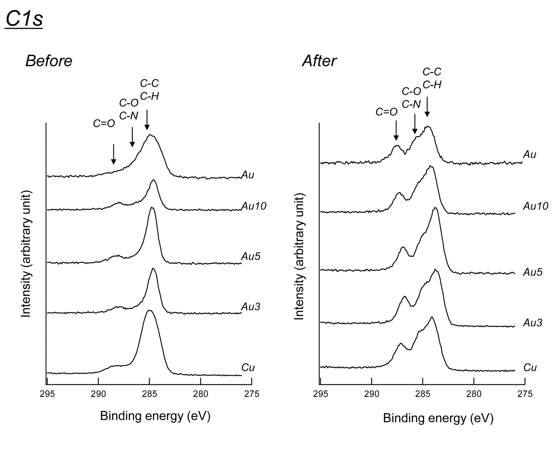
Analysis of C1s region demonstrated that carbonyl group (C=0) was detected in Cu plate, and the intensity of this peak increased during immersion in culture medium (Figure 4). The addition of increasing amounts of Au dose-dependently reduced the intensity of this peak, indicating that Au acted like an antioxidant.

XPS analysis of alloys before and after immersion of plates into culture medium. Cu plate and Au/Cu alloy containing 3, 5, or 10% of Au were analyzed by XPS before (A-C) and after (D-F) immersion for 90 min in culture medium. A, D: Survey spectra; B, E: Cu2p region; C, F: Cu Auger region.
Au reduced the elution of Cu ion. ICP analysis of the culture medium demonstrated that Cu ion was eluted from Au/Cu alloy. However, the inclusion of Au in the Cu plate dose-dependently reduced the elution of Cu into the culture medium. At 10% Au, the elution of Cu was reduced to approximately one-fourth to the one without Au (Figure 5).
Discussion
The present study demonstrates that the Au/Cu alloy containing more than 10% Au had no apparent cytotoxicity against HGF cells, indicating that Au effectively neutralized the cytotoxicity of Cu. We have previously suggested that Cu induced cytotoxicity by its oxidizing activity, based on the enhanced cystine consumption by Cu, and the neutralization of Cu cytotoxicity by N-acetyl-L-cysteine, a popular antioxidant (9) or by hypoxia (10). This is further supported by the present study that demonstrated that Cu enhanced carbonylation, an oxidative stress marker (13). The present study also demonstrated that Au effectively diminished cystine consumption (Figure 2) and retarded Cu oxidation (Figure 3), and carbonylation (Figure 4). This might suggest that Au protects cells by its antioxidant-like action.
There was a possibility that the most part of the cytotoxicity of Cu might be due to the Cu2+ eluted into the culture medium. We found that a high level of Cu2+ still remained in the culture medium, although the elution of Cu2+ was concentration-dependently inhibited by Au in the alloy (Figure 5). A marked elution of Cu has been reported in the presence of albumin (14), and at neutral pH in the presence of amino acids (15). On the other hand, Rae reported two actions of metals in particles against cells: the cytotoxic action by the soluble ions and organic metal compounds, and the direct interaction between the surface of the particle and the cellular membrane. Therefore, it may be possible that the environmental changes to the surface of the alloy might affect cell death induction (16). The present study demonstrates that 10% Au/Cu alloy was not cytotoxic to HGF cells, although it eluted significant amounts of Cu into the culture medium. This suggests that Cu-induced cytotoxicity cannot be determined solely by the eluted Cu ion, but may also be affected by the interaction between the cells and the metal. It remains to be investigated how contact with the culture medium modifies the surface of alloy and how it affects the elution of the Cu ion.

XPS analysis of C1s region before and after immersion in culture medium. Experimental conditions were the same as shown in Figure 3.

Elution of Cu into the culture medium. Cu plate and Au/Cu alloy containing 3, 5, or 10% of Au were immersed in the culture medium for 90 min, and the concentration of Cu eluted into the culture medium was determined by ICP. Each value represents the mean±S.D. from three independent experiments. *p<0.05 vs. control (0% Au).
Footnotes
-
This article is freely accessible online.
- Received April 12, 2012.
- Revision received June 11, 2012.
- Accepted June 11, 2012.
- Copyright © 2012 International Institute of Anticancer Research (Dr. John G. Delinassios), All rights reserved
References
In this issue


Jump to section
Related Articles
Cited By...
- No citing articles found.