
Main menu

  • Home
  • Content
    • Current
    • Archive
  • Info for
    • Authors
    • Subscribers
    • Advertisers
    • Editorial Board
  • Other Publications
    • Anticancer Research
    • Cancer Genomics & Proteomics
  • More
    • IIAR
    • Conferences
  • About Us
    • General Policy
    • Contact
  • Other Publications
    • In Vivo
    • Anticancer Research
    • Cancer Genomics & Proteomics

User menu

  • Register
  • Subscribe
  • My alerts
  • Log in
  • My Cart

Search

  • Advanced search
In Vivo
  • Other Publications
    • In Vivo
    • Anticancer Research
    • Cancer Genomics & Proteomics
  • Register
  • Subscribe
  • My alerts
  • Log in
  • My Cart
In Vivo

Advanced Search

  • Home
  • Content
    • Current
    • Archive
  • Info for
    • Authors
    • Subscribers
    • Advertisers
    • Editorial Board
  • Other Publications
    • Anticancer Research
    • Cancer Genomics & Proteomics
  • More
    • IIAR
    • Conferences
  • About Us
    • General Policy
    • Contact
  • Visit iiar on Facebook
  • Follow us on Linkedin
Research ArticleExperimental Studies

Effect of CO2 Laser Irradiation on Hormesis Induction in Human Pulp and Periodontal Ligament Fibroblasts

KENSUKE IWASAKA, ERI HEMMI, KAYOKO TOMITA, SACHIYO ISHIHARA, TADASHI KATAYAMA and HIROSHI SAKAGAMI
In Vivo September 2011, 25 (5) 787-793;
KENSUKE IWASAKA
  • Find this author on Google Scholar
  • Find this author on PubMed
  • Search for this author on this site
  • For correspondence: iwasaka@dent.meikai.ac.jp sakagami@dent.meikai.ac.jp
ERI HEMMI
  • Find this author on Google Scholar
  • Find this author on PubMed
  • Search for this author on this site
KAYOKO TOMITA
  • Find this author on Google Scholar
  • Find this author on PubMed
  • Search for this author on this site
SACHIYO ISHIHARA
  • Find this author on Google Scholar
  • Find this author on PubMed
  • Search for this author on this site
TADASHI KATAYAMA
  • Find this author on Google Scholar
  • Find this author on PubMed
  • Search for this author on this site
HIROSHI SAKAGAMI
  • Find this author on Google Scholar
  • Find this author on PubMed
  • Search for this author on this site
  • For correspondence: iwasaka@dent.meikai.ac.jp sakagami@dent.meikai.ac.jp
  • Article
  • Figures & Data
  • Info & Metrics
  • PDF
Loading

Abstract

Background: We have recently reported that a low level of CO2 laser irradiation induced growth stimulation (hormesis) of both human gingival fibroblast (HGF) and oral squamous cell carcinoma cell line (HSC-2), but the extent of hormetic response was much smaller than that previously reported for toxicants and radiation in other experimental systems. Here we investigated the extent of hormetic response induced by CO2 laser irradiation in human pulp cells (HPCs) and periodontal ligament fibroblast (HPLF). Materials and Methods: HPC and HPLF cells were established from the periodontal tissues of the first premolar extracted tooth. Cells were cultured for 24, 48 or 72 hours after exposure to various irradiation powers, and the viable cell number was determined by 3-(4,5-dimethylthiazol-2-yl)-2,5-diphenyltetrazolium bromide (MTT) method. Results: CO2 laser irradiation induced biphasic effects on the growth of both HPC and HPLF cells. The maximum hormetic response was less than 50%. The hormetic response was found within the energy density of 7.98-79.77 J/cm2, and cytotoxicity emerged at powers over 132.96 J/cm2. Combining with our previous report, HPCs showed the highest hormetic response, followed by HPLFs and then HGFs. Both HPLFs and HGFs showed similar time-course of hormesis response, increasing response with incubation time. Conclusion: The hormetic response may be the common survival mechanism by which cells escape from radiation-induced injury. Higher hormetic response of HPCs may reflect their potential for differentiation into one of the components in dentin.

  • CO2 laser irradiation
  • hormesis
  • cytotoxicity
  • pulp cell
  • periodontal ligament fibroblast

Different laser modalities have been used in the treatment of various disorders (1-4). However, as compared with Er:YAG laser therapy, there is insufficient evidence of safety and effects to support the clinical application of CO2, Nd:YAG, or diode laser (5, 6). Many toxicants, chemotherapeutic agents, hormones, metals and radiations have been reported to show bi-phasic growth-modulating effects: growth stimulation (hormesis) at lower concentrations and growth inhibition at higher doses (7, 8). Similarly, high level laser treatment induced cell membrane and DNA damage (9), whereas low level laser treatment induced growth stimulation and wound healing (10-12). It has been reported that hormesis may be detectable only under optimal condition (in terms of the dose and the treatment time) (7). On the other hand, we found that sodium fluoride (13), gefitinib (14), 2-aminotropone derivatives (15) and herbal extracts (16) induced little or no hormetic effects on cultured human oral normal cells [gingival fibroblast (HGF), pulp cell (HPC), periodontal ligament fibroblast (HPLF)] and human oral squamous cell carcinoma cell lines (HSC-2, HSC-3, HSC-4). These conflicting results suggest that the extent of hormesis induction may depend on the type of cell or the type of irradiation conditions. Most previous in vitro studies have been carried out with laser modalities other than CO2 laser. Based on this background, we initiated an investigation of the effect of CO2 laser irradiation on oral cells. We recently found that both the range and magnitude of hormetic response of human oral normal (HGF) and tumor (HSC-2) cells was very narrow and small (17). To explore more efficient application of CO2 laser irradiation in dentistry, here we investigated the extent of hormetic response induced by CO2 laser irradiation in HPC and HPLF cells.

Materials and Methods

Cell culture. HPC and HPLF cells were established from an extracted first premolar tooth in the mandible and periodontal tissues of a twelve-year-old girl, according to the guideline of the Intramural Board of Ethics Committee (no. A0808) (18). HPCs and HPLFs had an in vitro life-span of 43 and 43 cumulative cell population doubling level (PDL) (18). These cells were cultured in Dulbecco's modified Eagle's medium (DMEM) supplemented with 10% heat-inactivated fetal bovine serum (FBS), under a humidified 5% CO2 atmosphere. The cells at 10-15 PDL were used in this experiment.

Figure 1.
  • Download figure
  • Open in new tab
  • Download powerpoint
Figure 1.

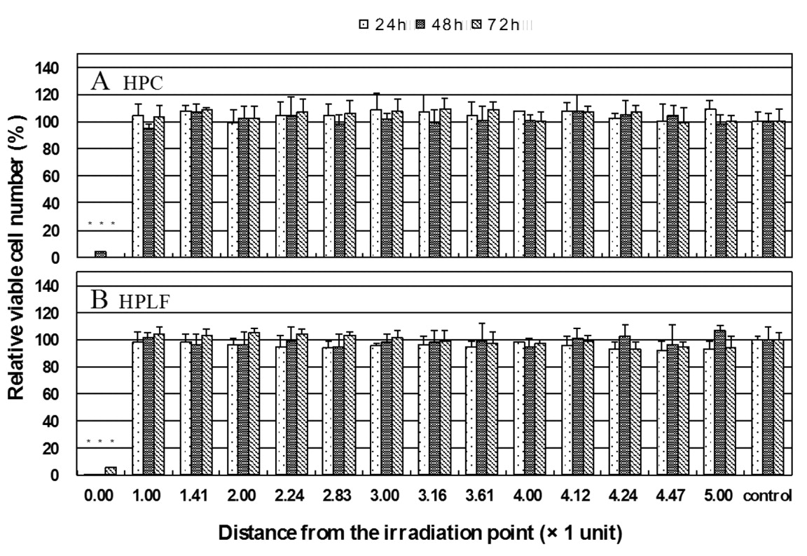
Ripple effect of laser irradiation on the viability of cells grown on the adjacent wells. HPC (A) and HPLF (B) cells were inoculated on a 96-microwell plate, and incubated for 48 hours to achieve complete attachment. A tip of CO2 laser (Opelaser PRO LA12) was set just 52 mm above the cell surface, and the cells were exposed to CO2 laser irradiation (5 W with dispatch mode CW, 30 s) without removing the medium (100 μl). After medium change, the cells were further incubated for 24 (left: dotted bars), 48 (center: gray bars) or 72 (right: slashed bars) hours, and the number of viable cells (expressed as percentage of un-irradiated control cells) was then determined by MTT method. The distance between one well to the adjacent well was defined as ‘1 unit’. Each value represents the mean±S.D. of 12 samples. *p<0.01.

Assay for cytotoxicity of laser irradiation. Cells (1.5×10³) were inoculated on 96-microwell plate (37.6 mm2) (Becton Dickinson, Franklin Lakes, NJ, USA), and incubated for 48 hours to allow complete attachment. The medium was changed for new medium, and the cells were placed at 52 mm distantce from the CO2 laser source (Opelaser PRO LA12; Yoshida, Tokyo, Japan) (17). All the cells were evenly irradiated with a specially manufactured tip, under the following conditions: irradiation power (0.5, 1, 2, 3 or 5 W), irradiation time (0.5, 1, 1.5, 3, 5, 10, 15, 20, 30 s) and dispatch mode (CW, SP1, SP2). After incubation for 24, 48 or 72 hours in fresh culture medium, the viable cell number was determined by the 3-(4,5-dimethylthiazol-2-yl)-2,5-diphenyltetrazolium bromide (MTT) method. In brief, cells were incubated for a further 4 hours with 0.2 mg/ml MTT reagent in DMEM/10% FBS at 37°C, and dissolved with 0.1 ml of dismethyl sulfoxide (DMSO) to determine the absorbance at 540 nm with a plate reader (17).

Statistical analysis. The mean values and standard deviations were calculated. The average values were compared by one-way ANOVA and Student's t-test.

Results

Ripple effect of CO2 laser irradiation. When either HPC and HPLF cells were exposed to laser irradiation (5 W, with a dispatch mode of CW, 30 s) from 52 mm distance, almost all of the cells under the irradiation point died after incubation for 24, 48 and 72 hours in regular culture medium (Figure 1). On the other hand, the cells that had been inoculated at the wells adjacent to those exposed to laser irradiation all appeared to survive, judging from cell growth comparable with that of control (unirradiated cells) (Figure 1). This finding made it possible for us to perform sequential CO2 laser irradiation without damaging the viability of the cells grown on the adjacent wells.

Effect on HPC cells. We investigated the effect of irradiation time and power on the viability of HPC cells (Figure 2). Lower power irradiation (0.5-1 W) for up to 30 s did not significantly affect the viability of HPC cells, regardless of dispatch mode and incubation time. Higher power irradiation [2 W (20-30 s); 3 W (15-30 s); 5 W (10-30 s)] reduced the viable cell number by more than 60%. The range of cytotoxicity induced by higher power irradiation is indicated by gray shading in Table I. Cytotoxicity of CO2 laser irradiation reached nearly a maximum level within 24 hours' incubation in culture medium (Figure 2), indicating the cytotoxicity induction by CO2 laser irradiation in HPC cells reached a plateau level within 24 hours.

Figure 2.
  • Download figure
  • Open in new tab
  • Download powerpoint
Figure 2.

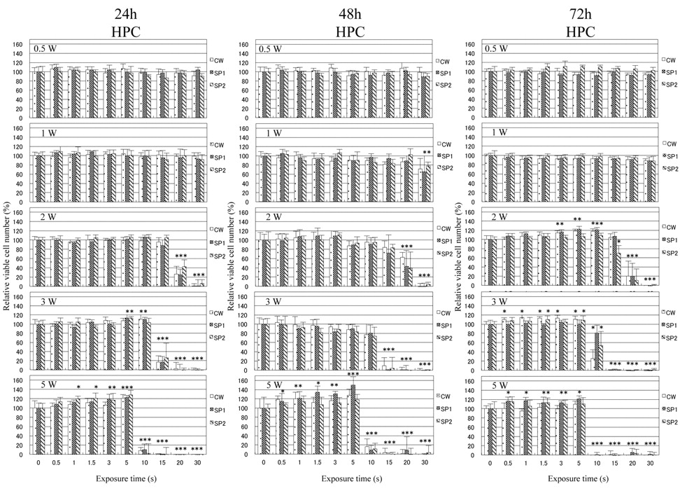
HPC cells were irradiated for 0 (control), 0.5, 1, 1.5, 3, 5, 10, 15, 20 or 30 sec at 0.5, 1, 2, 3 or 5 W with dispatch mode (CW, SP1, SP2), and then cultured for a further 48 hours in DMEM/10% FBS to determine the viable cell number. Each value represents the mean ±S.D. of 12 samples. *p<0.01.

Slight, but significant (p<0.01) growth stimulation was observed after incubation for 24-72 hours, using dispatch model of CW [2 W (72 hours), 3W (24 and 72 h), 5W (24 and 48 h)], SP1 [2 W (72 h), 3W (24 and 72 h), 5 W (24, 48 and 72 h)] and SP2 [2W (72 h), 3W (24 and 72 h), 5W (24, 48 and 72 h)] (Figure 2). The maximum hormetic response was 50.00%. The mean of the maximum hormetic response achieved with the use of either CW, SP1 or SP2 dispatch mode was 22.30, 31.6 and 20.38%, respectively, yielding the mean value of 24.79% (Table II).

Effect on HPLF cells. We next investigated the effect of irradiation time and power on the viability of HPLF cells (Figure 3). Lower irradiation power (0.5-1 W) for up to 30 s was not cytotoxic to HPLF cells, regardless of dispatch mode and incubation time. Higher power irradiation [2 W (20-30 s); 3 W (15-30 s); 5 W (10-30 s)] reduced the viable cell number by more than 60%. The range of cytotoxicity induced by higher power irradiation is indicated by gray shading in Table I. Cytotoxic effect of laser irradiation was slightly enhanced during prolonged incubation time (24, 48 or 72 h), indicating that the process of the cytotoxicity induction by CO2 laser irradiation in HPLF cells was slightly slower than that observed for HPC cells (Figure 3).

Little or no hormetic effect was found at the lower irradiation dose range (0.5-5W). The cell number was slightly, but significantly (p<0.01) increased, using dispatch mode of CW [2-3W (48 and 72 h)], SP1 [3-5 W (48 and 72 hours)] and SP2 [2-5W (48 and 72 h)] (Figure 3). The maximum hormetic response was 24.70%. The mean of the maximum hormetic response achieved with the use of either CW, SP1 or SP2 dispatch mode was 11.75, 13.61 and 17.68%, respectively, yielding the mean value of 14.35% (Table II).

Figure 3.
  • Download figure
  • Open in new tab
  • Download powerpoint
Figure 3.

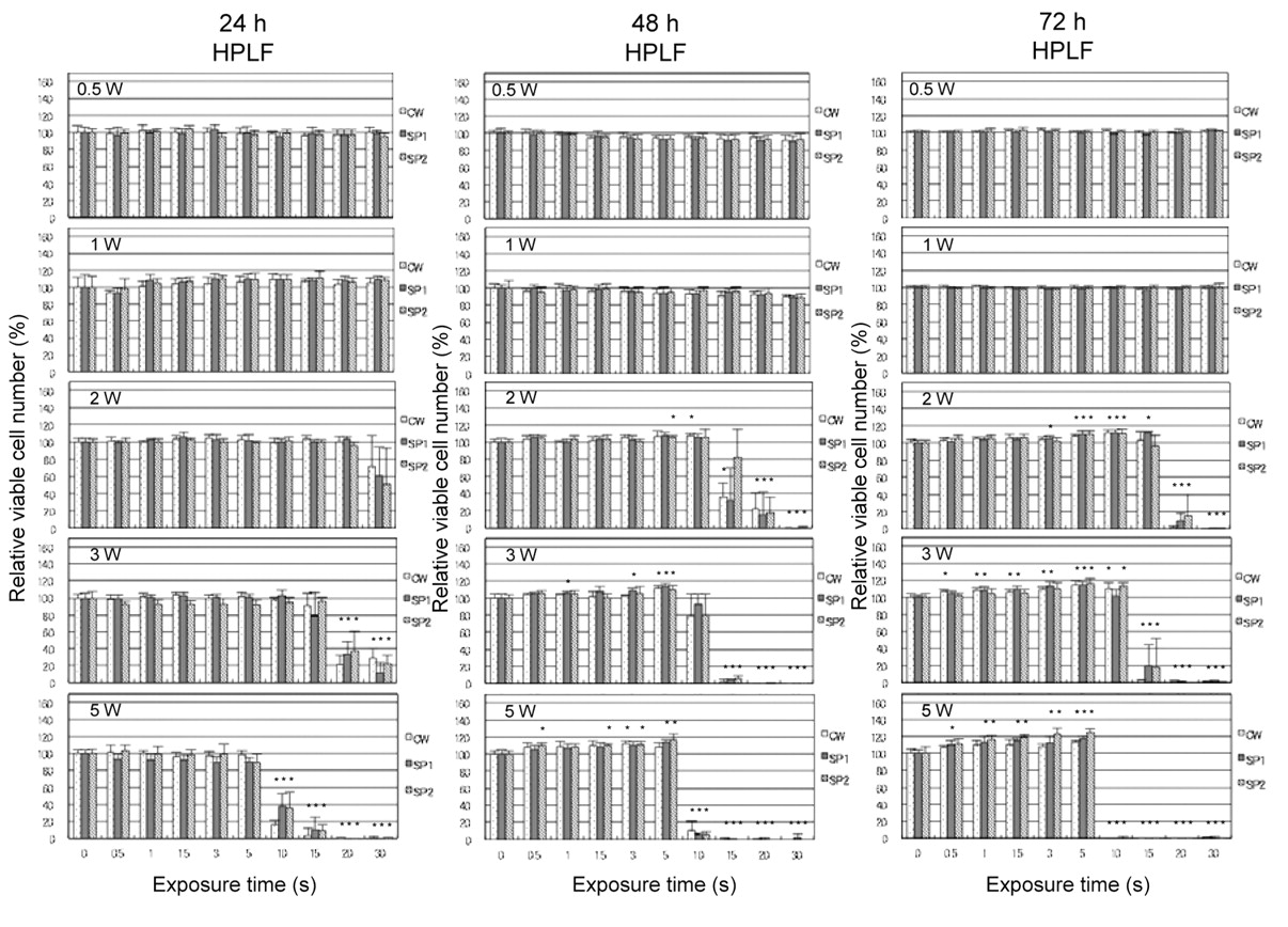
HPLF cells were irradiated for 0 (control), 0.5, 1, 1.5, 3, 5, 10, 15, 20 or 30 sec at 0.5, 1, 2, 3 or 5 W with dispatch mode (CW, SP1, SP2), and then cultured for a further 48 hours in DMEM/10% FBS to determine the viable cell number. Each value represents the mean ±S.D. of 12 samples. *p<0.01.

Discussion

The present study demonstrated that CO2 laser irradiation induced a very low level of hormesis (maximum: 24.79 and 14.35, respectively) in both HPC and HPLF cells (Table II). The hormetic response was found within the delivered energy density of 7.98-79.77 J/cm2, and significant cytotoxicity emerged at deliveries over 132.96 J/cm2 (Table I). This is consistent with our previous finding that the hormetic response induced by CO2 laser irradication in HGF cells was observed within a narrow range of irradiation doses (15.95-79.77 J/cm2), slightly lower than that of cytotoxic doses (106.37-398.87 J/cm2) (17). This suggests that the hormetic response may be the common survival mechanism by which cells escape from radiation-induced injury.

The present study demonstrated for the first time that HPCs showed the highest hormetic response among oral cells that we have investigated here and in our previous paper (17). Combined with our previous data (17), we found that the strength of the hormetic response observed during 24-72 hours' incubation was in the order of HPCs (24.79%) >HPLFs (14.35%) >HSC-2 (10.86%) >HGFs (8.76%). The higher hormetic response of HPC cells may reflect their potential for differentiation into dentin. Among three dispatch modes, SP1 was more effective for hormesis induction in HPC cells (31.69%), as compared with CW (22.30%) and SP2 (20.38%) (Table II)

Both fibroblast lines (HPLF and HGF) showed a similar time-course of hormesis response, with response increasing with the incubation time [10.23% (24 h)→. 14.19% (48 h) → 18.61% (72 h) in HPLFs; 5.70% (24 h) → 9.78% (48 h) → 10.81% (72 h) in HGFs]. Among three dispatch modes, SP2 was more effective in inducing the hormesis in both HPLF and HGF cells (17.68 and 10.71%) than SP1 (13.61 and 6.62%) and CW (11.75 and 8.95%, respectively).

It has been reported that low level laser treatment stimulated the growth of HGFs (19-23), HPCs (24) and HPLFs (25). Many possible mechanisms for this growth stimulation have been proposed: the induction of heat shock-proteins (10), production of collagen (26) and prostaglandin E2 via cyclooxygenase-2 expression (23), reduction of cyclic AMP production (27), and activation of PI3K/AKT (28) and MAPK/ERK (23) pathways, and modification of cytokine gene expression (29). It remains to be investigated whether hormesis induction by low level of CO2 laser irradiation in HPC and HPLF cells is mediated via these changes of intracellular events or not.

View this table:
  • View inline
  • View popup
  • Download powerpoint
Table I.

Energy density (J/cm2) as a function of irradiation time (0-30 s) and irradiation power (0.5-5 W).

View this table:
  • View inline
  • View popup
  • Download powerpoint
Table II.

Hormetic induction in normal oral cells by CO2 laser irradiation. The maximum hormetic response was determined as the highest value of hormetic response observed during 24-72 hours' incubation of HPC (Figure 2), HPLF (Figure 3), HGF (Figure 3 in ref. 17) and HSC-2 cells (Figure 4 in ref. 17).

Acknowledgements

The present study was supported in part by Miyata Research Fund-A, Meikai University School of Dentistry.

  • Received March 31, 2011.
  • Revision received May 24, 2011.
  • Accepted May 26, 2011.
  • Copyright © 2011 International Institute of Anticancer Research (Dr. John G. Delinassios), All rights reserved

References

  1. ↵
    1. Abergel RP,
    2. Meeker CA,
    3. Lam TS,
    4. Dwyer RM,
    5. Lesavoy MA,
    6. Uitto J
    : Control of connective tissue metabolism by lasers: recent developments and future prospects. J Am Acd Dermatol 11: 1142-1150, 1984.
    OpenUrl
    1. Coleton S
    : The use of lasers in periodontal therapy. Gen Dent 56: 612-616, 2008.
    OpenUrlPubMed
    1. Olivi G,
    2. Genovese MD,
    3. Caprioglio C
    : Evidence-based dentistry on laser paediatric dentistry: review and outlook. Eur J Paediatr Dent 10: 29-40, 2009.
    OpenUrlPubMed
  2. ↵
    1. Deppe H,
    2. Horch HH
    : Laser applications in oral surgery and implant dentistry. Lasers Med Sci 22: 217-221, 2007.
    OpenUrlPubMed
  3. ↵
    1. Schward F,
    2. Aoki A,
    3. Becker J,
    4. Sculean A
    : Laser application in non-surgical periodontal therapy: a systematic review. J Clin Periodontol 8(Suppl): 29-44, 2008.
    OpenUrl
  4. ↵
    1. Cobb CM,
    2. Low SB,
    3. Coluzzi DJ
    : Lasers and the treatment of chronic periodontitis. Dent Clin North Am 54: 35-53, 2010.
    OpenUrlPubMed
  5. ↵
    1. Calabrese EJ
    : Paradigm lost, paradigm found: The re-emergence of hormesis as a fundamental dose response model in the toxicological sciences. Environ Pollut 138: 379-412, 2005.
    OpenUrlCrossRefPubMed
  6. ↵
    1. Calabrese EJ,
    2. Staudenmayer JW,
    3. Stanek EJ III.,
    4. Hoffmann GR
    : Hormesis outperforms threshold model in national cancer institute drug screening database. Toxicol Sci 94: 368-378, 2006.
    OpenUrlAbstract/FREE Full Text
  7. ↵
    1. Hawkins DH,
    2. Abrahamse H
    : The role of laser fluence in cell viability, proliferation, and membrane integrity of wounded human skin fibroblasts following helium-neon laser irradiation. Lasers Surg Med 30: 74-83, 2006.
    OpenUrl
  8. ↵
    1. Yamasaki A,
    2. Ito H,
    3. Yusa J,
    4. Sakurai Y,
    5. Okuyama N,
    6. Ozawa R
    : Expression of heat shock proteins, Hsp70 and Hsp25, in the rat gingival after irradiation with a CO2 laser in coagulation mode. J Periodontal Res 45: 323-330, 2009.
    OpenUrlPubMed
    1. Powell K,
    2. Low P,
    3. McDonnell PA,
    4. Laakso E,
    5. Ralph SJ
    : The effect of laser irradiation on proliferation of human beast carcinoma, melanoma, and immortalized mammary epithelial cells. Photomed Laser Sur 28: 115-123, 2010.
    OpenUrl
  9. ↵
    1. Frigo L,
    2. Fávero GM,
    3. Campos Lim HJ,
    4. Maria DA,
    5. Bjordal JM,
    6. Joensen J,
    7. Iversen VV,
    8. Marcos RL,
    9. Parizzoto NA,
    10. Lopes-Martins RA
    : Low-level laser irradiation (InGaAlP-660 nm) increases fibroblast cell proliferation and reduces cell death in a dose-dependent manner. Photomed Laser Surg 28: 151-156, 2010.
    OpenUrl
  10. ↵
    1. Satoh R,
    2. Kishino K,
    3. Morshed SRM,
    4. Takayama F,
    5. Otsuki S,
    6. Suzuki F,
    7. Hashimoto K,
    8. Kikuchi H,
    9. Nishikawa H,
    10. Yasui T,
    11. Sakagami H
    : Changes in fluoride sensitivity during in vitro senescence of human normal oral cells. Anticancer Res 25: 2085-2090, 2005.
    OpenUrlAbstract/FREE Full Text
  11. ↵
    1. Chu Q,
    2. Amano O,
    3. Kanda Y,
    4. Kunii S,
    5. Wang Q,
    6. Sakagami H
    : Tumor-specific cytotoxicity and type of cell death induced by gefitinib in oral squamous cell carcinoma cell lines. Anticancer Res 29: 5023-5031, 2009.
    OpenUrlAbstract/FREE Full Text
  12. ↵
    1. Wakabayashi H,
    2. Narita T,
    3. Suga A,
    4. Sakagami H
    : Hormetic response of cultured normal and tumor cells to 2-aminotropone derivatives. In Vivo 24: 39-44, 2010.
    OpenUrlAbstract/FREE Full Text
  13. ↵
    1. Chu Q,
    2. Kobayashi M,
    3. Hashimoto K,
    4. Satoh K,
    5. Kanamoto T,
    6. Terakubo S,
    7. Nakashima H,
    8. Wang Q,
    9. Sakagami H
    : Antitumor potential of three herbal extracts against human oral squamous cell lines. Anticancer Res 29: 3211-3219, 2009.
    OpenUrlAbstract/FREE Full Text
  14. ↵
    1. Iwasaka K,
    2. Tomita K,
    3. Ozawa Y,
    4. Katayama T,
    5. Sakagami H
    : Effect of CO2 laser irradiation on hormesis induction in cultured oral cells. In Vivo 25: 93-98, 2011.
    OpenUrlAbstract/FREE Full Text
  15. ↵
    1. Kantoh K,
    2. Ono M,
    3. Nakamura Y,
    4. Nakamura Y,
    5. Hashimoto K,
    6. Sakagami H,
    7. Wakabayashi H
    : Hormetic and anti-radiation effects of tropolone-related compounds. In Vivo 24: 843-852, 2010.
    OpenUrlAbstract/FREE Full Text
  16. ↵
    1. Saygun I,
    2. Karacay S,
    3. Serdar M,
    4. Ural AU,
    5. Sencimen M,
    6. Kurtis B
    : Effects of laser irradiation on the release of basic fibroblast growth factor (bFGF), insulin like growth factor-1 (IGF-1), and receptor of IGF-1 (IGFBP3) from gingival fobroblasts. Laser Med Sci 23: 211-215, 2007.
    OpenUrl
    1. Azevedo LH,
    2. de Paula Eduardo F,
    3. Moreira MS,
    4. de Paula Eduardo C,
    5. Marques MM
    : Influence of different power densities of LILT on cultured human fibroblast growth: a pilot study. Laser Med Sci 21: 86-89, 2006.
    OpenUrl
    1. Pourzarandian A,
    2. Watanabe H,
    3. Ruwanpura SM,
    4. Aoki A,
    5. Ishikawa I
    : Effect of low-level Er:YAG laser irradiation on culture human gingival fibroblasts. J Periodontol 76: 187-193, 2005.
    OpenUrlPubMed
    1. Khadra M,
    2. Kasem N,
    3. Lyngstadaas SP,
    4. Haanaes HR,
    5. Mustafa K
    : Laser therapy accerelates initial attachment and subsequent behaviour of human oral fibroblasts cultured on titaniuim implant material. A scanning electron microscope and histomorphometric analysis. Clin Oral Implant Res 16: 166-175, 2005.
    OpenUrl
  17. ↵
    1. Pourzarandian A,
    2. Watanabe H,
    3. Ruwanpura SM,
    4. Aoki A,
    5. Noguchi K,
    6. Ishikawa I
    : Er:YAG laser irradiation increases prostaglandin E production via the induction of cyclooxygenase-2 mRNA in human gingival fibroblasts. J Periodontal Res 40: 182-186, 2005.
    OpenUrlPubMed
  18. ↵
    1. Miyata H,
    2. Genma T,
    3. Ohshima M,
    4. Yamaguchi Y,
    5. Hayashi M,
    6. Takeichi O,
    7. Ogiso B,
    8. Otsuka K
    : Mitogen-activated protein kinase/extracellular signal-regulated protein kinase activation of cultured human dental pulp cells by low-power gallium-aluminium-arsenic laser irradiation. Int Endod J 39: 238-244, 2006.
    OpenUrlPubMed
  19. ↵
    1. Kreisler M,
    2. Christoffers AB,
    3. Willershausen B,
    4. d'Hoedt B
    : Effect of low-level GalAlAs laser irradiation on the proliferation rate of human periodontal ligament fibroblasts: an in vitro study. J Clin Periodontol 30: 353-358, 2003.
    OpenUrlPubMed
  20. ↵
    1. Grbavac RA,
    2. Veeck EB,
    3. Bernard JP,
    4. Ramalho LM,
    5. Pinheiro AL
    : Effects of laser therapy in CO2 laser wounds in rats. Photomed Laser Surg 24: 389-396, 2006.
    OpenUrlPubMed
  21. ↵
    1. Taniguchi D,
    2. Dai P,
    3. Hojo T,
    4. Yamaoka Y,
    5. Kubo T,
    6. Takamatsu T
    : Low-energy laser irradiation promotes synovial fibroblast proliferation by modulating p15 subcellular localization. Lasers Surg Med 41: 232-239, 2009.
    OpenUrlPubMed
  22. ↵
    1. Zhang L,
    2. Xing D,
    3. Gao X,
    4. Wu S
    : Low-power laser irradiation promotes cell proliferation by activating PI3K/AKT pathway. J Cell Physiol 219: 553-562, 2009.
    OpenUrlCrossRefPubMed
  23. ↵
    1. Safavi SM,
    2. Kazemi B,
    3. Esmaeili M,
    4. Fallah A,
    5. Modarresi A,
    6. Mir M
    : Effects of low-level He-Ne laser irradiation on the gene expression of IL-1beta, TNF-alpha, IFN-gamma, TGF-beta, bFGF, and PDGF in rat's gingiva. Lasers Med Sci 23: 331-335, 2007.
    OpenUrlPubMed
View Abstract
PreviousNext
Back to top

In this issue

In Vivo: 25 (5)
In Vivo
Vol. 25, Issue 5
September-October 2011
  • Table of Contents
  • Table of Contents (PDF)
  • Index by author
  • Back Matter (PDF)
  • Ed Board (PDF)
  • Front Matter (PDF)
Print
Download PDF
Article Alerts
Sign In to Email Alerts with your Email Address
Email Article

Thank you for your interest in spreading the word on In Vivo.

NOTE: We only request your email address so that the person you are recommending the page to knows that you wanted them to see it, and that it is not junk mail. We do not capture any email address.

Enter multiple addresses on separate lines or separate them with commas.
Effect of CO2 Laser Irradiation on Hormesis Induction in Human Pulp and Periodontal Ligament Fibroblasts
(Your Name) has sent you a message from In Vivo
(Your Name) thought you would like to see the In Vivo web site.
CAPTCHA
This question is for testing whether or not you are a human visitor and to prevent automated spam submissions.
5 + 10 =
Solve this simple math problem and enter the result. E.g. for 1+3, enter 4.
Citation Tools
Effect of CO2 Laser Irradiation on Hormesis Induction in Human Pulp and Periodontal Ligament Fibroblasts
KENSUKE IWASAKA, ERI HEMMI, KAYOKO TOMITA, SACHIYO ISHIHARA, TADASHI KATAYAMA, HIROSHI SAKAGAMI
In Vivo Sep 2011, 25 (5) 787-793;

Citation Manager Formats

  • BibTeX
  • Bookends
  • EasyBib
  • EndNote (tagged)
  • EndNote 8 (xml)
  • Medlars
  • Mendeley
  • Papers
  • RefWorks Tagged
  • Ref Manager
  • RIS
  • Zotero
Reprints and Permissions
Share
Effect of CO2 Laser Irradiation on Hormesis Induction in Human Pulp and Periodontal Ligament Fibroblasts
KENSUKE IWASAKA, ERI HEMMI, KAYOKO TOMITA, SACHIYO ISHIHARA, TADASHI KATAYAMA, HIROSHI SAKAGAMI
In Vivo Sep 2011, 25 (5) 787-793;
del.icio.us logo Digg logo Reddit logo Twitter logo CiteULike logo Facebook logo Google logo Mendeley logo
  • Tweet Widget
  • Facebook Like
  • Google Plus One

Jump to section

  • Article
    • Abstract
    • Materials and Methods
    • Results
    • Discussion
    • Acknowledgements
    • References
  • Figures & Data
  • Info & Metrics
  • PDF

Related Articles

  • No related articles found.
  • PubMed
  • Google Scholar

Cited By...

  • Quantitative Structure-Activity Relationship Analysis of Cytotoxicity and Anti-UV Activity of 2-Aminotropones
  • Cytotoxic Activity of Benzo[b]cyclohept[e][1,4]oxazines
  • Cytotoxicity of Dental Compounds towards Human Oral Squamous Cell Carcinoma and Normal Oral Cells
  • Google Scholar

More in this TOC Section

  • Drug Screening of Potential Multiple Target Inhibitors for Estrogen Receptor-α-positive Breast Cancer
  • Combination Cancer Therapy of a Del1 Fragment and Cisplatin Enhanced Therapeutic Efficiency In Vivo
  • TBX15 rs98422, DNM3 rs1011731, RAD51B rs8017304, and rs2588809 Gene Polymorphisms and Associations With Pituitary Adenoma
Show more Experimental Studies

Similar Articles

In Vivo

© 2021 In Vivo

Powered by HighWire