


Abstract
Background: Several pharmacologically active substances have been isolated from orchid plants, but not from Odontoglossum Harvengtense ‘Tutu’. Whether MeOH extract fractions from Odontoglossum Harvengtense ‘Tutu’ bulb exert biological activity was investigated. Materials and Methods: The MeOH extract was stepwise separated by organic solvents into n-hexane, EtOAc, n-BuOH and H2O layer fractions. Cytotoxic activity against human tumor and normal cells was determined by MTT method. Nitric oxide (NO) was determined by Griess method. Osteoclastogenesis was monitored by tartrate-resistant acid phosphatase (TRAP) activity. Result: Among four fractions, the EtOAc fraction showed the highest tumor-specific cytotoxicity, and inhibited NO production by lipopoly-saccharide (LPS)-stimulated mouse macrophage-like cells and receptor activator for nuclear factor-κB ligand (RANKL)-induced osteoclastogenesis to the greatest extent. Conclusion: As compared with Odontioda Marie Noel ‘Velano’ bulbs, the anti-tumor and anti-inflammatory substances of Odontoglossum Harvengtense ‘Tutu’ are concentrated more exclusively into the EtOAc fraction.
- Odontoglossum Harvengtense ‘Tutu’ extracts
- cytotoxicity
- UV protection
- macrophage
- LPS
- RANKL
- NO
- osteoclastogenesis
Various pharmacologically active substances have been isolated from the stalks of some orchid species (Prosthechea michuacana, Dendrobium nobile, Bletilla striata, Dendrobium densiflorum, Cremastra appendiculata). These compounds have shown the radical-scavenging, antioxidative (1), antifibrosis (2), antitumor (3), antimicrobial (3, 4), antiplatelet aggregation (5), growth modulation of mouse T and B lymphocytes (6) and antimuscarinic activities (7). We recently found that MeOH extract fractions from Odontioda Marie Noel ‘Velano’ bulbs exerted several new biological activities: the tumor-specific cytotoxicity of the EtOAc fraction, UV protection activity of EtOAc and n-BuOH fractions, inhibition of macrophage activation by EtOAc and n-hexane fractions, and inhibition of osteoclastogenesis by all fractions (8). In this study, we investigated whether the MeOH fractions of another species, Odontoglossum Harvengtense ‘Tutu’ (Figure 1) display similar biological activity to those of Oda. Marie Noel ‘Velano’ bulbs. We roughly separated the MeOH extract of Odm. Harvengtense ‘Tutu’ into four fractions by stepwise partitioning with organic solvents, and investigated whether these separated fractions show the following biological activities: tumor-specific cytotoxicity, protection of cells from the cytotoxicity induced by ultraviolet (UV) irradiation, inhibition of nitric oxide (NO) production by lipopolysaccharide (LPS)-activated macrophage-like cells, and inhibition of receptor activator for nuclear factor-κB ligand (RANKL)-induced osteoclastogenesis.
Materials and Methods
Materials. The following chemicals and reagents were obtained from the indicated companies: Dulbecco's modified Eagle's medium (DMEM) (Invitrogen, Carlsbad, CA, USA); fetal bovine serum (FBS) (SAFC Bioscience, St. Louis, MO, USA); RPMI-1640 medium, α-minimal essential medium (α-MEM), 3-(4,5-dimethylthiazol-2-yl)-2,5-diphenyltetrazolium bromide (MTT), LPS from Escherichia coli (serotype 0111:B4) (Sigma-Aldrich, St. Louis, MO, USA); dimethyl sulfoxide (DMSO) (Wako Pure Chemical, Osaka, Japan); RANKL (R & D Systems, Minneapolis, MN, USA).
Sequential fractionation with organic solvents. Bulbs of Odm. Harvengtense ‘Tutu’ (Orchidaceae) were supplied from Nichirei Garden, Nagano Prefecture, Japan, during April of 2009. This specimen was proved and identified by Mr. H. Sumiyoshi (Nichirei Garden) and a voucher specimen (#20090618) was also deposited in the Medicinal Plant Garden of Josai University.

Photography of flower (A) and bulb (B) of Odontoglossum Harvengtense ‘Tutu’.
The air-dried bulbs of Odm. Harvengtense ‘Tutu’ (120 g) were extracted three times with MeOH under reflux for 3 hours. The MeOH solution (1500 ml) was added H2O (80 ml) then partitioned with n-hexane (1400, 700, 700 ml). The aqueous MeOH layer was evaporated, and the residual aqueous solution (100 ml) was added H2O (100 ml), then extracted with EtOAc (300, 150, 150 ml) and n-BuOH (300, 150, 150 ml), successively. The n-hexane and EtOAc layers were dried over anhydrous Na2SO4, and evaporated to dryness in vacuo to yield n-hexane (0.97 g) and EtOAc (1.51 g) fractions respectively. The n-BuOH and parts of the aqueous layer were also evaporated to give n-BuOH (1.94 g) and H2O fractions, respectively (Figure 2).
Cell culture. Human promyelocytic leukemia HL-60 cells were provided by Professor K. Nakaya, Showa University, Tokyo, Japan. Human oral squamous cell carcinoma cell lines (HSC-2, HSC-3, HSC-4) were provided by Professor M. Nagumo, Showa University. Normal human oral cells, gingival fibroblast (HGF), pulp cells (HPC) and periodontal ligament fibroblast (HPLF) were prepared from periodontal tissues, according to the guideline of the Intramural Ethic Committee (No. A0808), after obtaining informed consent from the 12-year-old patient at the Meikai University Hospital. Since normal oral cells have a limited lifespan of 43-47 population-doubling levels (PDL) (9), they were used at 8-15 PDL. Mouse macrophage-like RAW264.7 cells (10) were purchased from Dainippon Sumitomo Pharma, Osaka, Japan. The HL-60 cells were cultured in RPMI-1640 medium supplemented with 10% heat-inactivated FBS, 100 U/ml penicillin G and 100 μg/ml streptomycin sulfate under a humidified 5% CO2 atmosphere. The other cells were cultured in DMEM supplemented with 10% heat-inactivated FBS, 100 U/ml penicillin G and 100 μg/ml streptomycin sulfate. The normal cells were detached by 0.25% trypsin-0.025% EDTA 2Na in phosphate-buffered saline without Mg2+ and Ca2+ (PBS(−)) and subcultured at a 1:4 split ratio once a week, with a medium change in between the subculture. Tumor cell lines were similarly trypsinized and subcultured.

Sequential fractionation of MeOH with organic solvents.
Assay for cytotoxic activity. The cells (3×10³ cells/well, 0.1 ml/well) were seeded in 96-microwell plates (Becton Dickinson and Company, Franklin Lakes, NJ, USA) and incubated for 48 hours to allow cell attachment. Near-confluent cells were treated for 48 hours with different concentrations of the test compounds in fresh medium. The relative viable cell number of adherent cells (except for HL-60 cells) was then determined by the MTT method. In brief, control and sample-treated cells were incubated for 4 hours with 0.2 mg/ml of MTT in the culture medium. After removing the medium, the reaction product, formazan, was extracted with DMSO and the absorbance (the relative viable cell number) was measured at 540 nm by a microplate reader (Multiskan Bichromatic Labsystems, Helsinki, Finland). The viability of the suspended cells, i.e. HL-60, was determined by cell counting with a hemocytometer after staining with 0.15% trypan blue. The 50% cytotoxic concentration (CC50) was determined from the dose–response curve. The tumor-specificity index (TS) was calculated by the following equation: TS=mean CC50 (normal cells)/mean CC50 (all tumor cell lines).
Assay of UV protection. HSC-2 cells, which showed the highest sensitivity against UV irradiation among 6 adherent cell lines tested (8), were inoculated into 96-microwell plates (3×10³ cells/well, 0.1 ml/well) and incubated for 48 hours to allow cell attachment. The culture supernatant was replaced with PBS(−) that contained various concentrations of the test substances, placed at 21 cm distance from a UV lamp (wavelength: 253.7 nm) and exposed to UV irradiation (6 J/m2/min) for 1 min. The PBS were replaced with fresh DMEM containing 10% FBS, and the cells were incubated for a further 48 hours to determine the relative viable cell number by MTT method, as described above.
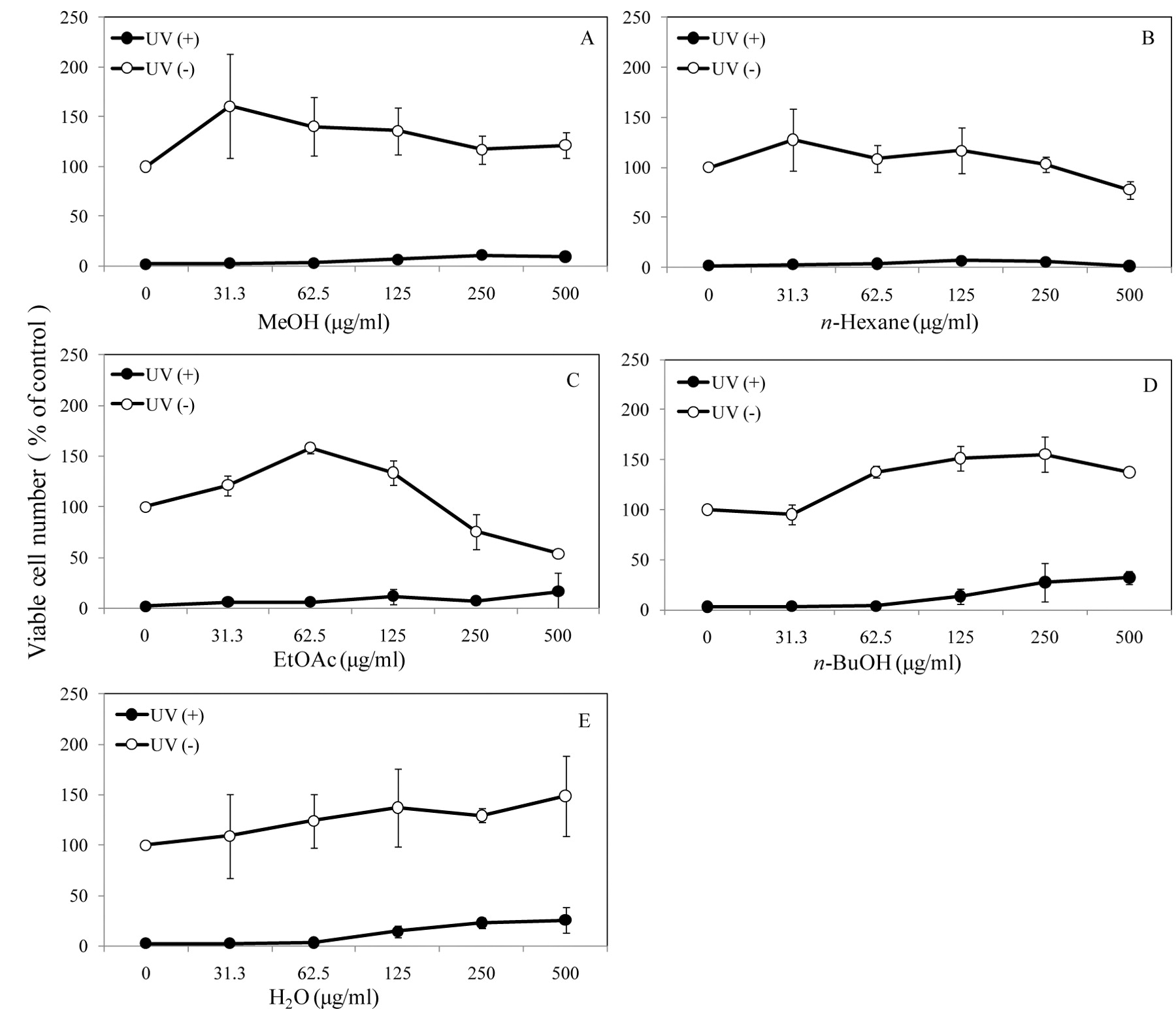
Protection of UV-induced cytotoxicity by Odm. Harvengtense ‘Tutu’ extracts. HSC-2 cells were exposed to UV irradiation (6 J/m2/min, 1 min) in 0.1 ml of PBS(−) that contained different concentrations of MeOH extract (A), or n-hexane (B), EtOAc (C), n-BuOH (D) or H2O (E) fractions of the MeOH extract of the bulb of Odm. Harvengtense ‘Tutu’. Cells were then incubated for 48 hours in 0.1 ml of fresh medium (DMEM+10% FBS) to determine the viable cell number (expressed as a % of control cells not exposed to UV irradiation). Each value represents the mean±S.D. of triplicate assays.
Effect on NO production by macrophage-like cells. RAW264.7 cells (6×10⁴ cells/ml, 0.1 ml/well) were inoculated into 96-microwell plates, and incubated for 3 hours. The medium was then replaced with phenol red-free DMEM containing 10% FBS and the indicated concentrations of test samples. After incubation for 24 hours, the NO released into the culture supernatant was measured by the Griess method. From the dose–response curve, the 50% effective concentration (EC50) was calculated. After removing the medium, the attached cells were stained with MTT reagent to determine the CC50, as described above. The selectivity index (SI) for the inhibition of NO production was determined by the following equation: SI=CC50/EC50 (11).
Osteoclastogenesis in cell culture. The RAW264.7 cells were suspended in α-MEM supplemented with 5% FBS and seeded at 2×10³ cells/well in 96-well plates (0.1 ml/well) in the presence or absence of RANKL (10 ng/ml) with or without test extract. After 4 days in culture, tartrate-resistant acid phosphatase (TRAP) activity of the medium was determined, and TRAP staining of the cells was performed.
Measurement of TRAP activity and TRAP staining. The culture media (30 μl) were incubated for 30 min at 37°C with 30 μl of 600 mM sodium acetate buffer (pH 5.5) containing L-ascorbic acid (17.6 mg/ml), sodium tartrate dehydrate (9.2 mg/ml), disodium 4-nitrophenylphosphate (3.6 mg/ml), Triton X-100 (0.3%), EDTA (6 mM), and NaCl (600 mM). The reaction was terminated by addition of 30 μl of NaOH (300 mM) and the absorbanced at 405 nm was measured by microplate reader. TRAP histochemical staining of the cells was performed using a leukocyte acid phosphatase kit (Sigma-Aldrich). The cultured cells were fixed with 100% MeOH for 1 min at room temperature and air-dried, then stained for TRAP activity.

Inhibition of NO production by MeOH extracts of Odm. Harvengtense ‘Tutu’ in LPS-stimulated RAW264.7 cells. RAW264.7 cells were incubated for 24 hours with the indicated concentrations of MeOH extract of the bulb of Odm. Harvengtense ‘Tutu’ (A), or n-hexane (B), EtOAc (C), n-BuOH (D), or H2O (E) fraction of the MeOH extract in the presence or absence of LPS (100 ng/ml). The viable cell number (●, with LPS; ○, without LPS) and extracellular NO concentration (bar) were then determined by MTT and Griess methods, respectively. Data of NO production only in the presence of LPS are shown, since not all extracts induced detectable amount of NO production. Each value represents the mean±S.D. of triplicate assays.
Statistical analysis. The difference between two groups was evaluated by Student's t-test.
Results
Cytotoxicity. The MeOH extract showed the highest cytotoxicity against HL-60 cells (CC50=105 μM), followed by HSC-4 (CC50=173 μM) > HSC-3 (CC50>210 μM) >HSC-2 (CC50>225 μM) (Table I). On the other hand, human normal oral cells (HGF, HPC, HPLF) showed lower sensitivity to the MeOH extract (CC50>224 μM), yielding a tumor-specificity index (TS) of 1.3 (Table I).
The MeOH extract of the bulb of Odm. Harvengtense ‘Tutu’ was separated into n-hexane, EtOAc, n-BuOH and H2O layers by partition with organic solvents (Figure 2). The EtOAc fraction showed the highest cytotoxicity, and the highest tumor-specificity (TS=2.8), followed by n-hexane (TS=1.5) > n-BuOH and H2O fractions (TS=1) (Table I). It should be noted that the order of sensitivity to the EtOAc fraction was again in the same order: HL-60 (CC50=14.3 μM) >HSC-4 (CC50=27.3 μM) >HSC-2 (CC50=53.9 μM) >HSC-3 (CC50=56.3 μM) >normal cells (HGF, HPC, HPLF) (CC50=74.6-122 μM) (Table I).
UV protection. One-minute exposure of HSC-2 cells to UV irradiation induced irreversible cell death after 48 hours incubation (indicated by black symbols in Figure 3). Addition of n-BuOH (Figure 3D) or H2O (Figure 3E) fraction to the irradiation buffer significantly protected the cells from the cytotoxicity induced by UV irradiation. The UV protective effect of n-BuOH or H2O fraction was increased with increasing concentrations up to 500 μg/ml, where no cytotoxicity was observed. The MeOH extract, n-hexane and EtOAc fractions displayed no UV protective effects (Figure 3A, B and C, respectively).

Effects of MeOH of Odm. Harvengtense ‘Tutu’ on osteoclast differentiation. RAW264.7 cells were cultured in the presence or absence of RANKL with the indicated concentration of MeOH extract, or n-hexane, EtOAc, n-BuOH, or H2O fraction of the MeOH extract. After 4 days in culture, TRAP activity of the medium and TRAP staining were performed. A: Representative morphology of the TRAP stained cells cultured with the indicated concentration of samples (μg/ml) with or without RANKL. B: TRAP activity of the medium with or without RANKL. All samples contain 0.2% DMSO as a vehicle control. The results are expressed as the mean±S.D. of quadruplicate assays. **p<0.01.
Cytotoxic activity of Odm. Harvengtense ‘Tutu’ extracts against cultured human tumor and normal cells. Cells were incubated for 48 hours with various concentrations of test samples, and CC50 and TS were determined as described in the Materials and Methods. Each value represents mean±S.D. of three independent experiments.
Inhibition of NO production in LPS-activated RAW264.7 cells by Odm. Harvengtense ‘Tutu’ extracts. Cells were incubated for 24 hours with various concentrations of test samples, and CC50, EC50 and SI values were determined from the data of Figure 4 as described in the Materials and Methods.
NO production by macrophages-like cells. Treatment of RAW264.7 cells with LPS (100 ng/ml) significantly elevated the extracellular concentration of NO from the background level to 8-16 μM (Figure 4), indicating the stimulation of NO production via induction of inducible NO synthase (iNOS) expression (12). Simultaneous addition of the EtOAc fraction (Figure 4C) inhibited the LPS-stimulated NO production (EC50=8.4 μg/ml, CC50=76.0 μg/ml, SI=9.0; Table II). The n-hexane (Figure 4B) and n-BuOH (Figure 4D) fractions also inhibited the LPS-stimulated NO production, but to a lesser extent (TS=6.0, >5.1). On the other hand, crude MeOH extract (SI=3.3) and H2O fraction (SI>1.1) showed much less or no inhibitory activity of NO production (Table II).
Effect on osteoclastogenesis. Treatment of RAW264.7 cells with RANKL stimulated the production of osteoclasts (Figure 5A), whereas the MeOH extract and its fractions were inactive in osteoclastogenesis (Figure 5B, upper panel). On the other hand, the EtOAc fraction significantly (p<0.01) inhibited the RANKL-stimulated osteoclastogenesis at higher concentrations (16 or 80 μg/ml; Figure 5B, lower panel). Other fractions and MeOH extract were inactive (Figure 5B, lower panel).
Discussion
The present study demonstrated for the first time that MeOH extracts of the bulb of Odm. Harvengtense ‘Tutu’ showed several new biological activities. As compared with Oda. Marie Noel ‘Velano’ bulbs, most of the biological activities were concentrated into the EtOAc fraction of MeOH extract of Odm. Harvengtense ‘Tutu’ bulbs. The EtOAc fraction of ‘Tutu’ showed higher cytotoxicity against human tumor cell lines (three oral squamous cell carcinomas and one promyelocytic leukemia) than against three human normal oral cells, yielding a TS value of 2.8 (Table I).
The present study also demonstrated that the EtOAc fraction inhibited both NO production by LPS-activated RAW264.7 cells (Figure 4, Table II) and RANKL-stimulated osteoclastogenesis (Figure 5). This suggests that the EtOAc fraction contains anti-inflammatory substance(s). This inhibitory activity was not due to the cytotoxic activity of this fraction, since the CC50 was much higher than the effective concentration (Table II and Figure 5). Further fractionation is necessary to separate the antitumor substance(s) from cytotoxic substance(s).
Acknowledgements
We are grateful to Nichirei Garden Corporation, Japan, for the kind supply of Odontoglossum Harvengtense ‘Tutu’.
- Received December 18, 2010.
- Revision received February 22, 2011.
- Accepted February 23, 2011.
- Copyright © 2011 International Institute of Anticancer Research (Dr. John G. Delinassios), All rights reserved
In this issue


Jump to section
Related Articles
Cited By...
- Cytotoxic Components Against Human Oral Squamous Cell Carcinoma Isolated from Andrographis paniculata
- Search of New Cytotoxic Crude Materials Against Human Oral Squamous Cell Carcinoma Using 1H NMR-based Metabolomics
- In Search of New Biological Activities of Isolates from Odontoglossum Harvengtense 'Tutu'
- Pilot Clinical Study of Sasa senanensis Rehder Leaf Extract Treatment on Lichenoid Dysplasia