


Article Figures & Data
Figures
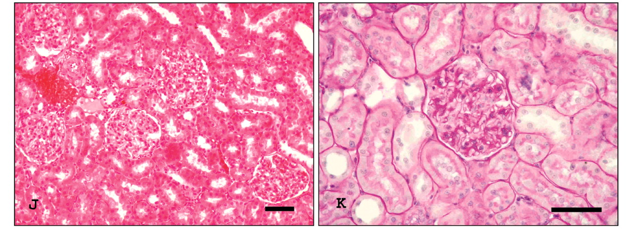
- Figure 1.
Representative photomicrographs of morphologic changes in rat kidneys. A, B: Control kidney. C, D: Irradiated rat kidneys at 8 weeks, showing glomerular tuft capsular adhesion, thickening of the basement membrane Bowman's capsule in glomeruli (arrowhead) and ballon-like cellular degeneration, cytoplasmic vacuolization and loss of luminal brush border in tubules (arrow). G, H: Mesangial sclerosis (curved arrow), segmental mesangiolysis (asterisk) and tubular atrophy (X) at 24 weeks postirradiation. E, F, J, K: Amifostine-pretreated rat kidneys at the relevant time point, showing marked reduction of glomerular and tubular damage. (A, C, G, E, J; H&E, B, D, H, F, K; PAS) Scale bar: 50 μm.
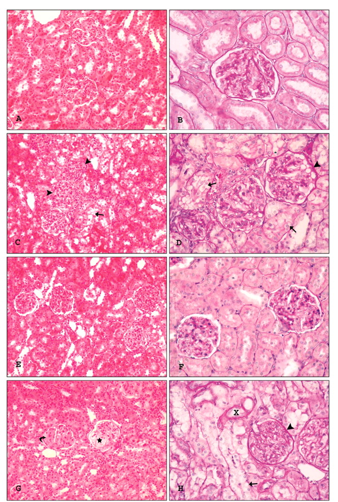
- Figure 2.
Representative photographs of Masson trichrome staining in the experimental groups. A: Control kidney. B: Irradiated rat kidneys at the 8 weeks (B) and 24 weeks (D) postirradiation. Interstitial fibrosis with inflammatory cell infiltrate (arrowhead) and increase of matrix in glomeruli (i.e., glomerulosclerosis; arrow) were observed in the irradiated kidneys. C, E: Fibrotic lesions were prominently ameliorated in amifostine-pretreated rat kidneys at the relevant time point. Scale bar: 100 μm.
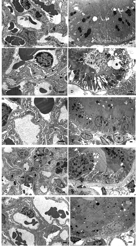
- Figure 3.
Representative electron micrographs of ultrastructural glomerular and tubular changes in rat kidneys. A, B: Control kidney, showing normal glomerular and tubular architecture. C, D: 15 Gy single-dose irradiated rat kidney at 8 weeks, showing focal fusion foot processes of podocytes (arrowheads) and vacuolization of the cytoplasm (asteriks), expansion in basal infoldings (curve arrows), swelling of epithelial cell and partial loss of luminal brush border (white arrow) in tubule cells. G, H: At 24 weeks postirradiation, kidney showing glomerular capillary wall thickening with subendothelial expansion (arrow), endothelial cell edema (E) and collapse of capillary loops (C) and tubular atrophy with thickening basement membrane (white arrowhead). E, F, J, K: Amifostine-pretreated rat kidney at the relevant time point, showing markedly improvement in tubular and glomerular ultrastructural lesions. Uranyl acetate and lead citrate. Scale bar: 1 μm.


Tables


In this issue


Jump to section
Related Articles
Cited By...
- No citing articles found.