


Abstract
Based on previous investigations on several hormones, 17α-hydroxyprogesterone (17α-HOPRG) was studied in respect to cancer initiation by its metabolites resulting from electron emission. The emission of electrons (e−aq) from its singlet excited state of 17α-HOPRG and HPLC-analysis of products were studied. Possible carcinogenicity of metabolites originating from 17α-HOPRG and the effect of progesterone (PRG) in this respect were studied in vitro. The results showed that 17α-HOPRG is very sensitive towards oxygen. The highest Q(e−aq) values were obtained by dissolution and UV-irradiation of substrate in airfree media. 17α-HOPRG metabolites showed a strong anticancer activity, which is, however, lower compared to that of PRG-metabolites. Mixture of both hormones, 17α-HOPRG and PRG, in respect to carcinogenicity showed a synergistic effect of PRG on 17α-HOPRG. Reaction mechanisms are presented.
It was recently discovered that sexual hormones, such as 17β-estradiol (17βE2), progesterone (PRG) (1) and testosterone (2), as well as other hormones and the phytohormone genistein (3-5), can emit electrons (e−aq) from their excited singlet state. The e−aq (solvated electrons) represent thermalized electrons (<10 eV energy), which are complexed with the positive dipoles of the surrounding water molecules. The formation of the salvation shell of the electrons occurs in ~10−12 sec. e−aq represents the basic form of the H atoms. It was also found that e−aq are partially scavenged by hormone molecules in ground state (reaction rate constants, k=108 to 3×10¹⁰ lmol−1s−1). The main share of e−aq, however, is transmitted to the brain receiving centres and subsequently to other biological systems (6). Obviously, the hormones act as ‘electron mediators’, communicating with each other and with various biological systems in the organism through electron transfer processes. Thereby, it was established that the yield of emitted e−aq strongly depends on the molecular structure and functional groups of the hormone molecules, as well as on the concentration of the given compound, pH, excitation energy and temperature. The highlights of numerous papers in this respect are presented in a review (7).
The short-lived hormone transients resulting from the electron emission process can react with compounds present in the medium, forming a number of metabolites depending on the hormone structure (4, 6). The determining factor for generation of a metabolite with carcinogenic properties is the reaction partner of the hormone-transient. Such a compound could originate from the environment, nutrition and certain impurities in drinking water, etc. The formation, determination and action of carcinogenic hormone metabolites is an important subject matter of studies in the last two decades (4, 5, 8-13).
The aim of the present work was an attempt to gain a deeper insight into the complicated reaction mechanism involved in the electron emission from 17α-hydroxyprogesterone (17α-HOPRG) and subsequent generation of metabolites and examination of their possible involvement in the initiation of cancer. 17α-HOPRG (17α-hydroxypregn-4-ene-3,20-dione; 4-pregnen-17α-ol-3,20-dione) is frequently designated as a metabolite of PRG. However, it possesses biological properties of a typical hormone, hence it is accepted as such.
Materials and Methods
UV-irradiation and electron emission. All solutions were prepared with highest purity chemicals (>99%, Sigma-Aldrich, Vienna, Austria) and triple-distilled water. Since 17α-HOPRG is not completely soluble in water, it was dissolved in a mixture of 40% water and 60% ethanol. Solutions of 1×10⁻⁵ to 5×10⁻⁵ mol/l 17α-HOPRG (pH~7,4) were UV-irradiated in the presence of 1×10⁻² mol/l chlorethanol used as specific scavenger of e−aq. Experiments showed that 17α-HOPRG is partly oxidized during dissolving in aerated water-ethanol mixture at room temperature. In order to examine this effect in respect to the electron emission process, two series of experiments were performed. Series A: The substrate and the electron scavenger were dissolved in aerated water-ethanol mixture. The solution was then saturated with argon for about 30 minutes in the irradiation vessel (Figure 1) and UV irradiated. Series B: The solvent mixture with added chlorethanol was first bubbled with argon in the irradiation vessel and then the substrate was added under continuous passing of argon. After dissolving of the substrate, UV-irradiation was started. Samples were taken after each desired UV-dose absorbed and analyzed. As a source for monochromatic UV-light (λ=254 nm; 4.85 eV/hν) a low-pressure Hg-UV lamp (HNS 12, OSRAM, 12 W) with incorporated VYCOR-filter for elimination of the 185 nm line was used in a special 4π-geometry irradiation double-wall vessel (Figure 1) (14). The desired temperature of the solution was kept constant during the experiment by a thermostat. The intensity of the lamp (I0=1×10¹⁸ hν ml−1 min−1), was determined by means of monochloric acetic acid as actinometer (15). The electrons (e−aq) emitted by 17α-HOPRG were scavenged by 1×10⁻² mol/l chlorethanol, where:
Analysis of hormone solution of both series before and after UV irradiation were performed by HPLC-method (Hewlett-Packard model 1046/1050; programmable detectors for fluorescence HP 1046; electrochemical measurements HP 1049 and absorption HP1100 with computer on line). Several columns were applied, but the most appropriate was Hypersil ODS 5 μm, 40×125 mm; injection sample: 15 μl, solvent mixture of 10% acetonitrile, 50% methanol and 40% water; flow rate: 1 ml/min.
Experiments in vitro. A Gammacell 220 (Nordion Corp., Canada) was used as a radiation source for production of free radicals. Its dose rate (Gy/min) was determined and controlled by modified Fricke dosimeter (17). The aim of the experiment was to investigate possible carcinogenicity of the metabolites from 17α-HOPRG, PRG and the mixture of both under the action of free radicals. As a model for living systems, Escherichia coli bacteria (AB1157) in aqueous media containing 4×10⁻² mol/l ethanol for better solubility of the hormones (pH~7.4) were used. The handling of the bacteria and the evaluation of the data have been previously reported (5). For purpose of comparison, 17α-HOPRG and PRG (each 2.5×10⁻⁵ mol/l) were applied as well as a mixture of both hormones. The toxicity of the media under these conditions amounted to 24% (N/N0-ratio=0.24, where N0=number of bacteria colonies before and N=colonies after treatment). Under these conditions the experiments in vitro were performed at various doses (Gy).
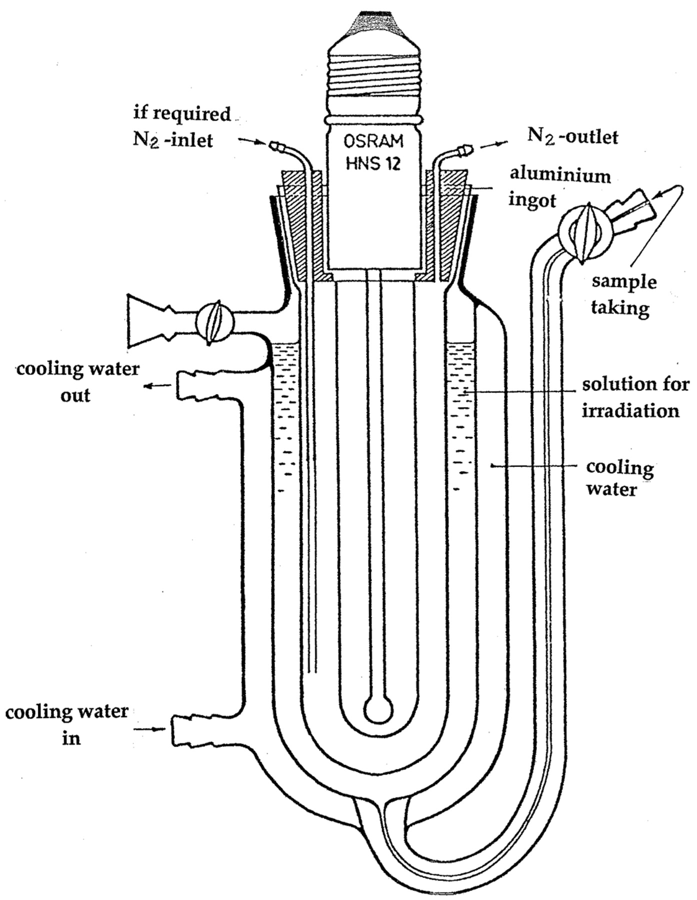
Irradiation apparatus for solutions using low pressure Hg lamp with incorporated VYCOR filter emitting monochromatic UV-light with λ=254 nm in 4π-geometry.
Results
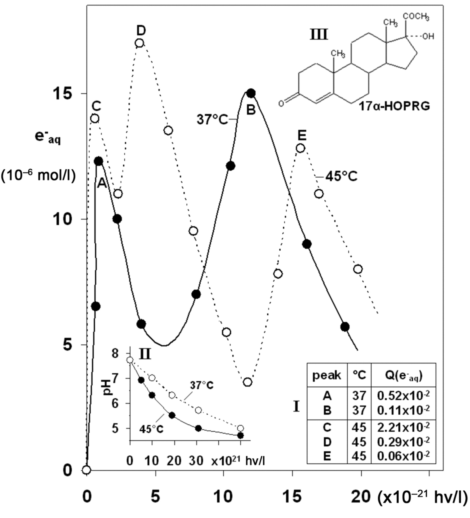
Electron emission. The emission of electrons from 17α-HOPRG was studied in a relatively wide range of substrate concentration as a function of absorbed UV dose. In Figure 2 the yield of e−aq (mol/l) emitted from 5×10⁻⁵ mol/l 17α-HOPRG (pH~7.4) for two series of experiments: at 37°C and 45°C, following series B procedure, is presented in dependence of absorbed UV quanta (hν/l). The observed curve at 37°C exhibits two maxima whereas at 45°C three maxima were registered within the same UV dose range. This indicates that the products resulting from the hormones are likewise able to emit electrons. The determined quantum yields, Q(e−aq), are given as inset I, Figure 2. The yield of emitted electrons decreases with UV dose at both temperatures. This effect was previously observed for 17βE2, PRG (1) as well as for TES (2) and was explained by formation of hormone associates (unstable complexes), which have poor solubility. Thereby a part of the emitted e−aq is reacting with substrate molecules in ground state. For example the reaction rate constant of PRG in order of k (e−aq + PRG) ~4×10⁹ lmol−1 s−1 (6). Simultaneously, with proceeding electron emission, a pH decrease of the medium occurs (inset II, Figure 2). This gives a hint to the reaction mechanism, expressed by equations 4 and 5. Similar observations were made upon the e−aq emission using higher 17α-HOPRG concentrations under the same experimental conditions. However, with increasing substrate concentration, the course of the observed electron emission curves indeed show three maxima, but the Q(e−aq) values decrease as shown in inset I, Figure 2 and Table I. Data obtained according to the working procedure series A and series B are given for comparison in Table I. Obviously, in the second case, the Q(e−aq) yields are much higher, demonstrating the oxygen effect during dissolving of the substrate in aerated media (series A). As expected, e−aq-yields were higher at 45°C .

Electron emission of 5×10⁻⁵ mol/l 17α-HOPRG, dissolved and UV-irradiated in an air-free mixture of 40% water +60% ethanol (pH~7.5), as a function of absorbed dose (hν/l) at 37°C 45°C. Inset I: Q(e−aq) values at the corresponding maxima according to series B procedure. Inset II: pH-change of the solvent mixture in dependence of absorbed UV dose at 37°C and 45°C. Inset III: structural formula of substrate.
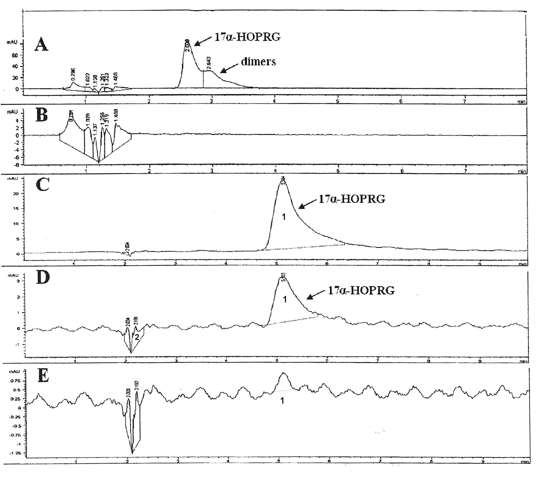
HPLC analysis. In order to examine the stability of 17α-HOPRG in aerated and air-free solvent mixture, as well as to provide the formation of metabolites, HPLC-analysis of unirradiatied and irradiated samples according to series A and B were performed. Surprisingly, it was observed that even during the dissolving process in aerated media and storage of the solutions for about 15 minutes at room temperature, oxidation of the substrate was remarkable, as shown in Figure 3, chromatogram (A). Under these conditions, about 48% 17α-HOPRG is oxidized resulting in the formation of various products. Their identification was not of interest in the frame of this work. Chromatogram (B) demonstrates complete photolysis of the hormone by an absorbed dose of 1.5×10²¹ hν/l. Based on these results, series B of experiments was performed (see Table I). Some results are presented in Figure 3, chromatograms C to E.
Q(e−aq) values obtained from various 17α-HOPRG concentrations in solvent mixture of 40% water and 60% ethanol (pH~7.4) at 37°C and 45°C. Series A: Substrate dissolved in aerated solvent, but UV irradiated in air-free media; Series B: Substrate dissolved and UV irradiated in air-free solvent mixture.
Experiments in vitro. The survival curves (N/N0 ratio) presented as a function of absorbed radiation dose, Gy (1 Gy=6.24×10¹⁵ eV/g absorbed energy) for each individual system are mean values of several series of experiments and are shown in Figure 4. Thereby the calculated ΔD37 values represent the radiation dose (Gy) at which N/N0=0.37, and were taken from the corresponding curves. The ΔD37(Gy) data were obtained by subtracting D37 buffer value from each measured individual D37 value, e.g. D37(Gy) sample–D37(buffer)=ΔD37(Gy) sample. The obtained positive ΔD37(Gy) data characterize the radiation protection ability of the system (scavenger of free radicals), whereas the negative ones indicate the antitumor property of the system. The corresponding data of the investigated systems are summarized and shown as an inset in Figure 4.

HPLC-chromatograms of 5×10⁻⁵ mol/l 17α-HOPRG dissolved in 40% water +60% ethanol. A: Sample dissolved in aerated media; unirradiated. (1) 17α-HOPRG, 52% remainder; (2) dimers, 18%; degradation products, 3%. B: Same solution first saturated with argon, then UV-irradiated (dose: 1.5×10²¹ hν/l). Substrate completely converted into products. C: Sample dissolved in air-free media (in the irradiation vessel); (1) substrate unchanged. D: Immediately UV irradiated (dose: 3.8×10²¹ hν/l); (1) 17α-HOPRG-remainder, 10.3%; (2) products. E: Irradiated sample (dose: 7.7×10²¹ hν/l); (1) 0.6% substrate remainder; (2) products.
Discussion
The observed oxidation of 17α-HOPRG during dissolving in aerated solution at room temperature shown by HPLC-analysis (Figure 3) is important for the handling of the substance. The Q(e−aq) decrease with increasing substrate concentration (Table I) is typical for all hormones investigated to date (1-6). The effect is explained by formation of associates (unstable complexes) in the ground state, which consume a part of the emitted e−aq. Figure 3 demonstrates also the fact, that during UV-irradiation a photolysis of the hormone takes place under emission of e−aq. This process is followed by a subsequent formation of metabolites. Based on previous results (1, 7, 18) it is assumed that the electron emission of 17α-HOPRG originates from ring A, leading to formation of a
 The resulting transients, R1•+ and R2 are highly reactive. Depending on the substances present in the media, they can lead to formation of metabolites with different biological properties. Simultaneously, the radical cation, R •+1, can regenerate 17α-HOPRG by reaction with water, as previously observed with β-carotene cation (18).
 The existence of the reactions shown in equations 4 and 5 is supported by the observed pH change of the media, occurring simultaneously with the electron emission (inset II, Figure 2).
A part of the emitted e−aq can be scavenged by carbonyl groups on 3- or 17-positions, resulting in the corresponding electron adducts, which can scavenge transients present in the actual media, as in the present case with the ethanol radical produced according to (equation 6):
 The aforementioned hormone free radicals (R •+1, R2) are also highly reactive species, leading to metabolite formation with the available •C2H4OH radicals:
 In the case that the reaction partner of the hormone-free radicals (R •+1, R2 etc.) in the organism is a polycyclic aromatic hydrocarbon, such as pyrene, the resulting metabolite will very likely initiate cancer (19). It might be mentioned that the metabolites are also able to eject electrons when excited in singlet state, appearing as second and third e−aq maxima shown in Figure 2 and Table I.
In organisms, reducing and oxidizing free radicals are permanently generated and consumed, and are responsible for dynamic biological processes (19). These in vitro experiments give an insight into the action and consequences of free radicals with 17α-HOPRG. For comparison, experiments with PRG and mixtures of both were also performed.
Under the given conditions the primary radiolytic products of air-free water (pH=6-8) are presented by cross reaction (equation 8), where the yields (G-values; number of species produced by 100 eV absorbed energy) are shown in brackets.
Simultaneously, the radiolysis of ethanol leads to formation of e−aq (solvated electron; G=2.0) in addition to several organic free radicals (20). Summing up, it can be stated that under the given conditions, the bacteria were attacked mainly by e−aq and radicals originating from ethanol radiolysis mostly by CH3•CHOH. Both types are reducing species. The obtained ΔD37(Gy) values have negative signs (Figure 4, insert). This fact indicates that the resulting 17α-HOPRG transients, similarly to those of PRG, have strong anticancer activity. However, the individual biological capacity of PRG in this respect is more than six-times higher in comparison to that of 17α-HOPRG. The PRG plus 17α-HOPRG mixture results in approximately the mean value of the corresponding ΔD37(Gy) data, indicating a synergistic action of 17α-HOPRG by PRG.

Survival curves of E.coli bacteria (AB1157): N/N0 ratio as a function of absorbed γ-irradiation dose (Gy) in air-free aqueous solution containing 4×10⁻² mol/l ethanol (pH~7.4) in the presence of: (A) buffer, (B) 5×10⁻⁵ mol/l 17α-HOPRG, (C) 5×10⁻⁵mol/l PRG and (D) 2.5×10⁻⁵ mol/L 17α-HOPRG + 2.5×10⁻⁵ mol/l PRG. Insert: D37(Gy) and ΔD37(Gy) values of the given systems.
Conclusion
The results of the present investigations on 17α-HOPRG embrace several subjects of biological importance in addition to a hormonal pathway of cancer initiation. The data can be summarized in the following points: 17α-HOPRG is rather sensitive towards oxygen in aerated solution. The oxygen effect depends on the temperature and duration of exposure. 17α-HOPRG is able to emit electrons (e−aq) from its excited singlet state in polar media like previously reported hormones (e.g. 1-6). The Q(e−aq) yield decreases with increase of substrate concentration, because of “associate” formation, where the hormone molecules in ground state consume a part of the ejected e−aq. The electron emission curves are passing several maxima, indicating that the generated metabolites also possess the ability to emit e−aq. HPLC-analysis proved that metabolites originating from 17α-HOPRG-transients and in media, available free radicals (in the present case, •C2H4OH, see equation 7) can likewise emit electrons, but with essential lower yield (Table I). This finding is of biological importance, showing that electron emission can still occur, even under a modified molecular structure of the hormone. The calculated ΔD37(Gy) values resulting from experiments in vitro using PRG, 17α-HOPRG and mixture of both in air-free media demonstrate that: (i) PRG metabolites show the highest antitumor activity, (ii) a similar effect is observed by 17α-HOPRG, but is six times lower, (iii) the mixture of PRG and 17α-HOPRG features the same property but showing a mean value of both. This fact indicates a synergistic effect of PRG on 17α-HOPRG in respect to formation of less carcinogenic metabolites. Possible reaction mechanisms for elucidation of the observed effects are also presented.
Acknowledgements
The authors greatly appreciate the financial support by FWF, Austria Science Fund, which enables to perform the research project “Free Radical Action on Sexual Hormones in Respect to Cancer”, contract No.P21138-B11.
- Received April 13, 2010.
- Revision received May 21, 2010.
- Accepted May 27, 2010.
- Copyright © 2010 International Institute of Anticancer Research (Dr. John G. Delinassios), All rights reserved
In this issue


Jump to section
Related Articles
Cited By...
- No citing articles found.