


Abstract
Novel atherosclerotic lesions were induced in the Microminipig (MMP, registered with the Japanese Ministry of Agriculture, Forestry and Fisheries as a novel variety of swine), the smallest pig available for experimental use, by feeding a high fat (12%) and high cholesterol (5%) diet (HFCD) with sodium cholate (SC, 0.7%) (HFCD/SC) for three months. Three MMPs were used: a male fed with normal diet (M-ND), and a male and an ovariectomized female fed with HFCD/SC (M-HFCD/SC and Fx-HFCD/SC). HFCD/SC induced hypercholesterolemia accompanied by an increase in serum total cholesterol (T-Cho), low-density lipoprotein cholesterol (LDL-C), high-density lipoprotein cholesterol (HDL-C) and cholesterol ester (CE) from the first week. Serum levels of T-Cho, LDL-C and CE reached a maximum in two to three weeks, and HDL-C gradually increased during the experimental period (duration). Serum lipoprotein analysis showed a dominant LDL-C fraction, as seen in humans, in all three MMPs. Body weight gain in the MMPs fed with HFCD/SC was greater than in the animal fed with M-ND. At the end of the experiment, computed tomography scans of conscious animals showed increases in subcutaneous and abdominal fat in those fed with HFCD/SC, suggesting the induction of obesity. Atherosclerotic lesions in systemic arteries (including external and internal iliac arteries, abdominal aorta, coronary artery, cerebral arterial circle), fatty changes, and foamy cell infiltration in the liver and spleen were histopathologically observed in the MMPs fed with HFCD/SC. Atherosclerosis and the pathological findings induced by HFCD/SC in MMPs were similar to the pathological changes associated with human atherosclerosis, suggesting that the MMP has the potential to be a suitable animal model for human atherosclerosis.
Atherosclerosis is the predominant contributor to mortality (approximately half of all deaths) and serious morbidity in the Western world (1). Westernization of the life-style, including diet, may account for the increased incidence of coronary and cerebral artery diseases (2-4) in Japan in recent years. This increase is strongly related to atherosclerotic lesions, the pathophysiology of which must be studied if the incidence is to be reduced. Accordingly, a number of models of atherosclerosis have been developed in animals such as the mouse, rabbit, and swine. Atherosclerosis is influenced by several genetic and environmental factors, and mice lacking apolipoprotein E (5, 6) and Watanabe heritable hyperlipidemic (WHHL) rabbits lacking low-density lipoprotein receptor (7, 8) have been reported as models of atherosclerosis with genetic abnormality. Nutritional manipulation by feeding high fat and high cholesterol has been used to develop animal models of atherosclerosis, including of swine (3, 9).
The wild boar, Sus scrofa, is said to be the ancestor of all modern breeds of swine, and although there is evidence of domestication in Europe some 3500 years ago, the cradle of the domesticated swine has been claimed as being China about 10000 years ago (10). Swine have been used extensively in biomedical research with a significant increase in recent decades (10), more than 60000 pigs having been used in a year in the EU (11). Because of their well-accepted physiological and anatomical similarities to humans, swine are becoming increasingly attractive toxicological and pharmacological models. Recently, minipigs, smaller than domestic swine, have been developed and there are several strains of these (Clawn, Göttingen, Yucatan, Sinclair, Hanford, and others) (12, 13). In general, in the minipig, atherosclerosis can be induced simply by control of diet, while this cannot be done in the mouse nor rabbit. Therefore, the minipig is thought to be an apposite experimental animal for research into atherosclerosis. However, many strains of minipigs are difficult to manage due to their large size, e.g. the Göttingen has an adult body weight of 30-40 kg under conditions of restricted diet (11). Very recently, Fuji Micra Inc. (Brand name, Microminipig: MMP; registered with the Japanese Ministry of Agriculture, Forestry and Fisheries as a novel variety of swine) has emerged as a possible experimental animal model for non-clinical pharmacological/toxicological use. The MMP has been established as a closed colony. The body weight of young mature MMPs is less than 7 kg, enabling ease of handling. For the use of the MMP in pharmacological experiments, enzyme activities of drug metabolism in the liver have been measured (14).
In this study, we first report atherosclerotic lesions in the MMP induced by feeding a high fat and high cholesterol diet. The results show the potential of this strain as a useful animal model for understanding the pathophysiology of human atherosclerosis.
Materials and Methods
Animals. MMPs aged three months were obtained from the breeder (Fuji Micra Inc., Shizuoka, Japan). We used two males and an ovariectomized female (Fx). The Fx animal was ovariectomized under inhalation anesthesia using sevoflurane two days before the start of the experiment. The animals were maintained in a filtered air laminar flow in a special room at Kagoshima University. The room temperature was maintained at 24±3°C and relative humidity at 50±20%, with a 12 h light/dark cycle. Tap water was available ad libitum. Food, 3% of body weight (BW)/day, was supplied twice a day and BW was measured once a week. One male was given normal diet (ND, Kodakara 73; Marubeni Nisshin Feed Inc., Tokyo, Japan), designated as M-ND. According to the maker, ND was composed of less than 9.0% (w/w) carbohydrate, greater than 15.0% (w/w) protein, greater than 2.0% (w/w) fat, and less than 9.0% fiber. The other male and the female were fed with a special diet for three months. The special diet was formed from a mixture of lard (refined lard, 12% w/w; Miyoshi Oil & Fat Co., Ltd., Tokyo, Japan) and cholesterol (5% w/w; Wako Pure Chemical Industries, Ltd., Osaka, Japan), and sodium cholate (SC, 0.7% w/w; Wako Pure Chemical Industries, Ltd) with ND. This special diet was designated as a high fat and high cholesterol diet with SC diet (HFCD/SC). The male and the ovariectomized female fed with HFCD/SC were designated as M-HFCD/SC and Fx-HFCD/SC, respectively. The use of animals in this research complied with all relevant guidelines set by Kagoshima University.
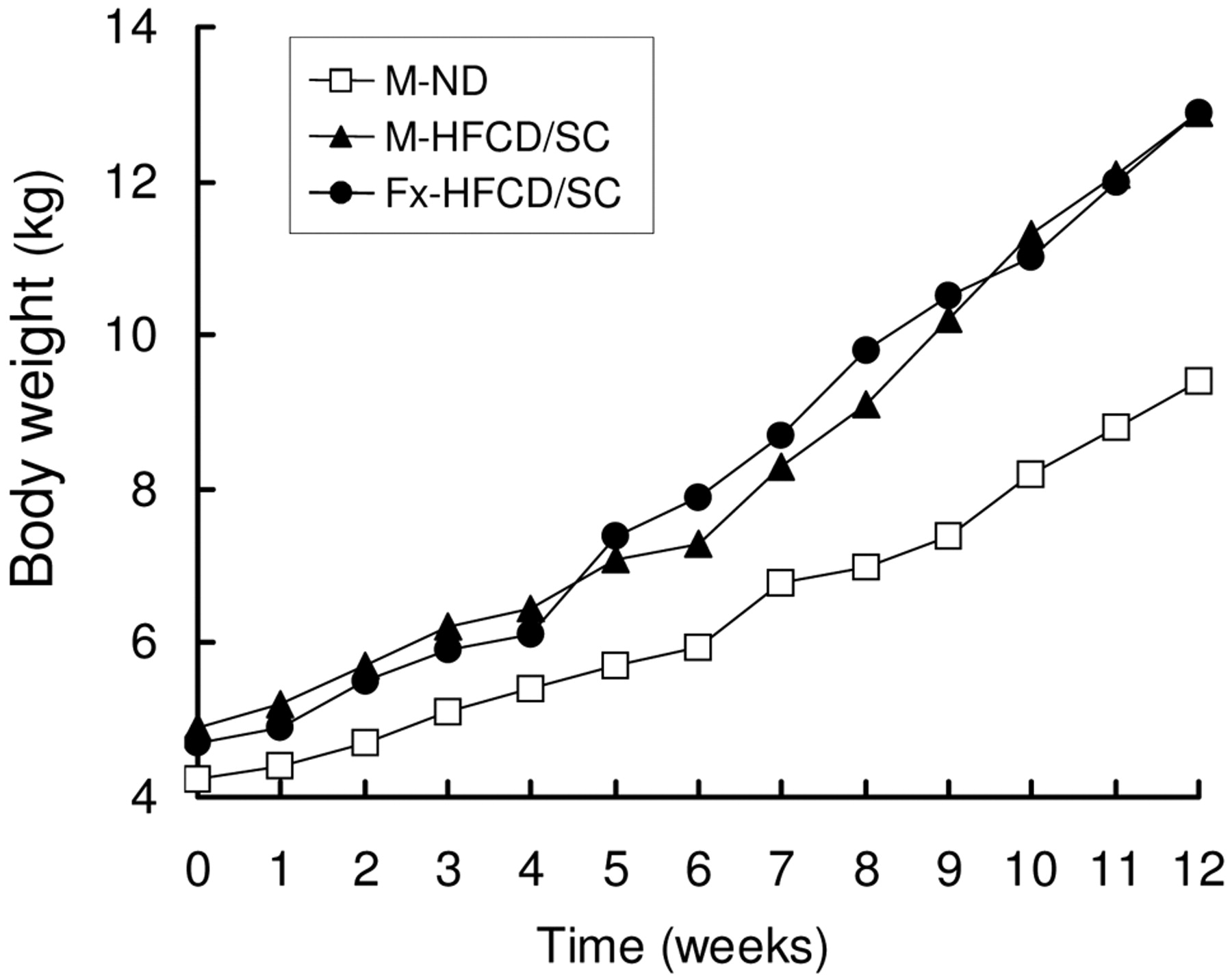
Body weight.
Blood chemistry. At the end of the experiment blood samples for hematological analysis were collected in tubes treated with heparin. White blood cell (WBC), red blood cell (RBC), and platelet (PLT) counts were determined using pocH-100iV (SYSMEX Co., Hyogo, Japan). Total protein (TP), glucose (Glu), aspartate aminotransferase (AST), alanine aminotransferase (ALT), blood urea nitrogen (BUN), creatinine (Cr), were measured using a biochemical analyzer DRI-CHEM 7000 (Fujifilm Corp., Tokyo, Japan). Serum estradiol (E2) and progesterone (Prg) were measured using mini-VIDAS (SYSMEX Co.). Blood samples for measuring biochemical parameters of fat metabolism were collected once a week. Serum was separated by centrifugation for 15 min at 3000 rpm. Total cholesterol (T-Cho), triglycerides (TG), and high-density lipoprotein cholesterol (HDL-C) were measured using DRI-CHEM 7000, as mentioned above. Low-density lipoprotein cholesterol (LDL-C) and free cholesterol (free-Cho) were analyzed at Clinical Pathology Laboratory, Ltd. (Kagoshima, Japan). Cholesterol ester (CE) was calculated as: CE=T-Cho–free-Cho. Serum cholesterol profile was determined by agarose-gel electrophoresis at Clinical Pathology Laboratory, Ltd.
Computed tomography (CT). Before the day of the necropsy, CT examinations of the thoracic, abdominal, and brain regions were performed for conscious MMPs using Toshiba Asteion Super 4 (Toshiba Medical Systems Corporation, Tochigi, Japan). In the abdominal region, a region of interest (ROI) within the liver on the CT scans of each animals was delineated and the attenuation values measured in Hounsfield units (HUs) of each ROI was obtained. For the thoracic CT, the thickness of subcutaneous fat was measured at the posterior region of the scapula.
Pathology. On the day following the final administration, all animals were preanesthetized by an intramuscular injection of ketamine (5 mg/kg), midazolam (0.2 mg/kg) and medetomidine (0.04 mg/kg) and then sacrificed by exsanguination through the bilateral common carotid arteries. At necropsy, 12 arteries [aortic arch, thoracic aorta, abdominal aorta, common carotid artery, pulmonary artery, renal artery, external and internal iliac arteries, right coronary artery (RCA), left anterior descending (LAD) artery, left circumflex (LCX) artery, and cerebral arterial circle], hearts, liver, kidneys, spleen, greater omentum, and abdominal fat, including the mesenterium, were removed from all the animals. The heart, liver, kidneys, spleen, greater omentum, and abdominal fat were weighed. All organs removed were fixed in 10% phosphate-buffered formalin (PBF), and embedded in paraffin. They were sectioned at 5 μm, stained routinely with hematoxylin and eosin (HE) and then examined histopathologically. All arterial sections were also stained with Masson trichrome (MT) and elastica van Gieson (EVG) to assess in detail changes in the elastic layers and extracellular matrix (ECM), respectively.

Biochemical parameters on fat metabolism.
Atherosclerotic arterial lesions were graded according to Stary Stage (I-VIII) (3, 15-17). Arteries, liver and spleen, fixed in 10% PBF, were embedded in OCT compound (Sakura Finetek Japan Co., Ltd., Tokyo, Japan) and frozen at −80°C. They were sectioned at 10 μm and stained with Sudan III and then examined histopathologically. For immunohistochemistry of the arteries, slides were deparaffinized in xylene and rehydrated through graded alcohol in accordance with a reported procedure (18). The primary antibodies and concentrations were as follows: mouse monoclonal HLA-DR (1:1000, clone TAL.1B5; Dako Cytomation Co., Ltd., Kyoto, Japan), mouse monoclonal α-smooth muscle actin (1:100, α-SMA; clone 1A4; Dako Cytomation Co.), mouse monoclonal vimentin (1:200, clone Vim 3B4; Novocastra Laboratories Ltd., UK), and rabbit polyclonal lysozyme (1:2000; Dako Cytomation Co.). The sections treated with primary antibody were incubated with the appropriate biotinylated secondary antibody with EnVision (Dako Cytomation Co.). Immunoreactivity was visualized with 0.075% 3,3′-diaminobenzidine tetrachloride (DAB). The sections were then washed, counter-stained, dehydrated, cleared in xylene, and mounted.

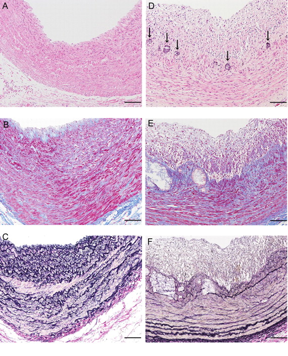
Microscopic appearance of atherosclerotic lesions in the external and internal iliac arteries. A-C: M-ND, D-F: M-HFCD/SC. The artery shows thickening of the intima and arterial media (D). All arrows show the internal elastic lamina (C, F). The thickest sections of the arteries of M-HFCD/SC show considerable foamy cell infiltration (G) and extracellular lipid accumulation (H) are observed. Fibrous cap is formed at the surface of the intima (E), at which collagen fiber proliferated (G) and elastic fiber organization is widely damaged (F). At the thickest part of the artery, hemorrhage is also observed (I). Bar=200 μm (D, I), 100 μm (A, C, E, F, G), 50 μm (B), 20 μm (H). HE stain: A, D, G-I; MT stain: B, E; EVG stain: C, F.
Results
Body weight. As shown in Figure 1, the animals fed with HFCD/SC showed higher body weight (BW) than did the M-ND. The final BW gains of M-ND, M-HFCD/SC, and Fx-HFCD/SC from 0 week (3 months old) to the 12th week were 5.15, 8.00, and 8.20 kg, respectively.
Biochemical analysis. As shown in Table I, PLT count in the animals fed with HFCD/SC was higher than that for M-ND. Glu and TP levels were similar in all three animals. Fx-HFCD/SC had higher levels of AST than M-ND and M-HFCD/SC. ALT, BUN, and Cre were similar in all three animals, and no marked liver or kidney damage from HFCD/SC was noted.
The E2 level was lower in Fx-HFCD/SC than in M-ND and M-HFCD/SC, resulting from ovariectomy; however, Fx-HFCD/SC had a higher Prg level than did M-ND and M-HFCD/SC.
Serum-related lipid metabolism parameters were analyzed (Figure 2). The animals fed with HFCD/SC had higher T-Cho, free-Cho, and CE levels than those for M-ND in the first week after HFCD/SC supplementation. At the second week, T-Cho, free-Cho, and CE levels almost reached maximum and were maintained throughout the experimental period. LDL-C followed a similar pattern to that of T-Cho, free-Cho, and CE in all three animals. Conversely, HDL-C for the animals fed with HFCD/SC was higher than that for M-ND at the first week after HFCD/SC supplementation, and gradually increased during the experimental period. TG level for Fx-HFCD/SC was higher than that for M-HFCD/SC and M-ND, and that for M-HFCD/SC was similar to that of M-ND four weeks after special diet supplementation. Serum in animals fed with HFCD/SC was grossly cloudy-white from the first week after special diet supplementation (data not shown). Serum lipoprotein analysis showed a dominant LDL-C fraction in all MMPs, as seen in humans (data not shown).
Organs. At six months of age, the animals were sacrificed and the organs weighed (Table II). The animals fed with HFCD/SC had higher body weight than that for M-ND probably due to the difference in calorie intake. Relative organ weight in the hearts and kidneys was similar in all three animals. The animals fed with HFCD/SC had a relatively higher weight distribution in the liver, spleen, greater omentum, and fat than M-ND. The animals fed with HFCD/SC showed white plaque in the abdominal aorta, internal iliac artery, pulmonary artery, renal artery, and three coronary arteries, and showed a yellow change in the liver, suggesting fatty liver.
CT. Before a day of sacrifice, the animals were examined by CT scanning under non-anesthetized conditions. In the abdominal region, the attenuation value of M-ND, M-HFCD/SC, and Fx-HFCD/SC were 81.3, 67.4,and 66.0 HU, respectively, suggesting that the animals fed with HFCD/SC suffered from fatty liver. The thickness of subcutaneous fat at the posterior margin of the scapula for M-ND, M-HFCD/SC, and Fx-HFCD/SC showed 5.2, 15.6, and 18.1 mm, respectively. Any abnormal finding of brain lesions in the animals fed with HFCD/SC was detected by CT scanning.
Evaluation of atherosclerotic lesions and pathology. As shown in Table III, the degree of atherosclerosis was evaluated by Stary classification. M-ND showed no findings of atherosclerosis in any artery examined. The animals fed with HFCD/SC had an almost similar degree of atherosclerosis in the abdominal aorta, coronary arteries, aortic arch, pulmonary artery, and renal artery. The degree of atherosclerosis in the external and internal iliac arteries and cerebral arterial circle of M-HFCD/SC was more severe than that for Fx-HFCD/SC, while that in the common carotid artery and thoracic aorta was more severe in the latter.
Blood examination at necropsy.
Absolute organ weights and relative organ weights (%) compared to body weight at necropsy.
Atherosclerosis score according to Stary classification.

Microscopic appearance of atherosclerotic lesions in the coronary artery. The artery shows the thickness of intima and arterial media (D). The thickest sections of the arteries of M-HFCD/SC show considerable foamy cell infiltration (G), and slight fibrous proliferation (E) and calcification in arterial media (D, arrows) are observed. Elastic fiber organization is also widely damaged (F). A-C: M-ND; D-F: M-HFCD/SC. Bar=100 μm (A-F). HE stain: A, D; MT stain: B, E; EVG stain: C, F.
The highest score for atherosclerosis was shown in the external and internal iliac arteries of M-HFCD/SC (Figure 3). The external and internal iliac arteries of M-HFCD/SC showed thickening of the intima and arterial media. The thickest sections of the arteries of M-HFCD/SC showed considerable foamy cell infiltration, and extracellular lipid accumulation and calcification were observed. Fibrous cap was formed at the surface of the intima, at which collagen fiber proliferated and elastic fiber organization was widely damaged. Similar atherosclerotic findings to those in the external and internal iliac artery of M-HFCD/SC were observed in the abdominal aorta of M-HFCD/SC and the common carotid artery of Fx-HFCD/SC. Atherosclerotic lesions in the coronary arteries of M-HFCD/SC and Fx-HFCD/SC were scored II-III by Stary classification (Table III, Figure 4). In particular, the cerebral arterial circle in M-HFCD/SC showed atherosclerotic lesions, scored III by Stary classification, in association with severe obstruction of the lumen (Figure 5).
Immunohistochemical examinations (Figure 6) showed positive expression of lysozyme, HLA-DR and vimentin in the subintimal areas of atherosclerotic lesions in M-HFCD/SC and Fx-HFCD/SC, suggesting that the infiltrated cells originated from macrophages. The severe atherosclerotic lesions corresponding to V and VI of Stary classification showed layered cell proliferation having positive expression for α-SMA, suggesting that the lesions were infiltrated by cells originating from smooth muscle.
In the liver, animals fed with HFCD/SC showed fatty change around the central veins and accumulation of particles stained with Sudan III in the hepatocytes (Figure 7A-D). Animals fed with HFCD/SC showed considerable foamy cell infiltration in the red pulp of the spleen (Figure 7E-H).
Discussion
The MMP, which is the smallest minipig available for experiments, showed a variety of atherosclerotic lesions induced by HFCD/SC in only three months. Atherosclerosis is the leading cause of morbidity and mortality throughout Europe and most parts of the world. Ischemic heart disease and cerebral stroke are prominently involved in atherosclerotic lesions in the relevant arteries (19). The prevention of atherosclerotic lesions is clinically essential, and an animal model for researching atherosclerosis is required. MMP showed diet-induced atherosclerotic lesions very similar to those seen in humans. The diet was composed of high fat, high cholesterol and SC. For establishment of atherosclerosis, more suitable fat, cholesterol and SC content should be re-evaluated. With a similar diet, many animals, such as rabbits and swine, have been reported to show similar atherosclerotic lesions in the external and internal iliac artery, coronary arteries, abdominal aorta, and common carotid artery (3-5, 20). However, the MMPs fed with HFCD/SC showed atherosclerosis in the cerebral arterial circle, which is related to cerebral stroke. This is an interesting finding, suggesting that the MMP is a potentially suitable animal for a cerebral stroke model based on atherosclerosis.
It is not clear whether there were sex differences in the formation of atherosclerosis because there are no data on the normal female MMP. Further experiments are required. In the present study, the degree of the atherosclerotic lesions in the Fx-HFCD/SC might have been expected to be more severe because E2 and Prg are known to play inhibitory and a promotive roles, respectively, in atherosclerosis (21-23). However, the Fx-HFCD/SC had a lower E2 level, and a higher Prg level than did the M-HFCD/SC, while the atherosclerotic lesions of the Fx-HFCD/SC was not even similar to that of the M-HFCD/SC. This may have been related to the observation period. The effect of ovariectomy on atherosclerosis should be evaluated for a longer period. However, the Fx-HFCD/SC showed atherosclerotic lesions, suggesting that the MMP is a potentially suitable animal model for human atherosclerosis in postmenopausal women (24).
The atherosclerotic lesions of the MMPs fed with HFCD/SC were evaluated pathologically. Severe lesions, which were evaluated by Stary classification, were thickening in the arterial media and stenosis of the intima from the accumulation of lipids and foam cell infiltration. Fibrous cap was formed at the surface of the intima, where elastic fiber organization was widely damaged. Immunohistochemistry confirmed that the infiltrated cells originated from macrophages and smooth muscle. These findings are similar to those in human cases (25).
T-Cho and LDL-C were increased for a short period after HFCD/SC supplementation. The increase in LDL-C preceded the increase in HDL-C. Therefore, compared with HDL-C, LDL-C is assumed to have been predominantly increased in the MMP. LDL-C metabolism in the MMP should be analyzed. TG level in the MMPs fed with HFC-SC showed greater variation during the experimental period. Atherosclerosis may be mainly influenced by cholesterol level and not by TG. This finding is similar to those in human cases (1).
The MMP is considerably smaller than other minipigs and atherosclerosis formed in a short period. Therefore, the amount of diet required to induce atherosclerosis was relatively small. This represents a cost benefit for such experiments. The animals were observed by CT scanning under non-anesthetized conditions, indicating the ease of handling of the MMP. These factors suggest that the MMP is a suitable animal model of atherosclerosis.

Microscopic appearance of atherosclerotic lesions in the cerebral arterial circle. The artery shows the thickness of intima and arterial media with severe obstruction of the lumen (stenosis: about 90%) (D). All arrows show the internal elastic lamina (C, F). The thickest sections of the arteries of M-HFCD/SC show considerable foamy cell infiltration (G). Fibrous cap is formed at the surface of the intima (E), where collagen fiber is proliferated. A-C: M-ND; D-F: M-HFCD/SC. Bar=100 μm (A-F). HE stain: A, D; MT stain: B, E; EVG stain: C, F.

Immunohistochemistry examination of atherosclerotic lesions in the external and internal iliac arteries. All arrows show the internal elastic lamina (A-H). Foamy cells infiltrated at the thickest part of the artery show positive expression of lysozyme (C), HLA-DR (D), α-SMA (G) and vimentin (H). A, B, E, F: M-ND; C, D, G, H: M-HFCD/SC. Bar=100 μm (A-H). Lysozyme: A, C; HLA-DR: B, D; α-SMA: E, G; vimentin: F, H.

Microscopic appearance of fatty liver and foamy cell accumulation in the spleen. Fatty change in centrilobular hepatocyte is observed in the liver of M-HFCD/SC (C, D). Accumulation of foamy cells including fat is observed in the red pulp of the spleen of M-HFCD/SC (G, H). A, B, E, F: M-ND; C, D, G, H: M-HFCD/SC. Bar=50 μm (A-H). HE stain: A, C, E, G; Sudan III stain: B, E, F, H.
Diet is as important a factor in atherosclerosis formation as are environmental factors, a number of which may be necessary to develop the pathology of atherosclerosis. Atherosclerosis reported in other animal models showed stable plaque (3-5, 20, 26). In human cases, the occurrence of organ damage involves a second step, such as the release of plaque and acceleration of inflammation. By using an animal model, the mechanism causing organ damage from atherosclerotic lesions can be examined. Related factors include stress from work, stimulation from changes in temperature, and arrhythmia. The swine is more suitable for analyzing the influence of environmental factors on atherosclerotic lesions than the mouse or rabbit because its feeding habits and life rhythm are similar to those of humans (10). The MMP, like normal swine, is diurnal (data not shown). It is preferable to evaluate the influence of night and day on atherosclerotic lesions in diurnal rather than nocturnal animals.
Finally, the MMP is a potentially suitable animal model for analysis of atherosclerosis for the following reasons: i) atherosclerosis in the MMP is very similar in location, pathophysiology and pathology to that in humans; ii) atherosclerosis is formed in a short period; iii) the MMP is very small and is easily handled.
Acknowledgements
This work was supported in part by the Suzuken Memorial Foundation. We are grateful to Dr. Yoshihiko Nakanishi (Kagoshima University), Mr. Masashi Omori and Mr. George Martin (Shin Nippon Biomedical Laboratories, Ltd.), Dr. Katzuhiko Itoh and Mr. Naoki Kaneko (Fuji Micra Inc.) for their advice and valuable technical assistance.
Footnotes
-
↵* These Authors contributed equally to this study.
- Received May 19, 2010.
- Revision received July 1, 2010.
- Accepted July 5, 2010.
- Copyright © 2010 International Institute of Anticancer Research (Dr. John G. Delinassios), All rights reserved
Reference
In this issue


Jump to section
Related Articles
Cited By...
- Development of a Microminipig Model of Atherosclerosis for the Evaluation of a HMGCR Inhibitor
- High Pathological Reproducibility of Diet-induced Atherosclerosis in Microminipigs via Cloning Technology
- NR6A1 Allelic Frequencies as an Index for both Miniaturizing and Increasing Pig Body Size
- Association Between HMGB1 and Thrombogenesis in a Hyperlipaemia-induced Microminipig Model of Atherosclerosis
- Diurnal Variation of Melatonin Concentration in the Cerebrospinal Fluid of Unanesthetized Microminipig
- Effects of Dietary and Lighting Conditions on Diurnal Locomotor Activity and Body Temperature in Microminipigs
- Corpus luteum Regression Induced by Prostaglandin F2{alpha} in Microminipigs During the Normal Estrous Cycle
- The Microminipig as an Animal Model for Influenza A Virus Infection
- Profiles of Reproductive Hormone in the Microminipig During the Normal Estrous Cycle
- Comparison of the Genomic Sequence of the Microminipig, a Novel Breed of Swine, with the Genomic Database for Conventional Pig
- Plasma Homocysteine Concentrations in Novel Microminipigs
- Investigation of Necessity of Sodium Cholate and Minimal Required Amount of Cholesterol for Dietary Induction of Atherosclerosis in Microminipigs
- Sex Differences of Serum Lipid Profile in Novel Microminipigs
- Coagulation Activity and White Thrombus Formation in the Microminipig