


Abstract
This study was aimed at studying the effect of contact with titanium alloy plates of different surface textures on the proliferative capability of mouse osteoblastic MC3T3-E1 cells. First, the proliferation characteristics of MC3T3-E1 cells were investigated. MC3T3-E1 cells showed a high capacity for proliferation and survived for a long period even under nutritionally starved conditions. During logarithmic cell growth, the consumption of Ser, Gln, Val, Ile and Leu increased time-dependently. Contact with an hydoxyapatite (HA)-coated titanium alloy plate resulted in the increase in the recovery of cells from the plate by trypsin, and an increase in the consumption of these amino acids, suggesting enhanced cell proliferation. On the contrary, contact with the sandblasted and anodized titanium alloy plates resulted in the reduction of the recovery of the cells from the plate, but a slight increase in the amino acid consumption, suggesting the tight adhesion of the cells to the plates. This study demonstrates that the present method, based on the amino acid consumption of the cells, is useful for monitoring the cell proliferative capability, without detachment of the cells from the plate. This method may be applicable to the study of the interaction between cells and metal plates.
Biomaterials used in medical treatment are required to have high functionality such as strong bonding to living tissues, osteoplastic activity, and bone conduction (1). Especially in the field of oral maxillofacial surgery, they are applied to artificial root and plate for osteosynthesis (2-5). The materials used for this purpose such as dental titanium implant need to have biocompatibility with bone, connective and epithelial tissues. There are many unsolved problems regarding the interface between the living tissues and dental materials, such as pellicle detachment due to fretting fatigue, although the coating technology of bioactive ceramics has improved the clinical achievements (6-8).
Titanium implants have played an important role in the clinic (9). However, the most effective surface treatment of titanium plate has not yet been achieved (10, 11). There is a possibility that the coating layer may deteriorate, be eluted and detach on prolonged implantation in the living body (12). This suggests the necessity to understand how different surface properties of titanium alloy affect cellular function. To address this question, the present study aimed to investigate the effect of contact with dental titanium alloy on the proliferation of mouse osteoblastic MC3T3-E1 cells cultured on plastic (control), and hydroxyapatite (HA)-coated, sandblasted, or anodized titanium alloy plates.
Materials and Methods
Materials. Titanium alloy plates (Ti-6Al-4V, 20×20×0.5 mm), hydroxyapatite (HA)-coated, sandblasted, or anodized were obtained from Nihon Medical Material Co., Ltd. (Osaka, Japan): Alpha minimum essential medium (α-MEM) was obtained from GIBCO BRL (Grand Island, NY, USA), Hank's balanced salt solution (HBSS) was from Sigma (St. Louis, MO, USA), fetal bovine serum (FBS) from JRH Bioscience (Lenexa, KS, USA), trichloroacetic acid (TCA) from Wako Pure Chem Co. (Osaka, Japan); plastic dishes, 6-well plates, 24-well plate and 96-microwell plates were provided by Falcon Becton Dickinson (Franklin Lakes, NJ, USA).
Cell culture. Osteoblastic MC3T3-E1 cells, established from the calvaria of C57BL/6 mice (13, 14), were cultured in α-MEM supplemented with 10% FBS, 100 U/ml penicillin G and 100 μg/ml streptomycin sulfate at 37°C under a humid 5% CO2 atmosphere. For the subculture, cells were washed with phosphate-buffered saline without Ca2+ and Mg2+ (pH 7.4) [PBS(−)] and detached by 0.25% trypsin-0.025% EDTA-2Na [in PBS(−)] for each experiment.

A, Viability of MC3T3-E1 cells and B, consumption of amino acids. MC3TC-E1 cells were incubated for the indicated times in α-MEM+10% FBS, and the viable cell number and the amino acids in the culture medium, and the consumption of amino acids were then determined. At the time indicated by the arrow, aliquots of the cells were replenished with fresh culture medium. Each value represents the mean±S.D. of 4 determinations. *Significant difference from the initial value (p<0.01).
To investigate the amino acid consumption during the growth, MC3T3-E1 cells (8×10⁴/ml, 1 ml) were seeded on 24-well plate, and incubated for 24 h to achieve complete adherence on the plate. Medium was changed with fresh medium (α-MEM + 10% FBS), and cells were further incubated for 0, 32, 52, 77, 102, 128 or 152 h, and the viable cell number and the amino acid concentration in the supernatant of the culture medium were then determined as described below. The consumption of the amino acids was calculated by subtracting the extracellular amino acid concentration at each time point from the initial value (at time 0) (Figure 1).
Induction of nutritional starvation. MC3T3-E1 cells (8×10⁴/ml, 1 ml) were seeded in 24-well plates, and incubated for 24 h. Cells were then washed twice with PBS(−), and cultured with α-MEM+10% FBS, α-MEM without serum, or HBSS. Cells were then incubated for the indicated times in a 5% CO2 incubator at 37°C to determine the viable cell number (Figure 2).
To investigate the intracellular amino acid concentration in autophagic cells, MC3T3-E1 cells (50×104/ml, 3 ml) were seeded in 6-well plates and incubated for 24 h to achieve the complete adherence to the dish. Cells were washed twice with HBSS, and incubated for 0, 3 or 6 h in the same solution. At each time point, cells were stained with acridine orange to detect the formation of acidic organelles. Aliquots of the cells were washed with cold PBS(−) and extracted with 5% TCA for the determination of intracellular amino acid concentration (Figure 3).
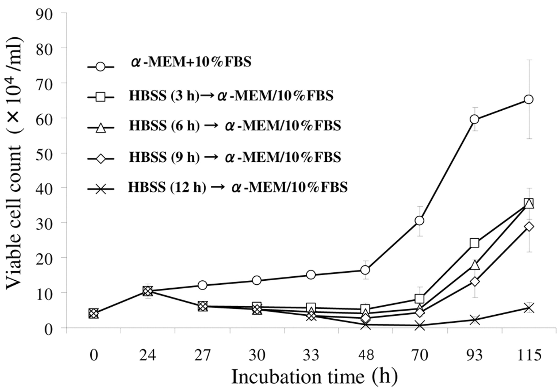
To investigate the resumption of cell growth by reculturing autophagic cells in regular culture medium, MC3T3-E1 cells (8×10⁴/ml, 1 ml) were inoculated on 24-well plate, and incubated for 24 hours. Cells were washed twice with HBSS, and incubated further for 0, 3, 6, 9 or 12 h in the same solution to induce autophagy, and the medium was changed from HBSS to normal medium (α-MEM+10% FBS). After incubation for the indicated times, the viable cell number was determined (Figure 4).
Contact to plates. MC3T3-E1 cells (25, 50, 75 or 100×104/ml, 1 ml) were inoculated on a plastic plate (20×20×0.5 mm) in a 35mm dish, incubated for 0, 24, 48 or 72 h in a 5% CO2 incubator at 37°C. Cells were washed with PBS(−), and detached by trypsin (0.2 ml). Medium (0.3 ml) was then added, and the viable cell number was determined (Figure 6).
MC3T3-E1 cells (25×104/ml, 0.5 ml) were seeded on plastic plate (control), and HA-coated, sandblasted, or anodized titanium alloy plates. After incubation for 48 h, the viable cell number, and the amino acid consumption during incubation were determined (Figure 7).
Assay for viable cell number. The viable cell number was determined by hemocytometer after staining the dead cells with 0.15% trypan blue solution.
Changes in the extracellular amino acid concentration during MC3T3-E1 cell growth, and comparison with the intracellular amino acid concentration.
Determination of free amino acids. Culture supernatant (medium fraction) was mixed with an equal volume of 10% TCA and stood on ice for 30 min. After centrifugation for 5 min at 10,000 ×g, the deproteinized supernatant was collected and stored at −40°C. The supernatants (20 μl) were subjected to an amino acid analyzer (JEOL JLC-500/V, Nihon Denshi, Tokyo) and amino acids were detected using the ninhydrin reaction.
Determination of intracellular amino acid concentration. Cells grown in 6-well plates were washed three times with cold PBS(−), and immersed in 5% TCA solution. Cells were then harvested by scraping with a rubber policeman into an eppendorf tube. The cell suspension was mixed for 10 s three times with a vortex mixer to ensure cell disruption, stood on ice for 30 min, and then centrifuged for 5 min at 10,000×g. The supernatant containing intracellular amino acids was collected and stored at −40°C. The consumption of each amino acid was calculated as the difference before and after incubation.
Detection of acidic organelles. Acridine orange is a pH-sensitive fluorescent pigment. When cells are induced to undergo autophagy, autophagolysosome (acidic organelle) stained by acridine orange as red granular dots is formed (15-17). Cells (5×10⁴/ml, 1 ml) were inoculated on a 24-well plate and incubated for 24 h. Then, medium was replaced by HBSS, and cells were further incubated for 3 or 6 h to induce autophagy. After staining for 20 min with acridine orange (1 μg/ml), the presence of cytoplasmic acidic organelles was assessed under confocal laser microscopy (excitation: 488 nm, emission: green 505-530 nm, red >650 nm).
Examination of titanium alloy surface. The surface of the titanium alloy plate was observed at a magnification of 1000-fold by scanning electron microscopy (JSM6360LV; JEOL, Tokyo). The extent of cell adhesion to the plate was assessed after fixing the cells with 2.5% glutaraldehyde and coating with 10 nm Au.

Growth potential of MC3T3-E1 cells under normal (A) or nutritionally starved (B, C) conditions. Each value represents the mean±S.D. of 4 determinations.
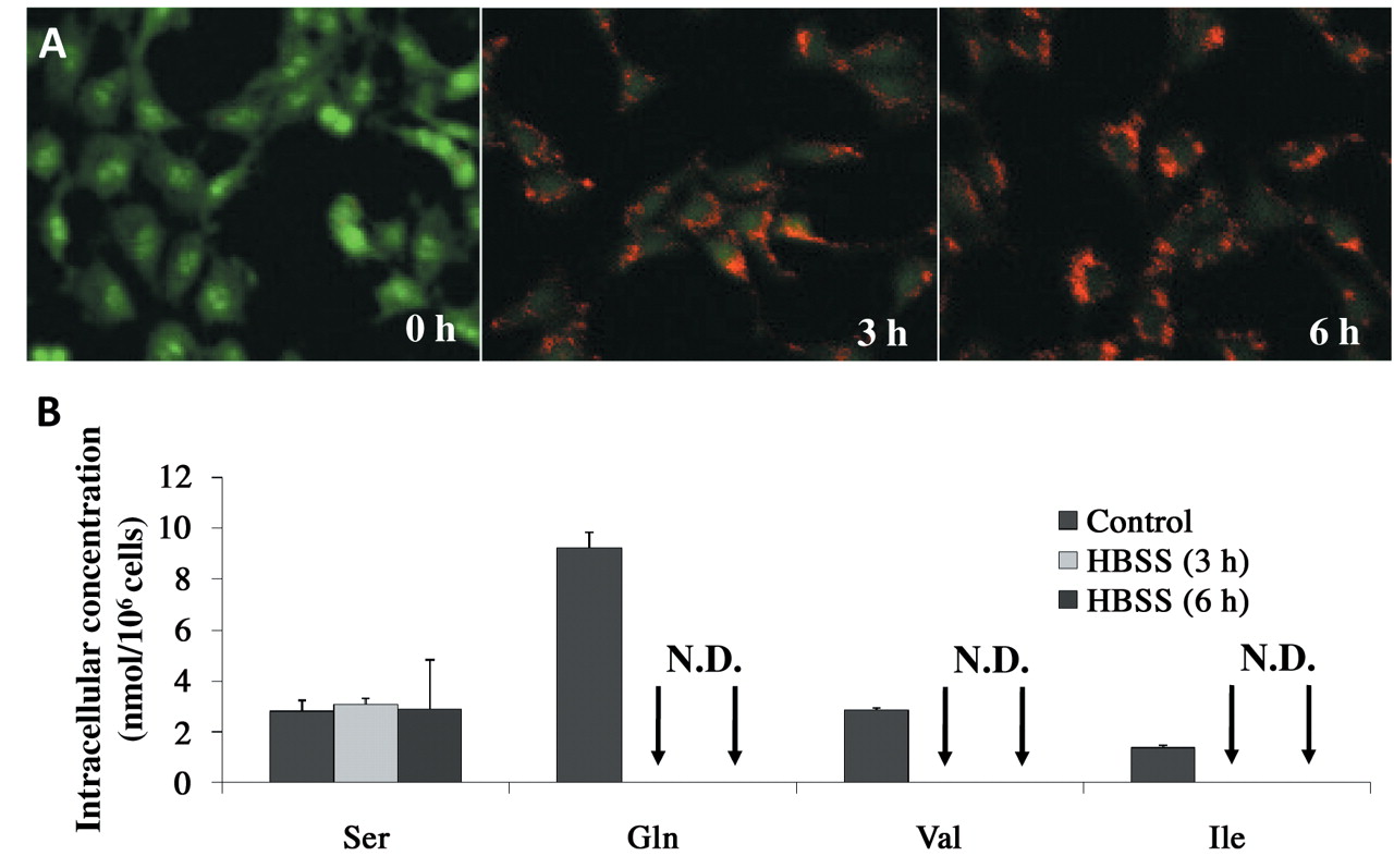
Induction of autophagy (A), as shown by staining with acridine orange to detect the formation of acidic organelles and changes in the intracellular amino acid concentration (B) during culture under starved conditions. Each value represents the mean±S.D. of triplicate determinations. N.D., Not detected.
Statistical treatment. Statistical analyses between two groups and for multiple comparisons were performed with Student's t-test or Scheffé test, respectively, taking significance at 5%.
Results
Amino acid consumption during cell growth. The doubling time of MC3T3-E1 cells during the logarithmic phase of cell growth was 19 h. Cells survived for approximately 6 days by changing into fresh culture medium. When the medium change was not performed, the viable cell number reached a plateau after approximately 4 days' culture (Figure 1A). The consumption of each amino acid was determined by subtracting its concentration at the indicated time point from the initial value. During the initial logarithmic cell growth (0-77 h after cell inoculation), Arg (429 nmol/ml/ 77 h) was consumed at the highest rate, followed by Gln (384)>Glu (371)>Asp (142)>Ile (103)>Leu (90), Ser (90)>Val (48)>Asn (13 nmol/ml/77h) (Table I). The consumption of Ser, Gln, Val, Ile and Leu significantly (p<0.01) increased with incubation time (Figure 1B).
The most abundant amino acid in the culture medium (α-MEM+10% FBS) was Gln, followed by Gly>Glu>Arg>Val> Thr>Leu>Ile>Ala>Pro>Asn>Ser>Asp>Tyr. On the other hand, Glu was the most abundant amino acid in the cells, followed by Asp>Gly>Gln>Ala>Pro>Thr>Tyr>Ser, Val> Arg>Asn>Ile>>Leu (not detectable). These data indicate that the intracellular amino acid concentration (intracellular amino acid pool) was 10 to 100-fold lower than that of the extracellular milieu, thus providing the rationale that amino acid utilization by the cells can be monitored by measuring the change in the concentration of extracellular amino acids.

Resumption of cell growth by reculturing autophagic cells in the regular culture medium. Each value represents the mean±S.D. of 4 determinations.

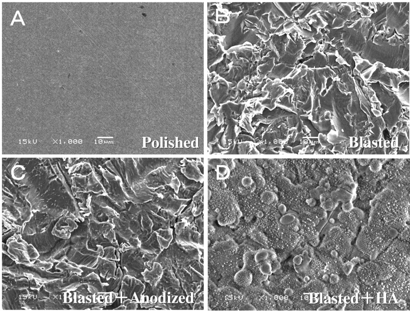
Surface texture of titanium alloy plates. The surface textures of 4 types of titanium alloys (A, polished; B, blasted; C, blasted + anodized; D, blasted + HA) were assessed using SEM at 1,000-fold magnification.
Cell proliferation and amino acid pool during nutritional starvation. MC3T3-E1 cells continued to proliferate up to 75 h even in the absence of serum. When cells were cultured in HBSS, a medium that is known to induce autophagy, cell proliferation was stopped, but nearly half of the cell population remained viable (Figure 2).
When MC3T3-E1 cells were cultured for 3 and 6 h in HBSS, cells produced acidic organelles stained with acridine orange, indicating the possible induction of autophagy (Figure 3A). Changes in the concentration of intracellular amino acids (amino acid pool) extracted with 5% TCA was next investigated. When autophagy was induced by incubating the cells in HBSS, the amino acid pool of Gln, Val and Ile was consumed, while that of Ser was unchanged (Figure 3B).
Cell proliferation resumed after about 24 h lag time when the autophagic cells (induced by 3 to 9 h culturing in HBSS) were recultured into normal medium (α-MEM+10% FBS) (Figure 4). However, the recovery was much delayed when the cells were incubated in HBSS for more than 12 h.

MC3T3-E1 cell proliferation pattern as a function of the number of cells inoculated on a plastic plate. Each value represents the mean±S.D. of 4 determinations.
Effect of contact with titanium plate on cell proliferation. The surface of the titanium alloy plate was examined by SEM (Figure 5). Polished plates appeared to have a quite smooth, uniform surface (Figure 5A). On the other hand, sandblasted (B) and anodized (C) titanium alloy plates appeared to be slightly rough with a flaky surface texture. The hydoxyapatite coating of titanium alloy gave the appearance of apatite-like crystals on the surface (D).

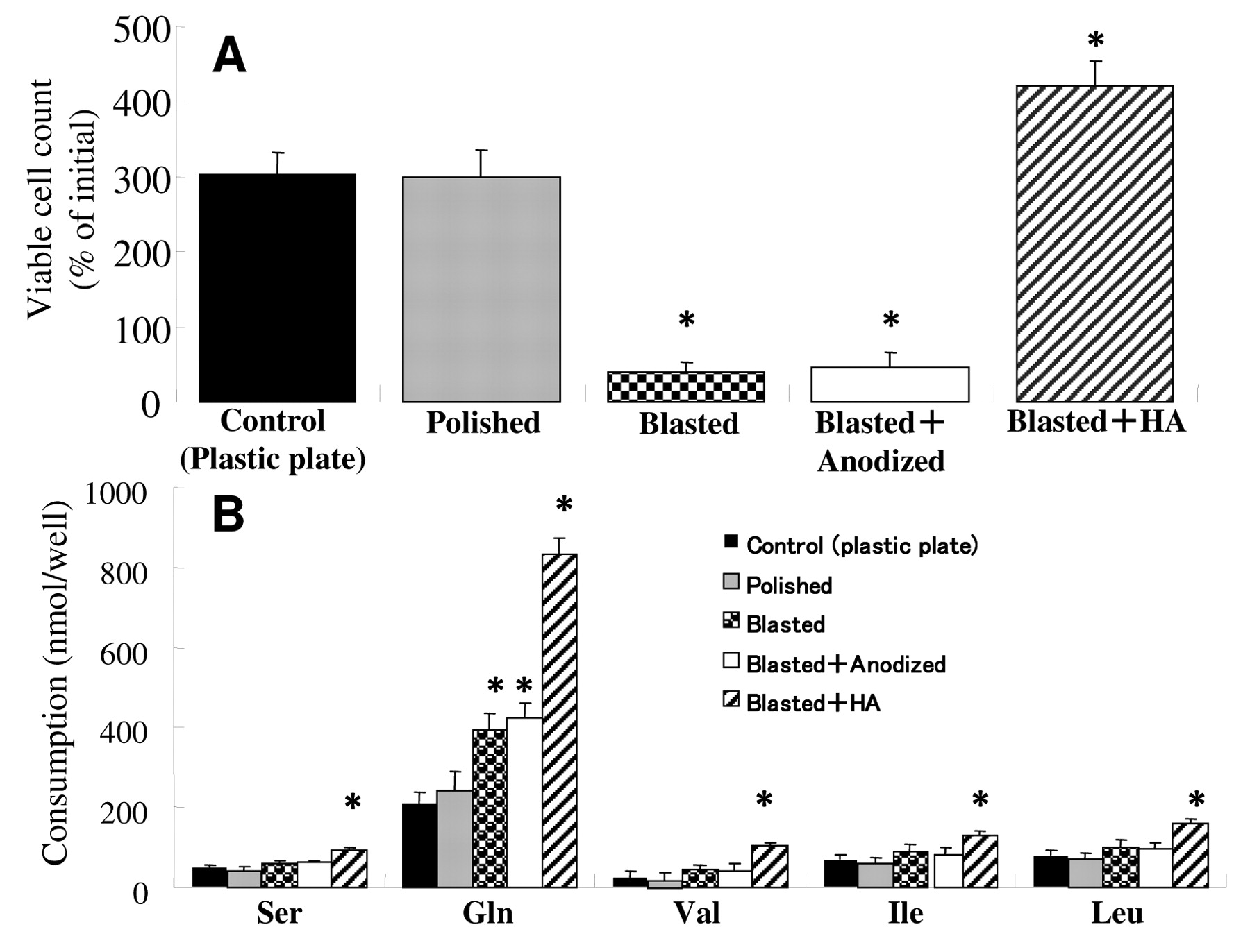
Effect of contact with titanium alloy plates on the proliferative activity (A) and amino acid consumption (B) of MC3T3-E1 cells. Each value represents the mean±S.D. of 5 determinations. *Significant difference from the control (p<0.01).
We first established the optimal conditions of cell inoculation for the subsequent cell—metal surface interaction experiments (Figure 6). The highest proliferation rate was found 48-72 h after cells were inoculated at 25×104/ml. The viable cell number peaked at 48 h and thereafter declined, when the inoculation cell density was 50, 75 or 100×104/ml. Based on these results, the inoculation cell density of 25×104/ml and the incubation time of 48 h that permitted logarithmic growth were used for the subsequent experiments.
We next investigated the effect of contact with different types of titanium plate on cell viability, using the plastic plates as control (Figure 7A). Contact with the HA-coated plate significantly (p<0.01) increased the number of cells recovered by trypsin treatment, as compared with the both plastic and polished titanium plates. On the other hand, contact with sandblasted or anodized titanium alloy plates significantly (p<0.01) reduced the number of cells recovered.
To investigate more quantitatively the effect of contact with different alloy plates on cell proliferation, the consumption of amino acids in the culture medium was investigated. The consumption of Ser, Val, Ile, Leu, and especially Gln was significantly (p<0.01) elevated by cells in contact with HA-coated titanium alloy. Contact with sandblasted or anodized titanium alloy also enhanced the consumption of Gln, as well as Ser, Val, Ile and Leu, but to a lesser extent (Figure 7B).
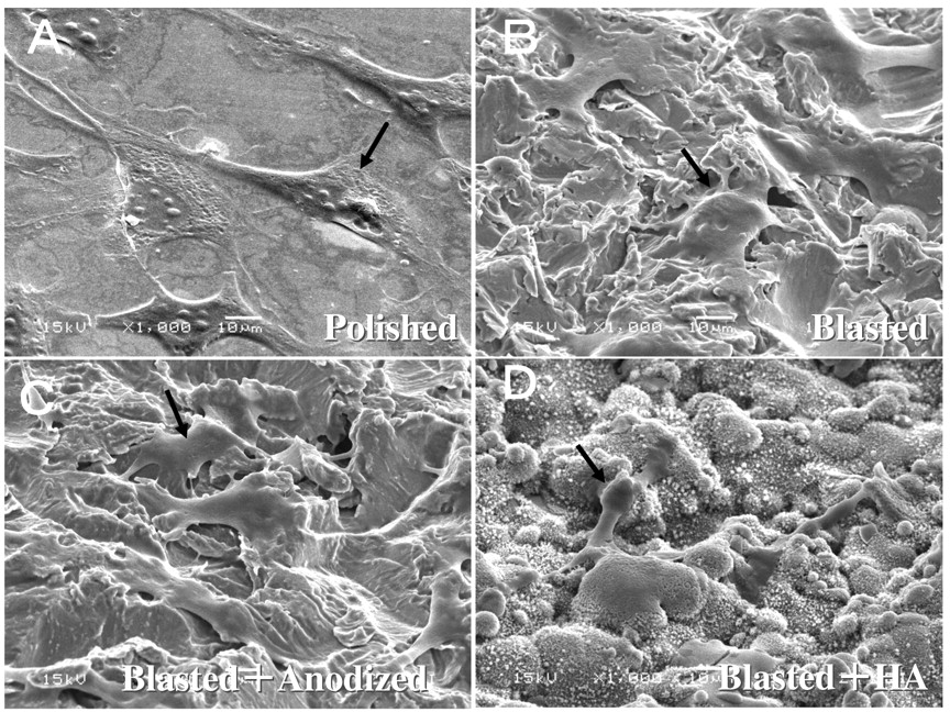
Cell morphology as related to plate surface. We investigated the morphology of cells in contact with four different titanium alloy plates using SEM (Figure 8). Contact with the polished titanium surface induced flat cell attachment (A). Contact with the sandblasted (B) and the anodized (C) titanium alloy plate induced cell spreading and protrusion, covering the roughened surface. The HA-coated plate allowed the cells to attach to the surface of apatite crystals with short cell protrusions (D).

Adhesion of MC3T3-E1 cells to titanium alloy plates (A, polished; B, blasted; C, blasted + anodized; D, blasted + HA) using SEM at 1,000-fold magnification. Morphological changes of cells noted are indicated by arrows.
Discussion
The present study demonstrated the high proliferative capability of mouse osteoblastic MC3T3-E1 cell line. MC3T3-E1 cells were able to survive for 4 days in regular culture medium without medium change and for 3 days even in the absence of serum. Cells were committed to undergo autophagy by culturing in HBSS, but resumed their logarithmic growth by re-culturing into regular culture medium, providing the incubation time in HBSS did not exceed 9 h.
Bone alkaline phosphatase, osteocalcin, type I collagen-C-peptide, type collagen-N-peptide are well known bone metabolic markers. Inactivation of mouse osteocalcin gene has recently been reported to elevate the amount of bone, but did not reduce the number of osteoclasts and bone resorptive ability (18). This suggests that it may not be essential for osteogenesis, nor reflect osteoblastic function during osteogenesis. Alkaline phosphatase was utilized to evaluate cell adherence to titanium plates, but it was difficult to completely recover the cells from the plate, suggesting that this method may not be suitable for the assessment of cell proliferative ability (19). Therefore, it was urgent to explore alternative methods by which the proliferative capability of cells (tightly bound to the plate even after trypsin treatment) could be measured. We therefore measured the proliferation of MC3T3-E1 cells by determining the consumption of amino acids in the culture medium. We found that the proliferation of MC3T3-E1 cells correlated well with the consumption of Ser, Gln, Val, Ile and Leu. We have previously reported that mouse macrophage-like cells (RAW264.7) (20) and human myelogenous leukemic cell lines (HL-60, ML-1, KG-1) that are differentiated into macrophages (21) extensively consume Ser, at much higher rates than most other amino acids except Gln. The present study shows that MC3T3-E1 cells consumed Ser at a much lower rate, there remaining approximately 43% (101 nmol/ml) of initial concentration even after 152 h, demonstrating for the first time that MC3T3-E1 cells does not show Ser requirement, quite different from the macrophage-lineage cells. The present study also demonstrates for the first time that MC3T3-E1 cells consume Arg at a higher rate than they do Gln, which is known as an energy source with glucose (22). We found that Arg was depleted from the culture medium at 4 days after cell inoculation when logarithmic growth nearly terminates, suggesting the necessity for adding extra Arg to α-MEM that is frequently used for the culture of osteoblasts.
Arg is used as a precursor for protein synthesis, but also for nitric oxide (NO) and citrulline (23). As yet, no paper has shown the relation of Arg consumption to the proliferation of osteoblasts. It remains to be investigated whether the prominent consumption of Arg is specific only for MC3T3-E1 cells, or characteristic of cells that are destined to differentiate into osteoblasts.
We next investigated the effect of contact with titanium alloy on cellular proliferation, based on the amino acid consumption. Our results showed that cell proliferation, as measured by amino acid consumption, on HA-coated titanium alloy, sandblasted and anodized titanium alloy plates was significantly higher than that on plastic plates. However, the number of cells recovered by trypsin treatment was lower in sandblasted and anodized titanium alloy plates as compared with plastic plates, in contrast to HA-coated titanium alloy plates. It has been reported that HA coating increased alkaline phosphatase (ALP) activity (24), and that cells adhered more strongly to sandblasted titanium alloy plates than to HA-coated ones (25). Similar findings have been reported for cells adhering to anodized titanium alloy plates (26). The present study demonstrated that contact with HA-coated plates induced growth stimulation of MC3T3-E1 cells with short protrusions while contact with sandblasted and anodized titanium alloy plates induced the formation of long protrusions. These results suggest that contact with sandblasted and anodized titanium alloy plates stimulates cell adhesion.
Human normal osteoblasts showed higher adherence and elongation in contact with regions of titanium alloy than with HA-coated regions (27). It is conceivable that HA coating may ideally replace the bone in the living body by remodeling cycle. In fact, HA in the living body was shown to stimulate osteogenesis, but was only marginally resorbed by osteoclasts, resulting in the poor replacement of bone (28). This suggests that cells bind to the HA-coated titanium plate via the HA layer, but not directly to titanium. There is the possibility in our study that trypsinization may have disrupted the bonding between the titanium plate and HA, resulting in the apparent increase in the recovered cell number.
HA and titanium implants utilize bio-integration (biochemical bonding via calcium bridge) and osseointegration (direct mechanical conjugation with bone), respectively. Since the surface treatment may reduce the original biocompatibility of titanium, the surface properties of implant should be selected according to the clinical purpose and bone condition (29, 30).
The present study recommends the use of HA coating in the case of deficiency of bone or for initial fixation during the implanting, and the use of sandblasted, or anodized titanium alloy in the case of immediate loading of an implant or stress caused by maxillo-fixation. Understanding the surface properties and characteristic of titanium alloy plate may improve clinical outcomes. It remains to be investigated whether the surface properties of titanium alloy plate may affect the differentiation toward maturing osteoblasts, and cell attachment to the surface of the plate.
Acknowledgements
We thank Drs. Shiro Kunii and Yoshiaki Ando, Laboratory of Electron Microscopy and X-ray Analysis, Meikai University School of Dentistry, for their technical assistance.
Footnotes
- Received July 2, 2009.
- Revision received October 22, 2009.
- Accepted October 27, 2009.
- Copyright © 2010 International Institute of Anticancer Research (Dr. John G. Delinassios), All rights reserved
References
In this issue


Jump to section
Related Articles
Cited By...
- Morphological Features of Osteoblasts Cultured on Ultraviolet-irradiated Titanium Plates
- Type of Cell Death Induced by Various Metal Cations in Cultured Human Gingival Fibroblasts
- Type of Cell Death Induced by Seven Metals in Cultured Mouse Osteoblastic Cells
- Quantification of Enhanced Osteoblastic Adhesion to Ultraviolet-treated Titanium Plate