


Abstract
Ampelopsis cantoniensis (AC) has been used as a folk medicine for reducing pain in the Taiwanese population. Our previous studies have shown that the crude extract of AC induced apoptosis in human promyelocytic leukemia HL-60 cells. In this study, the in vivo effects of AC on leukemia WEHI-3 cells and immune responses such as phagocytosis and natural killer (NK) cell activity were investigated. The weights of the livers and spleens were decreased in the AC-treated groups compared to the control groups. The AC treatment increased the percentage of CD3 and CD19 marker cells in WEHI-3-injected mice, indicating that the precursors of T and B cells were inhibited. The AC treatment promoted the activity of macrophage phagocytosis in the peripheral blood mononuclear cells (PBMC) and peritoneal cells. It was found that the NK cells from mice after treatment with AC can kill the YAC-1 target cells. Therefore, the AC treatment increased NK cell activity. In conclusion, AC can affect WEHI-3 cells in vivo and promote macrophage and NK cell activities.
In Taiwan about 2.1 per 100 thousand people die per year of leukemia from the reports of the “People Health Bureau of Taiwan” and this number seems to have increased when compared to ten years ago. So far, the treatments for human leukemia are not satisfactory. Epidemiological studies have shown that increased consumption of plant-based diet can reduce the risk of cancer, such as colon cancer (1, 2), and this observation has also been seen in animal studies (3), but the bioactive cancer preventives remain to be identified. Therefore, many investigators focused on naturally occurring dietary substances for cancer therapy and prevention.
Ampelopsis cantoniensis (AC), one of the native plants of Taiwan, has been used for reducing pain in folk medicine. However, scientific evidence is lacking, except that our laboratory was the first to show that the crude extract of AC induced apoptosis in human promyelocytic leukemia HL-60 cells (4). In order to further examine whether or not the crude AC extract can affect leukemia cells, its effects on WEHI-3 leukemia cells and on macrophage phagocytosis and natural killer (NK) cell activity in vivo was investigated.
Materials and Methods
Materials and reagents. The crude A. cantoniensis (AC) extract was produced from Dr. Tzu-Wei Tan in the Department of Pharmacology, China Medical University. All AC samples were dried and ground into fine powder, then extracted with methanol (MeOH) at room temperature. For MeOH extraction, 16 kg of the powdered samples were mixed with 2000 mL of methanol. Filtrate was collected twice by filter paper and MeOH was evaporated to dryness with a reduced pressure concentrator for approximately 12 h to obtain dry MeOH extracts. The extraction yield of the methanol extracts of AC is 8.75% . For in vivo studies, dry AC extracts were dissolved in DDW to the indicated concentrations of 50 and 100 mg/kg. RPMI 1640, fetal bovine serum, penicillin-streptomycin and glutamine were obtained from Gibco BRL (Grand Island, NY, USA).
Male BALB/c mice. Male BALB/c mice at the age of 8 weeks and about 22-28 g in weight were obtained from the Laboratory Animal Center, College of Medicine, National Taiwan University (Taipei, Taiwan).
Murine WEHI-3 leukemia cells. The WEHI-3 murine myelomonocytic leukemia cell line was obtained from the Food Industry Research and Development Institute (Hsinchu, Taiwan). Cells were cultured in RPMI-1640 medium supplemented with 10% fetal bovine serum, 1% penicillin-streptomycin (100 units/ml penicillin and 100 μg/ml streptomycin) and 1% L-glutamine and grown at 37°C under a humidified 5% CO2 atmosphere.
Drug treatment. Part I: Fifty BALB/c mice were randomly divided into 5 groups of 10 animals. One group was the control and another group was treated DDW. The other animals were injected with WEHI-3 cells (1×10⁵ cells in 100 μl PBS) and then treated or untreated with AC (50 or 100 mg/kg) in DDW. Part II: Another 30 BALB/c mice were divided into 3 groups of 10 animals. One was the control, another group was treated with distilled and deionized water (DDW; vehicle) and the third group was treated with 100 mg/kg of AC extract in DDW for 3 weeks. All the animals were given the above dose orally daily for up to 3 weeks before being weighed (5, 6).
Blood samples and immunofluorescence staining. Blood samples were collected (about 1 mL) from each animal in each group at the end of the experiments, immediately treated with ammonium chloride for lysing of the red blood cells then centrifuged for 15 min at 1500 rpm at 4°C. The isolated white blood cells were examined for CD3, CD19, Mac-3 and CD11b cell markers by staining with anti-CD3-FITC, CD19-PE, Mac-3-FITC and CD11b-PE antibodies (BD Pharmingen Inc). The cells peripheral blood mononuclear cell (PBMC) were stained for analysis of the cell marker levels by flow cytometry (FACS Calibur™, Becton Dickinson, NJ, USA) as described previously (5-7).
Liver and spleen samples. The animals were sacrificed and the livers and spleens were obtained and weighed individually (5).
Quantification of phagocytic activity of macrophages. A PHAGOTEST kit (ORPEGEN Pharma Gesellschaft für biotechnologische, Heidelberg, Germany) was used for measuring the phagocytosis. The cells were incubated for 1 h at 37°C with FITC-labelled E. coli (20 μL) according to the manufacturer's instructions. The reaction was stopped by the addition of ice-cold quenching solution (100 μL). At the completion of phagocytosis, the monocytes/macrophages were fixed and the DNA was stained according to the manufacturer's instructions. The cell preparations were then analyzed by flow cytometry (FACSCalibur, Becton Dikinson). Fluorescence data were collected on 10,000 cells and analyzed using CELLQUEST software (7, 8).
Quantification of NK cell activity. Approximately 1×10⁵ leukocytes from the spleens in 1 ml of medium were from the Part I groups containing control mice, those injected with WEHI-3 cells only and those injected with WEHI-3 cells and treated with AC (50 or 100 mg/kg/day), and Part II groups had control mice and mice treated with AC (100 mg/kg/day). Leukocytes were cultured in each well of a 24-well culture plate for 24 h. About 2.5×10⁷ YAC-1 cells were cultured in 15-ml tubes with serum-free RPMI-1640 medium and the PKH-67/Dilunt C buffer was added to the cells, mixed thoroughly for 2 min at 25°C then 2 ml PBS was added for 1 min. Then 4 ml RPMI-1640 was added for 10 min incubation followed by centrifugation at 1200 rpm of 25°C. About 2.5×10⁶ YAC-1 cells in 100 μl were placed of 96-well plates before the addition of the leukocytes to the wells for 12 h and determination of the NK cell activation by flow cytometry as described previously (9).
H&E stain and histopathology. Tissue samples from spleen were fixed in 4% formaldehyde and embedded in paraffin. Each section of 5 μm from each animal was stained with hematoxylin and eosin (H&E stain) according to standard procedures (5-7).
Statistical analysis. The results were expressed as mean±SD and the difference between groups was analyzed by Student's t-test. P<0.05 was taken as significant.
Results
Effect of crude extract on the weight of liver and spleen. Three weeks after the injection of WEHI-3 cells the animals treated with AC showed decreased spleen tumor size compared to the untreated mice (Figure 1A). The AC treatment decreased the weight of the compared to the untreated mice liver and spleen (Figure 1B). However, in the control animals that were treated with DDW or AC no significant differences were shown in these tissues (Figure 1C).
Effect of crude AC extract on the white blood cell surface markers. Figure 2A, B, C and D shows the percentage of CD3, CD19, CD11b and Mac-3 cells in the white blood cells of the control and the WEHI-3 injected AC treated or untreated mice. The animals not injected with WEHI-3 cells are shown in Figure 2E. The AC treatment increased the levels of CD3 (Figure 2A) and CD19 (Figure 2B) but did not affect the levels of CD11b (Figure 2C) and Mac-3 (Figure 2D) in the WEHI-3-injected animals. Furthermore, AC also promoted the levels of CD19 and Mac-3 (Figure 2E) in the animals, which were not injected with WEHI-3 cells.
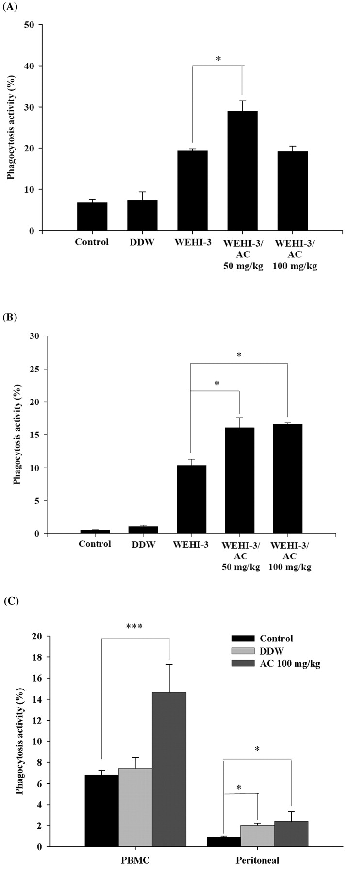
Effect of crude AC extract on phagocytosis. Figure 3A and B demonstrated that AC treatment promoted phagocytotic activity in the WEHI-3-injected animals at the low dose in the PBMC (Figure 3A) and both treatment doses led to increased phagocytotic activity in the WEHI-3 cells injected animals (Figure 3B). However, in the animals not injected with WEHI-3 cells AC (100 mg/kg) promoted phagocytotic activity in both the PBMC and the peritoneal cells.
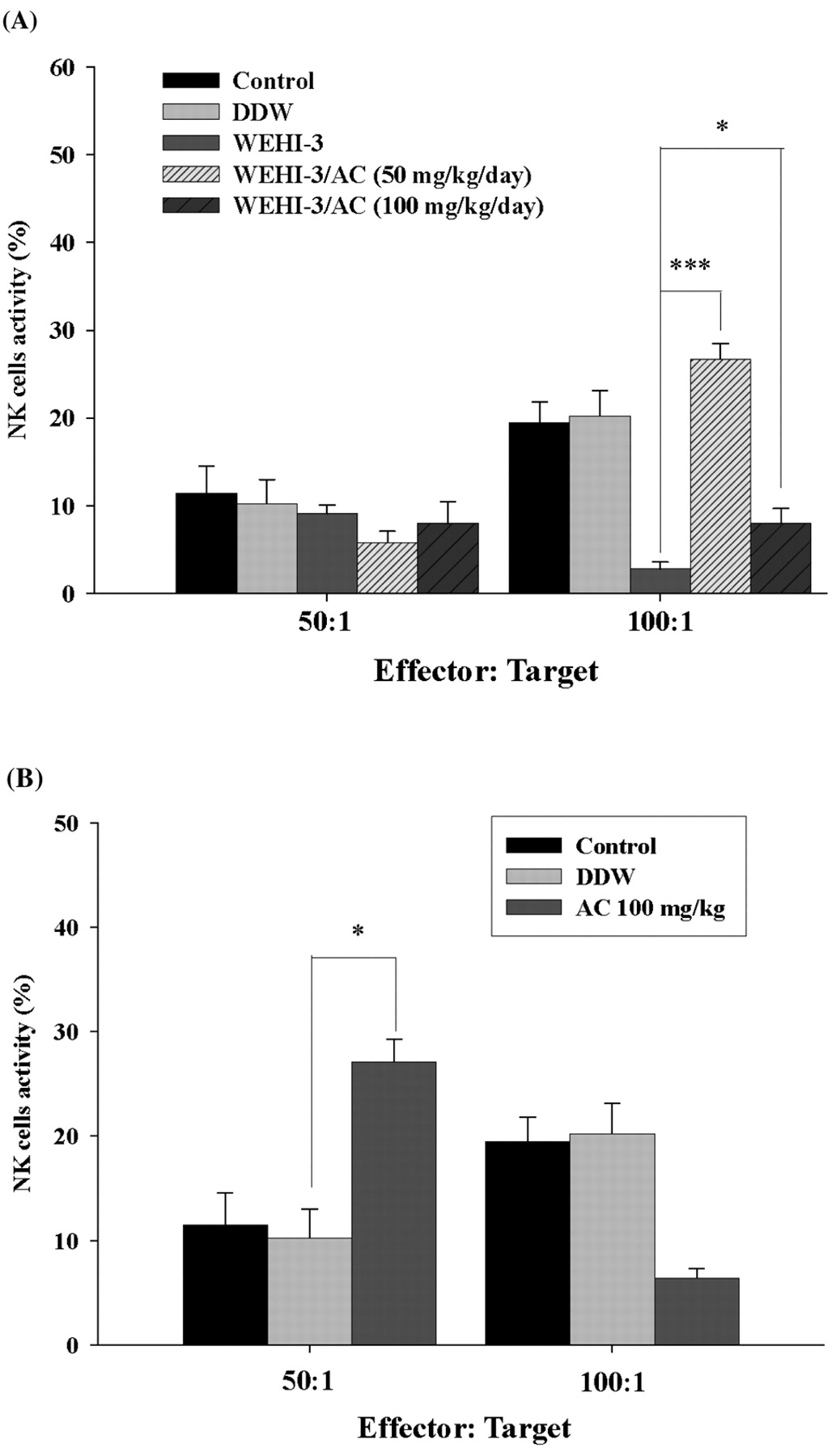
Effect of crude AC extract on NK cell activity. The YAC-1 target cells were killed by NK cells and the data demonstrated that the AC treatment promoted the activities of NK cells at both doses in target cells ratio of 100:1 in the WEHI-3 cells-injected animals (Figure 4A). However, in the animals not injected with WEHI-3 cells AC promoted NK cell activity only in low stimulation (effector: target=50:1) (Figure 4B).
AC extract affects the histopathology of spleen. Each spleen was isolated from all animals and was histopathologically examined. The representative results are presented in Figure 5 which showed that AC treatment affected the leukemia mice.

(A) Representative BALB/c mice 3 weeks after the injection of WEHI-3 cells untreated or treated with crude AC extract. Weights of spleen and liver from (B) WEHI-3 cells injected before AC treatment (C) or uninjected and then treated AC. *p<0.05.
Discussion
A model based on the murine host system was used due to the low cost and the ease and speed of establishment of the cancer, and finally its widely accepted experimental end-points (10, 11). The murine WEHI-3 leukemia cell line has the characteristics of myelomonocytic leukemia and has often been used for inducing leukemia in syngenic BALB/c mice and for evaluating the effects of drugs (12). Additionally WEHI-3 cells were originally derived from the BALB/c mouse (13). Several studies have used the WEHI-3 cell in vivo model for evaluating anti-leukemia activity including ATRA, adactinomycin A, interleukin-6 (IL-6), granulocyte-colony stimulating factor (G-CSF) and vitamin D3 induced in vitro differentiation of WEHI-3 cells in monocytic and granulocytic lineages (14-16). The present results indicated that AC decreased the weight of liver and spleen and increased the markers of CD3 and CD19 in the PBMC white blood cells.

White blood cell markers. (A) CD3; (B) CD19; (C) CD11b; (D) Mac-3 in WEHI-3 cells injected AC treated or untreated and control mice; (E) CD3, CD19, CD11b and Mac-3 in mice not injected with WEHI-3 cells. Each point is mean±S.D. of three experiments. *p<0.05; **p<0.01.

Macrophage phagocytosis. (A) PBMC and (B) peritoneal cells from WEHI-3 cells-injected mice and controls and (C) animals not injected with WEHI-3 cells. Each point is mean±S.D. of three experiments. *p<0.05, ***p<0.001.

Natural killer cell activity. Splenic leucocytes from (A) WEHI-3 cells injected and (B) normal not injected with WEHI-3 cells. Each point is mean±S.D. of three experiments. *p<0.05, ***p<0.001.
However, the important findings were that AC inhibited spleen leukemia tumor growth and also promoted macrophage phagocytosis and NK cell activity which may have lead to decrease in the spleen tumor size.

Histopathology of spleen from WEHI-3 cells-injected mice after treatment with AC extract. Each spleen was isolated from all animals and was histopathologically examined and the representative results are presented which showed AC extract affected the leukemia mice (→: infiltration of tumor cells into spleen red pulp).
Acknowledgements
This work was supported by grant CMU-94-122 from the China Medical University, Taiwan, R.O.C.
- Received November 25, 2008.
- Revision received February 18, 2009.
- Accepted May 20, 2009.
- Copyright © 2009 International Institute of Anticancer Research (Dr. John G. Delinassios), All rights reserved


