


Abstract
Background: Cytotoxic activity of saponins and phenolic compounds have been described in the literature, but no reports were found on their multidrug resistance (MDR)-modulating effects on human mdr1 gene-transfected mouse lymphoma cell line. Materials and Methods: Methylprototribestin, structurally related compounds and a mixture of 3 acetylated isomers of methylprotodioscin were investigated for antiproliferative effect and modulation of drug accumulation. Results: The growth inhibitory dose (ID50) of the compounds ranged from 12.64 to 20.62 μg/ml. Methylprototribestin was the most effective resistance modifier. However, methylprotodioscin, pseudoprotodioscin, prosapogenin A of dioscin, tribestin and 5-O-caffeoylshikimic acid showed moderate MDR reversal activity. In a checkerboard method, methyloprototribestin and the mixture of the 3 acetylated isomers enhanced the antiproliferative effects on MDR cells in combination with doxorubicin. Conclusion: Based on these results, methylprototribestin and the mixture of the 3 acetylated isomers can be recommended for further in vivo experiments in combination with anthracyclines in human MDR-cancer xenograft transplanted mice.
- Steroidal saponins
- Tribulus terrestris
- Smilax excelsa
- multidrug resistance
- MDR reversal
- efflux pump
- P-glycoprotein
Multidrug resistance (MDR) is associated with overexpression of the membrane protein P-glycoprotein (P-gp; 170 kDa) (1), which expels anticancer drugs from cells and thereby leads to the ineffectiveness of chemotherapy. To overcome this problem, various individual compounds of synthetic and natural origin have been screened for their ability to reverse MDR (2-4). Efforts are continuing in the search for lead compounds for the development of products effective in chemotherapy.
We have directed our attention to steroid saponins, which are widely distributed in the plant kingdom. They possess a range of biological activities including cytotoxicity against some cell lines from leukaemia and solid tumours (5-8). The ability of steroid saponins to act as MDR modulators has not been explored to date.
The medical plants Tribulus terrestris (Zygophyllaceae) and Smilax excelsa (Liliaceae) growing in Bulgaria are among the richest producers of saponins. Smilax species have long been used in traditional medicine in many countries for the treatment of infectious diseases, skin disorders and liver inflammation (9, 10). T. terrestris is mainly known for its effectiveness in libido disorders, impotence and infertility. Data have also been published on its cardiovascular, cytotoxic and antimicrobial activities (11). The steroidal saponins have been considered to be the main active principles of these plants, responsible for their numerous healing applications.
In the present study, 6 pure steroidal saponins, a mixture of 3 saponin isomers, and 2 phenolics were isolated from the saponin fractions of T. terrestris and S. excelsa. All these compounds were evaluated as MDR modulators on multidrug-resistant mouse lymphoma cells.
Materials and Methods
Tested compounds. The air-dried milled rhizomes of Smilax excelsa (2.0 kg) were extracted three times with MeOH (3×7l×24 h) at room temperature, which led after concentration to 213.2 mg of material in the extract, which was resuspended in a mixture of MeOH/H2O. This suspension was successively extracted with CHCl3 (3×500 ml) and n-BuOH (3×500 ml) to give the corresponding CHCl3 (6.0 g) and n-BuOH (27.5 g) extracts. From the n-BuOH extract, we succeeded in isolating methylprotodioscin (1); pseudoprotodioscin (3); dioscin (8); a mixture of 3 saponin isomers: (3-O-[2-O- (5), (3-O-[3-O- (6) and (3-O-[4-O (7) -acetyl-α-L-rhamnopyranosyl-(1→2)-{α-L-rhamnopyranosyl-(1→4) }-β-D-glucopyranosyl]-26-O-[β-D-glucopyranosyl]-22α-methoxy-(25R)-furost-5-ene-3β,26-diols; 5-O-caffeoylshikimic acid (10); and trans-resveratrol (11) were isolated (Figures 1 and 2) (12, 13).
Air-dried and powdered plant material (5.5 kg) of Tribulus terrestris was extracted with 70% EtOH (3×6l×24 h) at room temperature. The combined EtOH solutions were concentrated to a small volume and extracted in succession with CHCl3 (3×11×24 h) and n-BuOH (3×11×24 h). The n-BuOH layer was concentrated to dryness, giving the crude saponin extract (100.0 g). Liquid vacuum chromatography of this crude saponin extract (10.0 g) over silica gel (20.0 g) using CHCl3/MeOH/H2O (6:1:0.1 to 1:1:0.1) yielded fractions F1-F7. From fraction F4 (0.7 g), methylprotodioscin (1), methylprototribestin (2) and pseudoprotodioscin (3) were obtained (Figure 1) (14, 15). Fraction F2 (0.52 g) was subjected to medium-pressure liquid chromatography using gradient elution with MeOH/H2O (4:6 to 8:1); 15 fractions were collected. The fractions eluted with MeOH/H2O 7:3 and 4:1 were combined as subfractions R1 (0.11 g) and R2 (0.21 g). On slilica gel column chromatography with CHCl3/MeOH/H2O (6:1:0.1 to 4:1:0.1), R2 gave dioscin (8, 30 mg) and tribestin (9, 16 mg) (16). From R1 (0.11 g) by preparative thin-layer chromatography (CHCl3/MeOH/H2O, 6:1:0.1), prosapogenin A of dioscin (4, 9 mg) was obtained. Prosapogenin A of dioscin was identified on the basis of its mass, 1D and 2D NMR spectra by comparison with literature data (17-19). This identification was confirmed by direct comparison (co-thin-layer chromatography) with an authentic sample obtained by solvolysis of tribestin (9) (Figure 1) (16).
Cell cultures. L5178 mouse T-cell lymphoma cells (U.S. FDA, USA) were transfected with pHa MDR1/A retrovirus (20). The mdr1-expressing cell line was selected by culturing the infected cells with 60 ng/ml colchicine to maintain the expression of the MDR phenotype. L5178 (parental) mouse T-cell lymphoma cells and the human mdr1-transfected subline were cultured at 37°C in McCoy's 5A medium supplemented with 10% heat-inactivated horse serum, L-glutamine and antibiotics. The mouse lymphoma cell line was maintained in a 5% CO2 atmosphere.
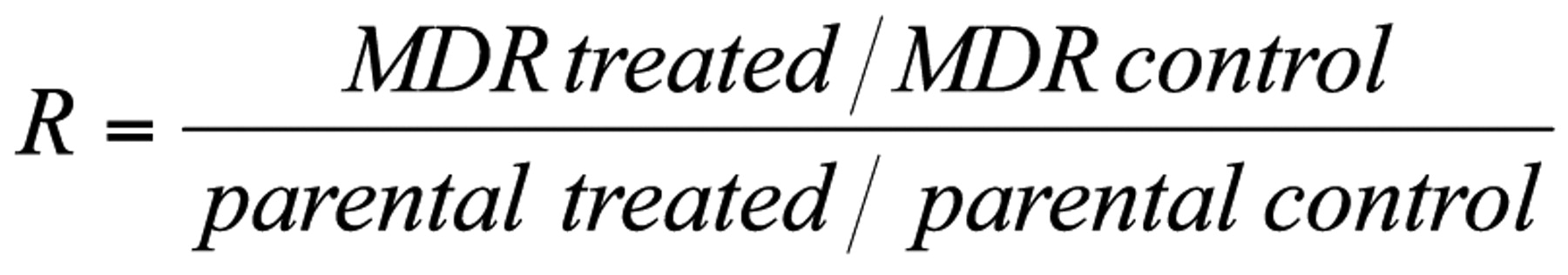
Assay for reversal of MDR in tumour cells. The cells were adjusted to a density of 2×10⁶ cell/ml, resuspended in serum-free McCoy's 5A medium and distributed in 0.5 ml aliquots into Eppendorf centrifuge tubes. The tested compounds were added at different final concentrations (4.0 and 40 μg/ml), and the samples were incubated for 10 min at room temperature. Ten μl (5.2 μM final concentration) of the indicator rhodamine 123 (Sigma, St Louis, MO, USA) was added to the samples and the cells were incubated for a further 20 min at 37°C, washed twice and resuspended in 0.5 ml phosphate-buffered saline (PBS) for analysis. The fluorescence of the cell population was measured with a FACS Star Plus flow cytometer (Beckton, Dickinson and Company, Franklin Lakes, NJ, USA). Verapamil (EGIS Phamaceuticals PLC, Budapest, Hungary) was used as a positive control in the rhodamine 123 exclusion experiments. The percentage mean fluorescence intensity was calculated for the treated MDR and parental cell lines as compared with the untreated cells. An activity ratio (R) was calculated via the following equation, on the basis of the measured fluorescence values:
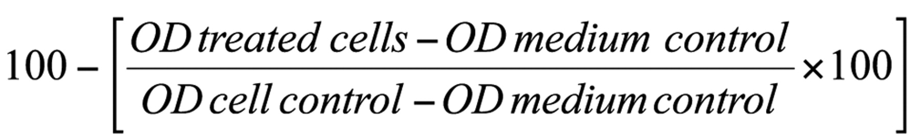
Assay for antiproliferative effect. The effects of increasing concentrations of the drugs alone and their combinations with resistance modifiers on cell growth were tested in 96-well flat-bottomed microtitre plates. The compounds were diluted in two-steps in a volume of 50 μl to a final concentration of 25 μg/ml. A total of 1×10⁴ cells in 0.1 ml of medium were then added to each well, with the exception of the medium control wells. The culture plates were further incubated at 37°C for 72 h, at the end of which 20 μl of MTT solution (thiazolyl blue solved in PBS to a final concentration of 5 mg/ml) were added to each well. After further incubation at 37°C for 4 h, 100 μl of sodium dodecyl sulphate (SDS) solution (10%) were measured into each well and the plates were further incubated at 37°C overnight. The cell growth was determined by measuring the optical density (OD) at 550 nm (ref. 630 nm) with a Multiscan EX ELISA reader (Thermo Labsystems, Cheshire, WA, USA). Inhibition of cell growth was determined as a percentage according to the formula:

A checkerboard microplate method was applied to study the effects of interactions between the resistance modifiers methylprototribestin (2) and the mixture of the 3 acetylated isomers of methylprotodioscin (5-7) and the anticancer agent doxorubicin on cancer cells as an in vitro model of combination chemotherapy. The dilutions of doxorubicin (A) were made in a horizontal direction and those of the resistance modifiers (B) vertically in the microtitre plate in a volume of 100 μl. The cell suspension in the tissue culture medium was distributed into each well in 50 μl containing 5×10⁴ cells. The plates were incubated in a CO2 incubator at 37°C for 48 h. The cell growth rate was determined after MTT staining and the intensity of the blue colour was measured on an Multiscan EX ELISA reader. Drug interactions were evaluated according to the following system:
FICA=ID50A combination/ID50A alone
FICB=ID50B combination/ID50B alone
where ID=inhibitory dose and FIC=fractional inhibitory concentration. FIX=FICA+FICB, where FIX=fractional inhibitory index. FIX<0.5 indicates synergism, 0.5<FIX<1 indicates an additive effect, 1<FIX<2 indicates an indifferent effect, and FIX>2 indicates antagonism between the anticancer drug and the potential MDR modifier.
Octanol/water partition coefficient (Log10 P) calculation. The theorecitcal Log10 P was determined by using the JME molecular editor (http://www.molinspiration.com/).
Results
Various biological activities of saponins and structurally related compounds have been extensively studied and several hundred structures have been elucidated. In our experiments, six pure saponins (1-4, 8, 9), 3 saponin isomers in a mixture (5-7) and 2 phenolics (10, 11) isolated from the saponin fractions of T. terrestris and S. excelsa were evaluated for their antiproliferative and multidrug resistance inhibitory effects on mdr1-transfected mouse lymphoma cells. In the antiproliferative experiment, the 50% growth inhibitory dose (ID50) values were found to be between 12.64 and 20.62 μg/ml after incubation at 37°C for 72h in a long-term experiment.

Chemical structures of the compounds (1-7).
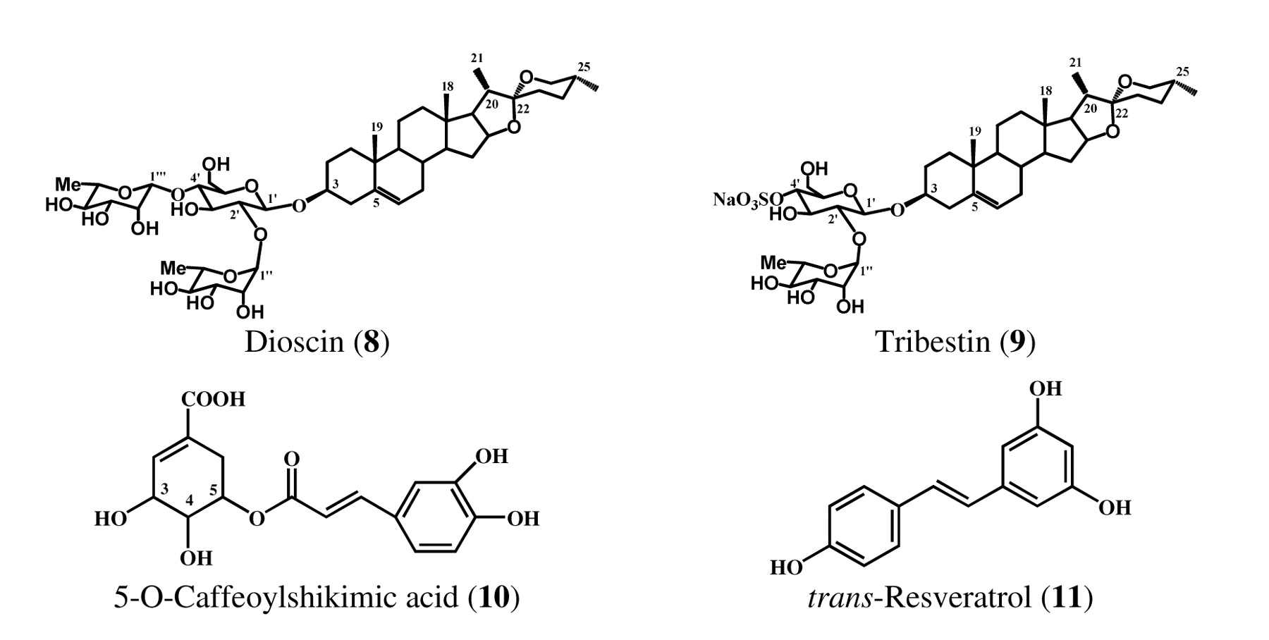
Chemical structures of tested compounds (8-11).
Then the effects of saponins and structurally related compounds isolated from S. excelsa and T. terrestris were studied on the rhodamine 123 (R123) accumulation on human mdr1-transfected mouse lymphoma cells. In this experiment, 1×10⁶ cells were exposed to 2 and 20 μg/sample for 10 min before R123 was added to the cells. The toxic effects of the compounds were checked with trypan blue test. Apparently, the compounds shown no toxic effects on 1×10⁶ cells at the applied 40 μg/ml concentration during the short incubation period. The majority of the saponin derivatives was ineffective on R123 accumulation when applied in 4 μg/ml concentration, but they were able to reverse multidrug resistance on the mouse lypmhoma cells in 40 μg/ml concentration incubated for 30 min. Verapamil was applied as a positive control. The results relating to MDR are shown in Table I.
The MDR reversal effects of the most effective compounds found in the antiproliferative assay were measured in combination with the anticancer drug doxorubicine in vitro. The activity of combinations of methylprototribestin (2) (Figure 3 and Table II) and the mixture of 3 acetylated isomers of methylprotodioscin (5-7) (Figure 4 and Table III) with doxorubicin were examined. Synergistic interaction between methylprototribestin (2) and doxorubicin (FIX=0.498) was found, and the interaction between the mixture of 3 acetylated isomers of methylprotodioscin (5-7) and doxorubicin (FIX=0.984) was found to be additive (Figure 4 and Table III). The results reveal that the compounds exhibit synergistic and addive effects in combination with doxorubicin.
The effects of compounds isolated from Smilax excelsa and Tribulus terrestris on rhodamine 123 accumulation in MDR mouse lymphoma cells: dose-response experiments.
Discussion
The emergence of MDR in cancer cells leads to the ineffectiveness of chemotherapy. Various compounds have been investigated for their ability to reverse MDR, including synthetic (21, 22) and naturally-occurring plant-derived compounds (23, 24).

The antiproliferative effect of methylprototribestin (MePt) and doxorubicin in combination.
Saponins are generally found in the roots, flowers and seeds of the higher plants and have long been used against various diseases, to enhance natural resistance, to restore the normal function of the body, to strengthen the constitution (25).
The present study was undertaken to access the inhibition of MDR of cancer cells by saponins and structurally related compounds. The resistance modifier compound 2 exhibited synergy, while compounds 5-7 had only additive effect in combination with doxorubicin, which is a well-known substrate of the P-gp-mediated efflux pumps. The activity of these saponins appears to correlate the position of sulphur-containing substituents, this may refer to the role of charge transfer complex formation in the binding of the compounds to P-gp.
The analysis of the structure activity relationship demonstrated the presence of sulphate and acetyl groups bound to the sugar moieties in compounds 2 and 5-7. The calculated partition coeffitients (Log10 p=0.09 and 1.41 for 2 and 5-7, respectively) indicate some differences in hydrophobicity of these compounds, as well. Obviously, other factors may also interfere with the ability of steroidal furostanol saponins to interact with the P-gp efflux pump.
The in vitro experimental data indicate that some of the saponins can be regarded as promising structures for rational drug design of P-gp inhibitors with a novel mechanism of action.

The antiproliferative effect of the mixture of 3 acetylated isomers of methylprotodioscin (3Ac) and doxorubicin in combination.
Evaluation of antiproliferative effect of methylprototribestin and doxorubicin in combination. *n.a.: non-applicable: in the concentration of 25 μg/ml, the cell proliferation was totally blocked by compound 2, therefore, the FIX value couldn't be calculated from the curve.
Evaluation of antiproliferative effect of the mixture of 3 acetylated isomers of methylprotodioscin and doxorubicin in combination. *n.a.: non-applicable: in the concentration of 25 μg/ml, the cell proliferation was totally blocked by compounds 5-7, therefore, the FIX value couldn't be calculated from the curve.
Acknowledgements
This work was supported in part by the Bulgarian National Science Fund (Contracts X-1312 and X-1601) and the Szeged Foundation for Cancer Research.
- Received December 9, 2008.
- Revision received May 12, 2009.
- Accepted May 20, 2009.
- Copyright © 2009 International Institute of Anticancer Research (Dr. John G. Delinassios), All rights reserved
References
In this issue


Jump to section
Related Articles
Cited By...
- No citing articles found.