


Abstract
Prostate cancer is a major public health problem in the world. Molecular studies are necessary for the development of prognostic markers in prostate cancer. There is a great interest in mucin studies in treatment development of human malignancies, including prostate cancer. Nevertheless, their expressions in prostate cancer need further investigation. Materials and Methods: Mucin 1 (MUC1) expression was examined in 100 prostate biopsies and were compared with prostate carcinoma cell lines (DU-145, PC-3, LNCaP) by immunohistochemistry. Results: Biopsies were healthy, tumor, peritumor or presented an intraepithelial neoplasia. Staining of MUC1 was exihibited in PC-3 cells, was higher in DU-145, and was not expressed by LNCaP. Tumor sections presented more positive expression of MUC1 than non-tumor sections. Conclusion: MUC1 expression is correlated with the histological degree of malignancy.
In 2002, prostate cancer was the fifth most common cancer in the world and the second most common cancer among men, with 679,000 new cases (1). The prostate was the most frequent cancer site among men accounting for 35% of the cases. In 2002, in France, more than 3.5% of men over 74 years old were living with a prostatic cancer diagnosed within 5 years (2). The incidence of prostate cancer is rising worldwide and is particularly increasing in the elderly population. The increased number of cases is identified following prostate specific antigen (PSA) testing (3). Nevertheless, the use of PSA to detect prostate cancer is clinically imprecise since benign and malignant prostate disease can cause elevations in PSA. The biological behaviour and the natural course of prostate cancer are poorly understood (4). Many criteria are used to establish the diagnosis, but new biomarkers are required. A useful diagnostic marker detectable by immunohistochemistry could be Mucin 1 gene. Mucin 1 (MUC1) is a Type I membrane glycoprotein of the mucin family having an extensive extracellular domain consisting of hundreds of tandem repeat units, a single transmembrane domain, and a C-terminal cytoplasmic tail (5-7). There is growing interest in mucins as treatment targets in human malignancies including prostate, cancer; however, the role of their expression in the progression of prostate cancer is unclear. MUC1, which is expressed at the apical cell surface of many normal secretory epithelial cells, has been suggested to prevent adhesion and to promote development of metastatic disease (8). In prostate cancer, overexpression of MUC1 in tissue has been correlated both with a higher Gleason grade and an advanced tumor stage (9). In the present study, the hypothesis that the degree of prostate cancer malignancy is correlated with immunohistochemical expression of MUC1 is tested.
Materials and Methods
Cell culture. The prostate cancer cell lines DU-145, PC-3, and LNCaP were obtained from the American Type Culture Collection (ATCC, USA). DU-145 and PC-3 are known to be negative for androgen receptor (AR), and LNCaP cells were positive for AR. DU 145 were cultured in Eagle's minimum essential medium (EMEM), PC-3 cells were cultured in F-12K media, and LNCaP were cultured in RPMI 1640. All cultures were supplemented with 1% glutamine (Sigma®), 0.1% gentamycin (Sigma®) and 10% FBS (fetal bovine serum, Life Technologies) and were grown in a humidified atmosphere at 37°C containing 5% CO2.
Patients. This study was undertaken with the participation of 100 patients. These 100 men had a prostate biopsy by an urologist to establish a diagnosis. They were hospitalized at the CHU of Clermont-Ferrand, France. They accepted, in a letter of consent, to give a sample for Research. This study was counselled by the PPC (People Protection Committee) within the framework of the constitution of biological samples collection.
A transrectal ultrasound-guided prostate biopsy was performed with a spin and in double sextant. With the agreement of the patient, a small section was used for research (Figure 1). This examination was made with a local anaesthesia on the recumbent patient. The identity of each patient was confidential and with the anatomopathological examination, the stage of development of cancer was known and correlation was noted.
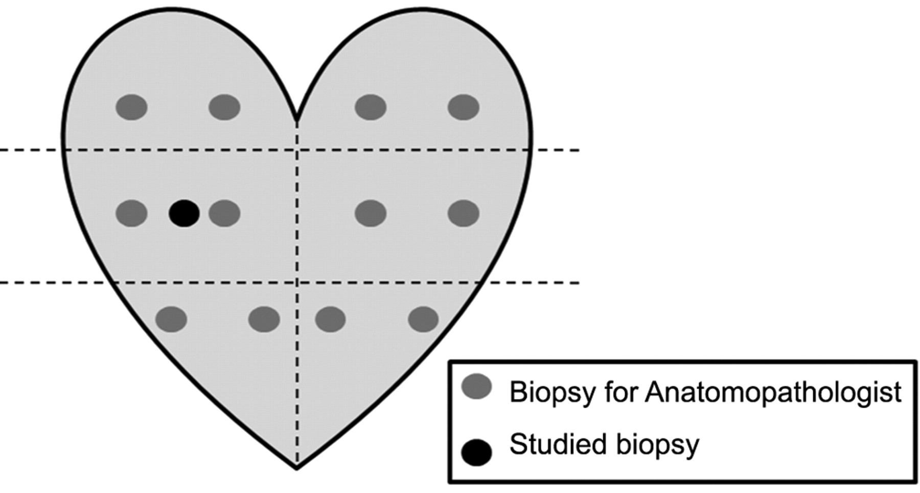
12-core biopsy scheme.
Grading and pathologic evaluation. The tissues were fixed in 10% buffered formalin, which was routinely processed, and whole-mount-embedded. Hematoxylin and eosin-stained sections were reviewed, tumor foci identified, circled in ink, and then graded. The tumor grade was assigned using the Gleason score grading system (10).
Immunohistochemistry. Four micrometer thick, alcohol-formalin-acetic acid-fixed and paraffin-embedded, sections were cut using a microtome, mounted on silanized glass slides (Starfrost), and dried overnight at 37°C. Graded alcohols (100% and 70%) and distilled water were used to deparaffinizate and rehydrate them. A heat-induced antigen retrieval method was then used, which included a 3-min incubation in citrate buffer, pH 5.9, in a pressure cooker, followed by a 15-min cooldown period in a water bath. Further processing was performed with a NexES automated immunostainer using an AEC kit (Ventana Medical Systems Inc.; Tucson, AZ, USA). Slides were then incubated at 37°C for 32 min with anti-Muc-1 (Mouse Monoclonal antibodies, [MBC-2], Abcys®, France) primary antibody. The antibodies were used at 1: 20 dilution. Subsequent incubations with a biotinylated secondary antibody, avidin-conjugated peroxidase complex, were carried out in a Ventana NexES immustainer in accordance with the manufacturer's protocol.
Slides were then counterstained with hematoxylin for 3 min, rinsed in distilled water, and coverslipped with an aqueous Faramount mounting media (DAKO; Glostrup, Denmark). The primary monoclonal antibody was omitted and replaced with PBS as a negative control.
Results
MUC1 staining in prostate cell lines. MUC1 was found positive in the cytoplasm of PC-3 cells, but with DU-145 cells, the staining was more intense. In addition, MUC1 was not expressed in LNCaP cells (Figure 2).
Location of mucin 1 (MUC1) protein in prostate biopsy sections with monoclonal antibody at the dilution 1/20 against MUC1 (MBC-2).
Staining intensity of mucin 1 (MUC1) in different grades of prostate cancer according to Gleason score with monoclonal antibody MBC-2.
MUC1 immunohistochemistry in prostate biopsy sections. One hundred biopsies were included in the study (Table I). Twenty four samples were non-malignant (NM), 18 presented a prostate intra-epithelial neoplasia (PIN), 26 were peri-tumoral tissue (PTT), and 32 were adenocarcinomas (ADC). MUC1 location was detected in cytoplasm. MUC1 staining was observed in 28.1% of adenocarcinoma, 44.4% of intraepithelial neoplasia, 23.1% of PTT and 4.2% of healthy tissue. To summarize, 22% of all samples exhibited MUC1 and the majority of these stained tissues were malignant. Only 1 healthy biopsy exihibited MUC1 as compared to 21 malignant tissues, 28% of tumor sections showed MUC1 expression as compared to 22% of non-tumor sections (Figure 3). The majority of the MUC1 staining in prostate biopsies was found in the cytoplasm (Figure 4).
MUC1 immunohistochemical intensity in prostate cancer biopsies according to Gleason score. For the 32 adenocarcinomas, 9/32 (28.1%) expressed MUC1, of which 7/9 (78%) were high-grade tumors (Gleason score 6 or more), 1/9 (11%) of positive cases were strongly positive while 8/9 (89%) were weaker. The expression of MUC1 was quite uniform in the majority of the prostate cancer cells. The majority of staining ranged from moderate to strong intensity for high-grade tumors (Table II).

Immunohistochemical staining of human prostate cell lines with paraffin-embedded sections (a, PC-3 cells; b, DU-145 cells; c, LNCaP cells (×20); and d, negative control (×40)). Antibodies against MUC1 (MBC-2, 1/20), exhibited cytoplasmic staining (arrow).
Discussion
Mucins are high molecular weight glycoproteins. They are found almost exclusively on the apical surface of many glandular epithelia including the gastrointestinal, respiratory and reproductive tracts (11). Lubrification and hydration of epithelium and protection from microbial attack are the primary functions of MUC1 (12).
There has been significant focus on the role of these proteins in the development and progression of cancer (13). MUC1 is upregulated and aberrantly glycosylated in many carcinomas (14). It is a transmembrane mucin that is highly expressed in various cancers and correlates with malignant potential (15). Therefore, many studies have been interested in MUC1 as a biomarker. MUC1 expression was studied in prostate cancer cell lines (PC-3, DU-145 and LNCaP). Results confirmed a precedent study in which the expression of MUC1 protein was present in PC-3, was highly expressed in DU-145 cells, and was not expressed by LNCaP (16). Mitchell et al. determined whether AR activation regulates the expression of MUC1. They clustered prostatic cell lines according to AR and MUC1 protein expression: PC-3 and DU 145 were AR- and MUC1+ and LNCaP were AR+ and MUC1-. This corroborated the presented results. The MUC1 overexpression has been correlated with poor prognostic features in adenocarcinomas. The androgen-dependent regulation of MUC1 may be important in AR+ tumors as well as normal physiological processes and warrants further relevant study (17).

MUC1 expression in human prostate tissues. Biopsies were stained with MBC-2 antibodies and scored as described in Materials and Methods. Lighter bars indicate MUC1-negative sections, while darker bars reflect MUC1-positive sections. Numbers on each bar indicate the number of sections scored as positive or negative as compared to the total number of sections in both non-tumor and tumor categories.
Increased mucin production occurs in many adenocarcinomas, including cancers of the pancreas, lung, breast, ovary, colon, prostate, etc. MUC1 and MUC4 membrane mucins have been studied extensively in relation to their pathological implication in the disease process (18-20). Yamada et al. provided the first report that MUC1 gene expression is regulated by DNA methylation and histone H3 lysine (15). In breast adenocarcinoma and in some epithelial tumors, their transcription is dramatically upregulated. MUC1 is particularly relevant to breast cancer and steroid hormones which also stimulate its expression (21). MUC1 seems to be a potential target in breast cancer immunotherapy since it is over-expressed in breast cancer and is absent, or expressed in low levels, in normal mammary glands (22). Smith et al. used mucin immunohistochemistry in the diagnosis and mapping of extra-mammary Paget's disease (23). The same conclusion has been drawn in colorectal cancer, where a positive expression of MUC1 and MUC3 were seen in 32% and 74% of colorectal tumors respectively. Many of these results have shown a suggestive role for MUC1 in colorectal cancer development, possibly through its effects on cell adhesion (13). Andren et al. reported that the MUC1 gene is associated with prostate cancer death (24). These results are hereby corroborated since this study showed that MUC1 was more expressed in malignant tissues when compared to normal tissues. In this study, only one non-malignant biopsy exhibited MUC1 (4.16% of whole healthy resections) and 21 malignant biopsies were positive for MUC1 (26.9% of whole malignant biopsies). MUC1 was expressed more intensively in PIN and malignant prostate glands than in benign glands. This was confirmed by Garbar et al. in a study of MUC1 expression in benign prostatic glands, high-grade prostate intraepithelial neoplasia and malignant prostatic glands (25). MUC1 expression was significantly more frequent in patients with a Gleason score of 7 points or higher. This indicates that increased expression of MUC1 is mainly associated with late events in the developement of prostate cancer and its expression level is up-regulated during cancer progression (26). It is also hereby reported that the Gleason score and the MUC1 staining intensity are correlated.

Immunohistochemical staining of human prostate biopsies with paraffin-embedded sections (a, b and c were adenocarcinoma, d, control with an irrelevant first layer antibody). Monoclonal antibodies against MUC1 (MBC-2, 1/20), exhibited cytoplasmic staining (arrow) (×40).
In conclusion, evidence has been provided for receptor-mediated up-regulation of MUC1 protein expression by androgen in in vitro model of hormone-responsive prostate cancer. Previous findings that tumor prostate tissues expressed intensive MUC1 staining as compared to normal tissues have also been confirmed. Moreover, the Gleason score seemed to be correlated with MUC1 staining intensity in prostate cancers. It is concluded that MUC1 could become a good biomarker for prostate cancer screening.
Acknowledgements
We thank C. Picard, E. Brunet and D. Perrone for technical assistance. N. Rabiau is recipient of a grant “CIFRE” from Soluscience S.A., Clermont-Ferrand, France. This study was supported by “La Ligue Nationale Française de Lutte Contre le Cancer” (Puy-de-Dôme, Allier et Cantal). We thank T. H. Gunnels for assisting with the English translation of this study.
- Received November 6, 2008.
- Revision received December 4, 2008.
- Accepted December 24, 2008.
- Copyright © 2009 International Institute of Anticancer Research (Dr. John G. Delinassios), All rights reserved
References
In this issue


Jump to section
Related Articles
Cited By...
- A Single Point Mutation Resulting in Cadherin Mislocalization Underpins Resistance against Bacillus thuringiensis Toxin in Cotton Bollworm
- Soy Phytoestrogens Modify DNA Methylation of GSTP1, RASSF1A, EPH2 and BRCA1 Promoter in Prostate Cancer Cells
- MUC1 oncoprotein is a druggable target in human prostate cancer cells