


Abstract
The aim of the present study was to investigate the anticancer properties of five alkaloids isolated from Amaryllidaceae, including the inhibitory effect on P-glycoprotein (P-gp) and the apoptosis-inducing capacity. The tested alkaloids were evaluated for their multidrug resistance (MDR)-reversing activity on human MDR1-gene-transfected L5178 mouse lymphoma cells, using the rhodamine-123 (Rh-123) assay. Trisphaeridine and pretazettine increased the intracellular Rh-123 concentration 30- and 50-fold, respectively, as compared to the non-treated cells, and 2-O-acetyllycorine and trisphaeridine were demonstrated by means of the checkerboard method to enhance the antiproliferative activity of doxorubicin on L5178 MDR mouse lymphoma cells. The MTT assay revealed that pretazettine, trisphaeridine and 2-O-acetyllycorine displayed excellent antiproliferative effects on both the human and the mouse cell lines. The apoptosis-inducing activities of selected agents (2-O-acetyllycorine and trisphaeridine) were measured via acridine orange and ethidium bromide dual staining and flow cytometry of the subG1 population.
One of the most serious problems in the treatment of cancerous diseases is the development of multidrug resistance (MDR) during the therapy. This can occur for a variety of reasons, e.g. the activation or the up-regulation of the ATP-binding cassette (ABC) proteins, leading to a decreased sensitivity of the cancer cells to cytostatic agents (1). To date, 49 members of the ABC family have been discovered in the human genome, with similar sequences and structural elements (2); on the basis of their structural features, they are classified into 7 subfamilies (from ABCA to ABCG). Three of the human ABC proteins (ABCB1, ABCC1 and ABCG2) are generally accepted as transporters responsible for the development of MDR during the chemotherapy of cancers. ABCB1 (also known as P-glycoprotein or P-gp) was first recognized and is currently the most widely studied member of this transporter family.
Antitumor agents can exert their cytostatic effects in many ways but all of the mechanisms lead to one of the two distinguishable basic forms of cell death: apoptosis and necrosis. Necrosis is the consequence of an intensive physical or chemical insult, resulting in rapid degradation of the cell and the liberation of inflammatory mediators which deteriorate the surrounding tissues. On the other hand, apoptosis is the strictly regulated and programmed self-demolition of the cell; this is a general feature of multicellular organisms which is responsible for the elimination of damaged cells and plays a physiological role during embryonic development. This mode of death is characterized morphologically by cellular shrinkage, nuclear condensation and increased membrane permeability, and biochemically by the internucleosomal cleavage of DNA, leading to an oligonucleosomal “ladder”, phosphatidylserine externalization and proteolytic cleavage of a number of intracellular substrates (3).
Plants and natural products have long played a crucial role in the treatment of various illnesses. They offer an invaluable source of compounds with a wide variety of chemical structures and biological activities and provide important prototypes for the development of novel drugs (4-6). It is difficult to overrate the importance of natural extracts as potential sources of new drugs. It is estimated that the plant kingdom comprises about 250,000 species, of which merely approximately 6% have been studied for biological activity, and about 15% phytochemically (7, 8).
The Amaryllidaceae family, consisting of some 1,000 species in 65 genera, is found mainly throughout the tropics. The alkaloids of the family are mainly of 7 structural types: compounds of lycorine, crinine, narciclasine, galanthamine, tazettine, lycorenine and montanine type (Figure 1) (9). Excelling among the compounds are galanthamine, which is used in the treatment of Alzheimer's disease, and pancratistatin, which possesses a cytostatic effect (10, 11). The aim of the present study was to investigate the anticancer properties of a set of alkaloids isolated from Amaryllidaceae species, including their P-gp-inhibitory effect and their apoptosis-inducing capacity.
Materials and Methods
Plant-derived compounds. The compounds were isolated from 3 species of the Amaryllidaceae family. 2-O-Acetyllycorine (a) and homolycorine (b) were from Leucojum vernum L., pretazettine (c) from Sprekelia formosissima (L.) Herb. and trisphaeridine (d) and ismine (e) from Hymenocallis × festalis Hort. ex Schmarse (Figure 2). All these compounds were isolated, purified and identified at the Department of Pharmacognosy, University of Szeged (12, 13). Stock solutions (30 mM) of the tested materials were prepared with dimethyl sulfoxide (DMSO). Other substances were purchased, if not otherwise specified, from Sigma-Aldrich, Budapest, Hungary.
Cancer cell lines. Human cancer cell lines (MCF7, HeLa and A431 isolated from breast, cervix and skin epidermoid carcinoma, respectively) were maintained in minimal essential medium supplemented with 10% foetal bovine serum and 1% non-essential aminoacids and an antibiotic-antimycotic mixture.
A mouse lymphoma cell line (L5178) transfected with the human MDR1 gene was used for measurement of MDR reversal. The T-cell line lymphoma cells were transfected with the pHa MDR1/A retrovirus, previously described by Pastan et al. (14). The sensitive (denoted PAR for parental) and the resistant cells (denoted MDR) were maintained in McCoy's 5A medium supplemented with 10% heat-inactivated horse serum, 1% L-glutamine and antibiotic-antimycotic. The medium of the MDR cells contained colchicine (60 ng/ml) to ensure P-gp activity. All cell types were cultured at 37°C in a humidified CO2 incubator.
MTT assay. The cytostatic effects of the tested compounds were measured in the MTT (3-(4,5-dimethylthiazol-2-yl)-2,5-diphenyltetrazolium bromide) assay, which is often used in screening studies. On the first day, 5,000 and 10,000 cells/well were seeded (human and mouse cell line, respectively), after which the medium was changed to the compound-containing medium. After 72 h, 20 μl of MTT solution were added to the medium (15). The yellow water-soluble tetrazolium salt was converted by the active mitochondria to insoluble purple formazan, which was dissolved in DMSO by 1-h shaking for the adherent cells and in sodium dodecylsulfate for the mouse lymphoma cells. The absorbance was read at 545 nm with an ELISA reader and IC50 was calculated by means of GraphPad Prism 4 (GraphPad Software, San Diego, CA, USA). In vitro experiments were carried out on 2 microplates with at least 5 parallel wells, doxorubicin being used as a positive control.
Acridine orange-ethidium bromide double staining. The morphological changes due to apoptosis were detected on HeLa cells after a 24-h incubation by means of fluorescence microscopy with acridine orange (AO) and ethidium bromide (EB) staining (0.1 mg/ml for both AO and EB in phosphate-buffered saline (PBS)) (16). After 10 min, the wells were washed with PBS and the cells were viewed with a Nikon Eclipse inverted microscope at ×200 magnification with a 500/20 nm excitation filter, a cut-on 515 nm LP dichromatic mirror and a 520 nm LP barrier filter (Chroma Technology; Rockingham, VT, USA). Pictures were taken with a Nikon D50 digital camera.
Rhodamine-123 exclusion assay. The MDR reversal effect was investigated in rhodamine-123 (Rh-123) exclusion assays with a flow cytometer on L5178 mouse lymphoma cells. A total of 106/ml cells were incubated with the tested compounds (40 or 400 μM) for 10 min then 10 μl of the substrate Rh-123 (1 mg/ml) were added to the samples and the cells were incubated for 20 min at 37°C, washed twice and resuspended in 0.5 ml of PBS for flow cytometric analysis with a FACStar (Becton-Dickinson, Mountain View, CA, USA). Verapamil (final concentration 40.6 μM) was used as a positive control. The results of the tests were calculated for the treated MDR and PAR cell lines as compared with the untreated cells. The fluorescence activity ratio (FAR) was calculated as the ratio of the fluorescence of the treated and untreated MDR cells.
Combination study with doxorubicin. To study the interactions between these acridones and doxorubicin, the checkerboard method was applied (17, 18). A series of 2-fold dilutions of the acridones was tested in combination with 2-fold dilutions of doxorubicin. The dilutions of doxorubicin (A) were made in the horizontal direction and those of the resistance modifiers (B) vertically in a 96-well microplate. The cell growth rate was determined by MTT assay. IC50 values were calculated for both agents (A and B) applied alone and in combination also. Drug interactions were evaluated according to the system indicated in Table I.
Detection of the hypodiploid population. For the measurement of cellular DNA content, flow cytometric analysis was used after a 24-h treatment with 2-O-acetyllycorine (a) or trisphaeridine (d), as described previously (19). Briefly, HeLa cells were washed with PBS and permeabilized by detergent treatment (0.1% Triton® X-100 in PBS) for 30 min. DNA was stained with propidium iodide (PI, 10 μg/ml) in the presence of RNase (50 μg/ml). The samples were then analysed with FACStar. In each analysis, the percentage of the cells in the different cell-cycle phases (G1, S and G2/M) was calculated by using winMDI2.8. The subdiploid subG1 fraction was regarded as the apoptotic cell population (20).
Data analysis. All studies were conducted in triplicate; results are expressed as means±standard errors. Differences between the results of different treatments were analysed by analysis of variance (ANOVA), followed by the Dunnet assay. All statistical analyses were carried out with GraphPad Prism 4.0 (GraphPad Software Inc., San Diego, USA).
Results
Cytostatic effect. The antiproliferative effects of the tested alkaloids are listed in Table II. Compound c was the most potent on the human cancer cells, its effect being comparable with that of cisplatin. Compounds a and d had substantially weaker effects, while b and e exhibited no cytostatic effect on the human cancer cell line and b only modest action against murine cells.
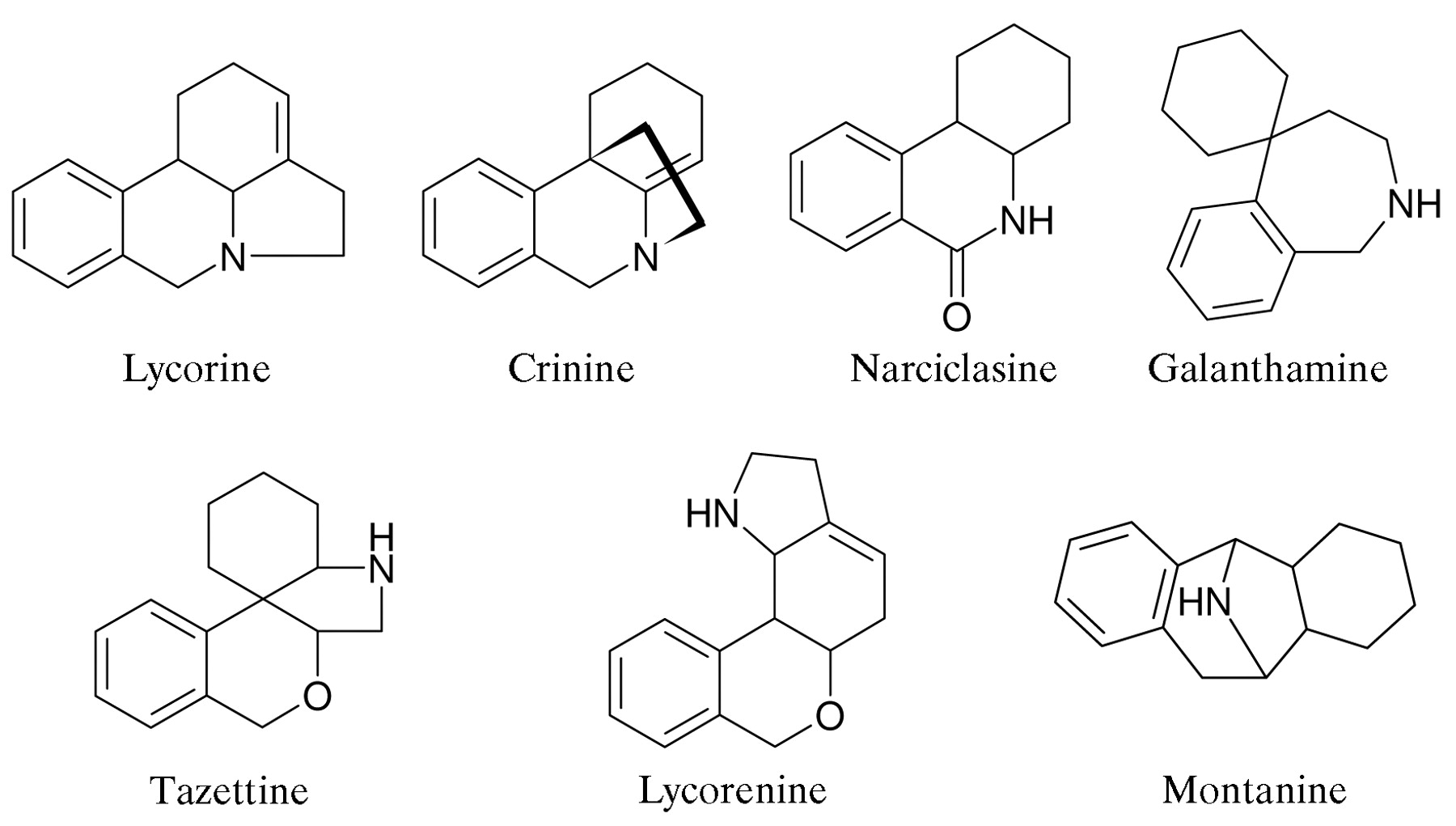
Amaryllidaceae alkaloid skeletal types.
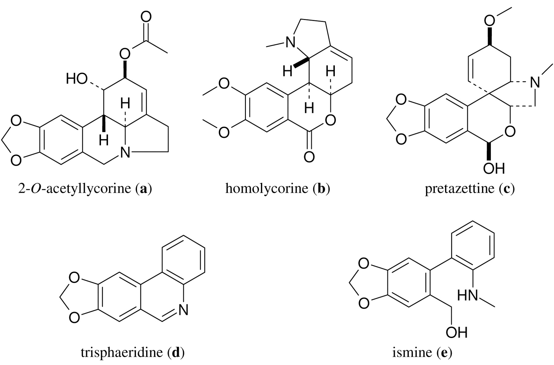
Structures of the tested Amaryllidaceae alkaloids.
The sequence of cytostatic potency on the human MDR1 gene transfected and non-transfected mouse lymphoma cells was c >a>d>b>e. Hohmann et al. previously demonstrated the cytostatic effects of lycorine and tazettine on L5178 MDR cells (0.82 and 23.68 μM, respectively) (13). These data allow the conclusion that the acetylation of lycorine decreases, whereas introduction of a hydroxy group onto tazettine increases, the cytostatic effect of the given compound.
Evaluation of combination experiments.
The parental cell line was more sensitive than the MDR1-gene transfected cells to these substances, the differences proving substantial for b and c. From these data, it may be speculated that these drugs are substrates for P-gp.
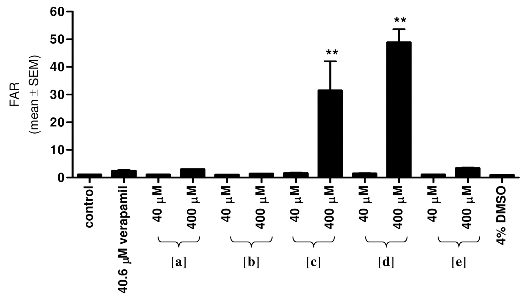
Reversal of MDR. To evaluate their ability to inhibit Pgp-mediated drug efflux, the investigated alkaloids were tested in the Rh-123 accumulation assay with the L5178 MDR mouse lymphoma cell line (Figure 3). At 40 μM, all of the tested compounds had a lower effect than that of the positive control, verapamil (40.6 μM). At higher concentration (400 μM), compounds c and d displayed a significantly increased Rh-123 accumulation (FAR values: 31.40 and 48.75, respectively) as compared with the untreated control cells. At 400 μM, a and e exhibited modest effects (2.92 and 3.32, respectively), while b had no pump-blocking effect. Hohmann et al. studied related Amaryllidaceae alkaloids but no P-gp blocking effect by tazettine, lycorine, haemanthamine and haemanthidine was detected (13).
Interaction with doxorubicin. To assess the effects of the Amaryllidaceae alkaloids on doxorubicin-induced toxicity, MTT colorimetric assays were performed to determine the interactions between the alkaloids and doxorubicin, using the checkerboard method (Table III). Compounds a and c were able to potentiate the antiproliferative activity of doxorubicin on the L5178 MDR cell line, as an indication of synergism between these two compounds. Although compound d was the most effective agent in the MDR reversal test, surprisingly the combination with doxorubicin resulted in an additive antiproliferative effect. An additive interaction was also observed for compound b. The discrepancy between the results of the Rh-123 accumulation and checkerboard experiments reflects the basically different contitions of the two assays. The inhibition of P-gp in an acute situation can be compensated during a longer exposure, as in the case of the MTT assay. On the other hand, a long-lasting functional deterioaration of P-gp could develop through expressional modulation of the protein, instead of its direct inhibition, as in the case of ecteinascidin-743, a tetrahydroisoquinoline alkaloid isolated from a marine tunicate (21).
Calculated IC50 values of the tested compounds.
Results of the combination study with doxorubicin. The calculated fractional inhibitory index (FIX) indicates the character of the combination. Each result is the mean±SEM of data from three experiments.
Apoptosis induction. AO/EB double staining is useful for the parallel detection of apoptosis and necrosis in a cell population seeded onto a 96-well plate. AO permeates the intact cell membrane and stains the nuclei green; EB is taken up only when the cytoplasmatic membrane integrity is deteriorated, and stains the nucleus red. Intact cells therefore exhibit homogeneous green nuclei, whereas apoptotic cells contain condensed or fragmented chromatin. Late apoptotic or necrotic cells have orange nuclei. HeLa cells treated with a or d exhibited apoptotic morphological changes, including cellular shrinkage and granulation in the nucleus (Figure 4). The homogenous green staining for the control cells denotes AO intercalated into the DNA. The progression of chromatin condensation and nuclear fragmentation (green dots) and the red fluorescence of EB were visible in the treated groups.
In parallel with the morphological changes, the biochemical features of apoptosis can be approached. The degradation of nuclear DNA was detected by flow cytometry with PI staining. Cells with a subdiploid DNA content were regarded as an apoptotic population. While the subG1 population of intact cells was less than 5%, a significant and concentration-dependent increase in DNA fragmentation was observed in HeLa cells after 24-h treatment with a or d. At the highest concentration, the content of subG1 cells was about 25-35% (Figure 5).

Effects of the Amaryllidaceae alkaloids on L5178 MDR mouse lymphoma cells in the Rh-123 accumulation assay. Each result is the mean±SEM of data from three experiments. **Denotes p<0.01 as compared with the control value.
Discussion
Plants of the Amaryllidaceae family, are amongst the top 20 in the most widely used medicinal plant families. As primary constituents, nearly 500 structurally diverse alkaloids have been isolated, most of which possess significant biological activity (22). Extracts from the plants are applied in ethnopharmacology for different diseases. One of the most important compounds is galanthamine, an inhibitor of acetylcholinesterase, which is registered as a drug for Alzheimer's disease (10, 23-25). As a result of further research, a wide range of pharmacological action has been attributed to the constituents of these plants, including antimalarial (26), sedative (27), antiviral (28), analgesic (29) and antihypertensive effects (30).
The antitumor properties of the Amaryllidaceae alkaloids are well known. The first-described compound with a cytostatic effect was lycorine (31), which has been shown to exhibit antitumor activities (32-35), e.g. it can suppress leukaemia cell growth and reduce cell survival by arresting the cell cycle and inducing the apoptosis of tumour cells (36) and it can induce apoptosis and cell cycle arrest in a pre-B lymphoid cell line (KM3) (37). Other types of Amaryllidaceae alkaloids likewise exhibit cytostatic effects, e.g. those of tazettine or crinine type (38, 39). However, the most promising candidates are the narciclasin type pancratistatin and narciclasine. These molecules reached the stage of preclinical development, but exhibit low water-solubility and cannot be formulated for in vivo use (40-42). The apoptosis-inducing capacity of the crinine type alkaloids crinamine and haemanthamine has been confirmed (43).
In the present set of experiments, pretazettine (c) proved to be the most active compound on both human and murine lymphoma cells; such a cytostatic effect has also been recognized by other groups (39, 44). The MDR-reversal effect of the Amaryllidaceae alkaloids has not been reported previously. Pretazettine (c) and trisphaeridine (d) significantly increased the intracellular concentration of Rh-123.
In order to assess the effects of the Amaryllidaceae alkaloids on doxorubicin-induced toxicity, MTT colorimetric assays were performed to determine the interactions between the alkaloids and doxorubicin, using the checkerboard method. Compounds a and c enhanced the antiproliferative activity of doxorubicin on the L5178 MDR cell line; synergism was detected for these two compounds. Although d was the most effective agent in the MDR reversal test, its combination with doxorubicin surprisingly resulted in an additive antiproliferative effect. An additive interaction was also observed for compound b.
An apoptosis-induction capacity rather than necrosis induction is accepted as a key feature of a potential antitumor drug. Accordingly, the apoptotic potentials of the tested agents were investigated for two selected alkaloids (a and d) (45). The morphological changes were detected by AO/EB double staining with fluorescence microscopy. Typical markers, including cellular shrinkage, nuclear condensation and an increased membrane permeability, were observed after a 24-h treatment. In parallel with the morphological changes, the biochemical features of apoptosis were detected and the degradation of nuclear DNA was measured by flow cytometry after PI staining. While the subG1 position of the vehicle-treated cells was less than 5%, the treatment with a or d resulted in a concentration-dependent increase in the proportion of subG1 cells.
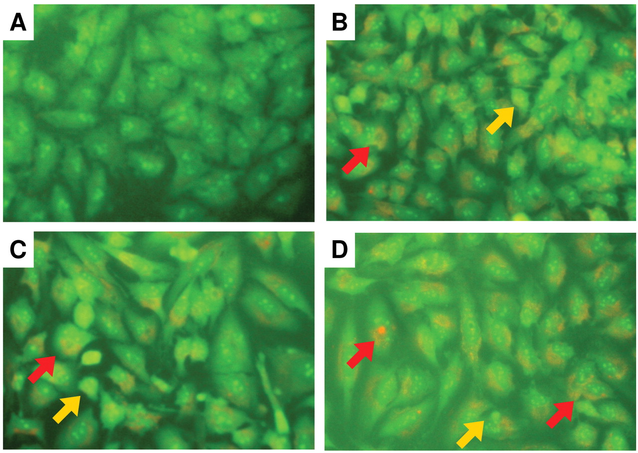
Fluorescence microscopy images of double staining with AO and EB: control HeLa cells (A); treatment with 15 or 30 μM 2-O-acetyllycorine (B, C), or 15 μM trisphaeridine (D), respectively, for 24 h. Red arrows indicate the fluorescence of EB; yellow arrows indicate cellular shrinkage and nuclear granulation. Initial magnification ×200.

DNA fragmentation assay. SubG1 population of HeLa cells after 24-h treatment with 2-O-acetyllycorine or trisphaeridine. *,**Denote p<0.05 and p<0.01, respectively, as compared with the control value.
Taken together, these data indicate that the tested Amaryllidaceae alkaloids exhibit a combination of anticancer activities, including a direct cytostatic effect and a P-gp-inhibiting property. With the exception of pretazettine, the calculated IC50 values of the agents are not outstanding; however, two selected agents (2-O-acetyllycorine and trisphaeridine) were able to induce apoptosis in HeLa cells. Pretazettine inhibited the activity of P-gp and also increased the effect of doxorubicin against resistant murine lymphoma cells. Overall, it is suggested that some of the Amaryllidaceae alkaloids can be regarded as promising starting structures for the development of future anticancer agents.
Acknowledgements
This work was supported by Hungarian Scientific Research Fund (OTKA K72771). I. Zupkó is grateful for support from a Bolyai János Postdoctoral Fellowship.
- Received July 7, 2008.
- Revision received December 1, 2008.
- Accepted December 10, 2008.
- Copyright © 2009 International Institute of Anticancer Research (Dr. John G. Delinassios), All rights reserved
References
In this issue


Jump to section
Related Articles
Cited By...
- No citing articles found.