


Abstract
Background: Chemo-mechanical caries removal eliminates the outermost portion of the infected layer, leaving behind healthy dentine surfaces, with scarce dental tissue damage; however, the safety of caries solvents has not been established. The aim of the present study was to investigate the possible cytotoxicity of two popular chemo-mechanical caries removal agents. Materials and Methods: The cytotoxicity of Carisolv, Papacarie Duo and control vehicle solution (0.155-20% v/v) against human oral squamous cell carcinoma cells (HCS-2, HSC-3, HSC-4, Ca9-22) human gingival fibroblast (HGF), pulp (HPC) and periodontal ligament fibroblast (HPLF) was determined by 3-(4,5-dimethylthiazol-2-yl)-2,5-diphenyltetrazolium bromide method. Prostaglandin E2 (PGE2) was quantified by enzyme-linked immunosorbent assay. Changes in fine cell structure were assessed by transmission electron microscopy. Results: Carisolv exhibited neither cytotoxicity nor hormetic growth stimulation. Papacarie Duo significantly reduced the viable cell number within 30 min. HSC-4 exhibited the highest sensitivity, followed by HSC-2>HSC-3>HPLF>Ca9-22>HPC>HGF cells. Interleukin-1β (3 ng/ml) stimulated HGF, but not HPC cells to produce PGE2 in the culture medium. Papacarie Duo stimulated HGF cells to produce PGE2 in synergistic fashion with interleukin-1β. Conclusion: Carisolv had acceptable biocompatibility with both normal and cancerous oral cells. On the other hand, Papacarie Duo had a rapid but slight cytotoxicity and pro-inflammatory action against oral cells, suggesting the importance of careful application of this agent.
Although current ‘minimally-invasive’ dentistry has been well-developed for the detection, diagnosis, interception and treatment of dental caries at the microscopic level (1), inherent fundamental drawbacks of the drilling approach (such as unpleasantness to patients, need for local anesthesia, and potential adverse effects on pulp due to heat and pressure) remain. Chemo-mechanical caries removal, introduced almost three decades ago, is considered to be a non-invasive alternative for the removal of carious dentine. The main objective of chemo-mechanical caries removal is to eliminate the outermost portion of the infected layer so as to re-mineralize and repair the de-mineralized dentin (2, 3). In essence, this technique applies a chemo-mechanical solution onto the caried tissue, inducing a partial degradation of collagen therein by chlorination and disrupting hydrogen bonding (4). A chemo-mechanical system using 5% sodium hypochlorite was first introduced in 1975 (5), followed by GK-101 (6), the Caridex system (7) and then Carisolv (8). Due to disadvantages, such as a short half-life, high corrosiveness, requirement for specialized instruments, and high cost, a new formula known as Papacarie was introduced in Brazil in 2003 (9). Papacarie consists mainly of papain, which has bacteriostatic, bactericidal and anti-inflammatory effects.
Carisolv and Papacarie Duo are the two most popular chemo-mechanical removal caries systems. Their caries-removing efficacy, as compared to conventional rotatory caries removal, has been confirmed by scanning electron microscopy (10, 11), residual number of cariogenic bacteria (12, 13), tissue microhardness (14, 15), shear bond strength of adhesive materials (16), residual dentin composition (15, 17, 18) and long-term clinical evaluation (19-21). We previously reported that the efficacy of chemo-mechanical caries removal by Carisolv was comparable to that of conventional rotatory caries removal, judging from the remaining Vickers microhardness and composition of surface tissue (unpublished data).
Despite worldwide use of chemo-mechanical caries removal systems, means to combat their adverse effects have not yet been found. To our knowledge, only a few reports have dealt with their cytotoxic effects on cultured cells: The contact of Carisolv and Papacarie with mouse embryonic fibroblast (NIH-3T3) (22) and mouse mammary carcinoma (FM3A) (23) cell lines did not affect cell viability.
We investigate herein the cytotoxicity of two popular chemo-mechanical caries removal agents, Carisolv and Papacarie Duo, against human pulp cells (HPC), gingival fibroblast (HGF), periodontal ligament fibroblast (HPLF), oral squamous cell carcinoma (OSCC) (HSC-2, HSC-3, HSC-4) and gingival carcinoma (Ca9-22) cells, and their effect on the pro-inflammatory prostaglandin E2 (PGE2) production and the changes in fine cell structure by transmission electron microscopy.
Materials and Methods
Materials. The following chemicals and reagents were obtained from the indicated companies: Dulbecco's modified Eagle's medium (DMEM), GIBCO BRL (Grand Island, NY, USA); fetal bovine serum (FBS), JRH Bioscience (Lenexa, KS, USA); Carisolv gel, single mix, MediTeam (Göteborg, Sweden); Papacarie Duo, Formulae Acao (San Pablo, Brazil); 3-(4,5-dimethylthiazol-2-yl)-2,5-diphenyltetrazolium bromide (MTT), Aldrich (St. Louis, MO, USA); PGE2 assay kit, Cayman Chemical Co. (Ann Arbor, MI, USA), culture plastic dishes, 6-well and 96-mircowell plates, Becton Dickinson (Franklin Lakes, NJ, USA); dimethyl sulfoxide (DMSO), Glutamic acid, leucine, lysine, NaCl and NaOH, Wako Pure Chem Ind. (Osaka, Japan); NaOCl, Honcho Chemical Inc. (Noda, Chiba, Japan).
Cell culture. Human OSCC (HSC-2, HSC-3, HSC-4) and gingival carcinoma cell lines (Ca9-22) were obtained from Riken Cell Bank (Tsukuba, Japan). Normal human oral cells (HGF, HPC, HPLF) were prepared from periodontal tissues, as previously reported (24). Cells were cultured at 37°C in DMEM supplemented with 10% heat-inactivated FBS, 100 units/ml penicillin G and 100 μg/ml streptomycin sulfate under a humidified atmosphere with 5% CO2. Cells were then harvested by treatment with 0.25% trypsin-0.025% EDTA-2Na in phosphate-buffered saline without calcium and magnesium [PBS(−)] and either subcultured or used for experiments.
Sample preparation. Carisolv gel, single mix was prepared as per the manufacturer's instructions. Carisolv and Papacarie Duo were dissolved in fresh culture medium by vortexing for 3 min. We used a control vehicle solution following the Carisolv composition (0.5% of NaOCl, 0.1 M each of glutamic acid, leucine, lysin, NaCl and pH adjusted to 11 with NaOH). The control solution was similarly dissolved in culture medium and vortexed for 3 min.
Assay for cytotoxic activity. Cancer cells (2×10⁴ cells/0.1 ml) and normal cells (1:3 ratio) were inoculated into each well of 96-microwell plates and incubated for 48 h to achieve the complete cell adherence. Carisolv, Papacarie Duo or control solutions were added at 0, 0.155, 0.31, 0.62, 1.25, 2.5, 5, 10 and 20% (v/v). After incubation for a further 48 h, the relative viable cell number was then determined by the MTT method. The culture medium was replaced with MTT (0.2 mg/ml) dissolved in DMEM, and cells were incubated for a further 4 h at 37°C. The formazan product was dissolved with DMSO, and the absorbance of the lysate at 540 nm, which reflects mitochondrial activity, was determined using a Multiskan microplate reader (Biochromatic Labsystem, Osaka, Japan). The 50% cytotoxic concentration (CC50) was determined from the dose–response curve and the mean CC50 (±S.D.) value of each solution was calculated in triplicate from three independent experiments.
Prostaglandin E2 (PGE2) production. Near-confluent HPC and HGF cells were treated with different concentrations of Papacarie Duo (0, 1.25, 2.5, and 5% v/v) for 30 min in the fresh culture medium. Cells were induced to produce PGE2 with interleukin (IL)-1β (3 ng/ml) (R&D Systems, Minneapolis, MN, USA) for a further 24 h. The concentration of PGE2 released into the culture medium was determined by enzyme-linked immunosorbent assay (ELISA) (Cayman Chemical Co., Ann Arbor, MI, USA) with the PGE2 assay kit, according to the manufacturer's recommended procedures.
Fine cell structure. HPC, HPLF and HGF cells were treated with 0, 0.62, and 1.25% (v/v) of Papacarie Duo for 3 h. The cells were washed three times with cold PBS(−) and fixed for 1 h with 2% gultaraldehyde in 0.1 M cacodylate buffer (pH 7.4) at 4°C, scraped with a rubber policemen, post-fixed for 90 min with 1% osmium tetraoxide-0.1 M cacodylate buffer (pH 7.4), dehydrated and then embedded in Araldite M (CIBA-GEIGY Switzerland; NISSHIN EN Co., Ltd., Tokyo, Japan). Thin sections were stained with uranyl acetate and lead citrate, and were then observed under a JEM-1210 transmission electron microscope, Japan Electron Optics Laboratory Co., Ltd. (Akishima, Tokyo, Japan) (magnification: ×5,000) at an accelerating voltage of 100 kV (25).
Statistical analysis. Data are expressed as the mean±standard deviation (SD). Statistical analysis was performed by paired t-test and ANOVA post-hoc Tukey test using SPSS (Statistical Package for the Social Sciences, Chicago, IL, USA). Differences were considered significant at p<0.05.
Results
Cytotoxic activity of caries solvents. We investigated the cytotoxicity of chemo-mechanical caries removal agents (Carisolv, Papacarie Duo) and control solution against a total of seven normal and cancer oral cells (Table I). Carisolv showed little or no apparent cytotoxicity nor hormetic growth stimulation (Figure 1). Papacarie Duo led to some cytotoxic effects at high concentrations, reducing the number of viable cancer cells more efficiently than of normal cells. Repeated experiments (n=3) confirmed that the CC50 values of Carisolv and control solution were greater than 20% (v/v) against normal cells, without statistical difference between them (p>0.05) (Table II). Papacarie Duo significantly reduced the viable cell number. HSC-4 cells had the highest sensitivity against Papacarie Duo, followed in order by HSC-2, HSC-3, HPLF, Ca9-22, HPC and HGF (Table II). Cancer cell lines were generally more sensitive (p<0.05) to Papacarie Duo than to Carisolv.
Cytotoxic activity (% v/v) of Carisolv, Papacarie Duo and control solution against human oral normal and cancer cells. Values represent mean±S.D. of three independent experiments.
PGE2 production. IL-1β (3 ng/ml) did not affect the number of viable HGFs, but induced an approximately 10-fold increase of PGE2 release into the culture medium [from 0.43 ng/ml (control) to 5.10 ng/ml (IL-1β)]. Although Papacarie Duo alone reduced HGF viability (Figure 2C), it enhanced PGE2 production slightly, but significantly at 5% (v/v) (control 0.43 ng/ml vs. Papacarie Duo 0.63 ng/ml, p<0.01). Furthermore, Papacarie Duo synergistically enhanced IL-1β-stimulated PGE2 production (Figure 2D).
IL-1β also did not affect the viable cell number of HPCs (Figure 2A), but dramatically enhanced the release of PGE2 into the culture medium. Papacarie Duo was more cytotoxic to HPCs, and reduced IL-1β-stimulated PGE2 production (Figure 2B), possibly due to its higher cytotoxicity (Figure 2A).
Change in fine cell structure. As compared with control HPCs, HPLFs and HGFs cells (Figure 3A, D and G), cells treated for 3 h with 0.62% (Figure 3B, E and H) and 1.25% (Figure 3F and I) (v/v) of Papacarie Duo exhibited somewhat irregular morphology of cell membrane, cytoplasm and nucleus, with many vacuoles containing a flocculent and granular material, but having morphologically normal Golgi apparatus and mitochondrial structures without any pathological change. On the other hand, HPCs and HGFs treated with Papacarie Duo at 1.25% (v/v) exhibited slight membrane damage (Figure 3C and I).

Effect of chemo-mechanical caries removal agents on the viable cell number. Near-confluent human pulp cells (HPC), periodontal ligament fibroblast (HPLF), gingival fibroblast (HGF), oral squamous cell carcinoma (OSCC) (HSC-2, HSC-3, HSC-4) and gingival carcinoma (Ca9-22) cells were incubated for 24 h with Carisolv (A, B), Papacarie Duo (C, D) and control solution (E, F) at different concentrations (v/v). After incubation for a further 48 h, the relative viable cell number was determined by the 3-[4,5-dimethylthiazol-2yl]-2,5-diphenyltetrazolium bromide assay. Each value represents the mean±S.D. of triplicate assays.
Discussion
Chemo-mechanical caries removal systems have been used globally, not only in the field of pediatric dentistry, but also in operative dentistry, since they provide comfortable caries elimination and preservation of healthy dental structures. The present study demonstrated, to our knowledge for the first time, that two solutions used for chemo-mechanical caries removal had different magnitudes of cytotoxicity towards oral normal and cancer cells. Carisolv and control solution had little or no cytotoxicity against all cells tested. On the other hand, Papacarie Duo significantly reduced the viable cell number of both normal and cancer human oral cells, the cancer cells being more sensitive.

Synergistic stimulation by Papacarie Duo and interleukin (IL)-β on PGE2 production. Near-confluent HGF or HPC cells were pre-treated with different concentrations (v/v) of Papacarie Duo for 30 min, and then stimulated with IL-1β (3 ng/ml) or not for a further 24 h. The viable cell number (A, C) and the concentration of PGE2 in the culture medium (B, D) were then determined by the 3-[4,5-dimethylthiazol-2yl]-2,5-diphenyltetrazolium bromide assay and enzyme-linked immunosorbent assay (ELISA), respectively. Each value represents the mean±S.D. by triplicate assays. *p<0.05, **p<0.01 paired t-test.
Cytotoxic concentration (CC50) of Carisolv, Papacarie Duo and control solution against normal and cancer oral cells. Values represent the mean±S.D. of three independent experiments.
The present study also demonstrated for the first time that Papacarie Duo induced slightly, but significantly PGE2 production in HGFs, and the PGE2 production was synergistically enhanced in the presence of IL-1β. On the other hand, Papacarie Duo did not have such a synergistic effect of PGE2 production with IL-1β in HPCs, possibly due to severe damage (as demonstrated by the TEM study, Figure 3C). We recently found that TiO2 nanoparticles synergistically enhanced IL-1β-stimulated PGE2 production in HGFs (26). These results suggest that dental materials such as Papacarie Duo and TiO2 nanoparticles may induce or aggravate the gingivitis.
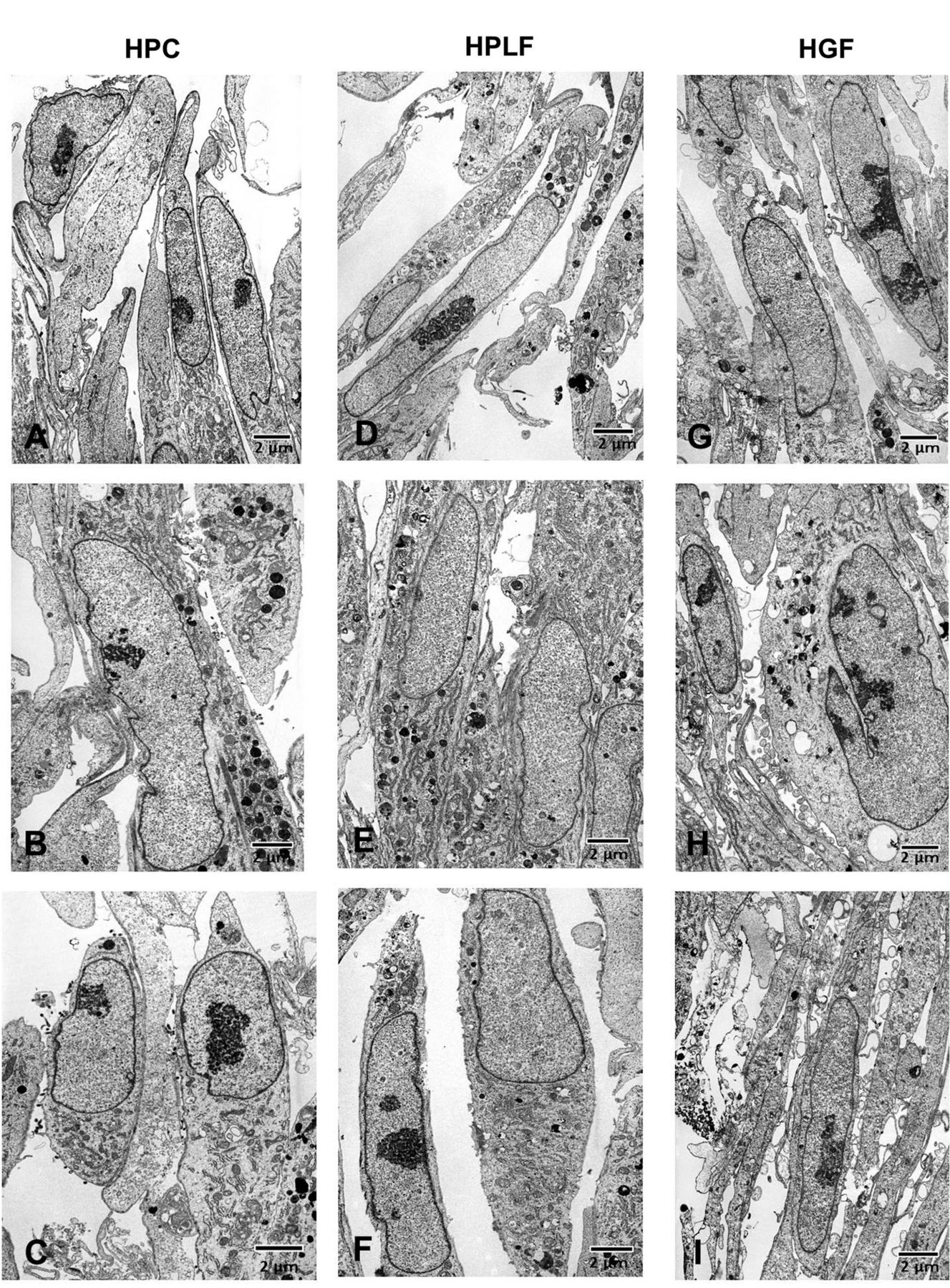
Effect of Papacarie Due on fine cell structure. Near-confluent human pulp cells (HPC) (A-C), periodontal ligament fibroblast (HPLF) (D-F) and gingival fibroblast (HGF) (G-I) cells were treated with Papacarie Duo at 0 (A, D, G), 0.62 (B, E, H), or 1.25% (C, F, I) (v/v) for 3 h. The cells were then washed three times with cold phosphate-buffered saline without calcium and magnesium [PBS(−)], fixed and post-fixed for observation under transmission electron microscopy.
It has been reported that the contact to Carisolv and papain gel (Papacarie) for 12 h did not affect the viability of mouse embryonic fibroblast (NIH-3T3) (22) and mouse mammary carcinoma (FM3A) (23). This is not in agreement with the results of the present study with seven human cell types that were all sensitive to Papacarie Duo. As for Carisolv, studies have shown different effects, depending on the targets. Certain in vivo and in vitro studies have not reported any toxic effect of Carisolv (23, 27), whereas short exposure to Carisolv of oral mucosa caused inflammatory reactions for a further 24 h (28). Carisolv in contact with pulp tissue induced the alkaline hydrolysis of cellular components, but did not influence the distribution or neuropeptide expression of sensory fibers, nor decomposed the collagenous tissue components (29, 30). This is in agreement with the present finding that Carisolv did not affect the cell viability of three human oral normal cell types and four OSCC cell lines, suggesting acceptable biocompatibility.
In contrast to the extensive study of Papacarie Duo on caries removal efficiency (31), morphological and chemical remaining dentine (32) and number of residual bacteria (13), study of the biocompatibility and safety of Papacarie Duo is limited. The present study demonstrated that Papacarie Duo exhibited some cytotoxicity and enhanced PGE2 production in HGFs. Clinically, the use of Papacarie Duo should be limited to special treatments for delicate or compromised sick patients since its mishandling may result in significant damage to pulp and mouth tissues.
In conclusion, the present study suggests that chemomechanical caries removal with Carisolv may have acceptable biocompatibility against oral cells. Papacarie Duo rapidly reduced the viability and enhanced inflammatory responses in HGFs, suggesting the importance of careful application in the dental practice.
Footnotes
-
↵* Ph.D. student of Health Science, Autonomous University State of Mexico.
- Received January 29, 2014.
- Revision received April 1, 2014.
- Accepted April 2, 2014.
- Copyright © 2014 International Institute of Anticancer Research (Dr. John G. Delinassios), All rights reserved
References
In this issue


Jump to section
Related Articles
Cited By...
- No citing articles found.