


Abstract
Background/Aim: The banana flower is used for ameliorating urinary disturbance. However, there is limited evidence to support the efficacy or mechanism of action of banana flower against benign prostatic hyperplasia (BPH). In the present study, the anti-BPH activity and mechanisms of banana flower extracts were investigated in vitro and in vivo. Materials and Methods: The banana flower extract is a water-soluble extract obtained by sonication. MTT assay was used to examine whether banana flower extract exhibited cytotoxic effects on BPH-1 cells. The effect of banana flower extract on cell-cycle distribution was examined by flow cytometry. The expression of cell-cycle-regulatory molecules was determined by western blot analysis. Testosterone propionate (TP)-induced rat model of BPH was used to evaluate the anti-BPH activity of banana flower extract in vivo. Results: Banana flower extract reduced epithelial cell line BPH-1 cell viability through cell-cycle arrest at G1 phase. Moreover, banana flower extract reduced the expression of cyclin D1 and cyclin-dependent kinase 6, while it increased the expression of p53 and p27. Interestingly, banana flower extract suppressed BPH-related inflammatory responses through suppressing cyclo-oxygenase-2 expression and prostaglandin E2 production. Finally, banana flower extract administered orally to male rats reduced prostatic weight and serum dihydrotestosterone level, and improved prostate gland morphology. High-performance liquid chromatography revealed that banana flower extract contains citric acid, taurine, pantothenic acid and nicotinic acid components. In summary, banana flower extract may be used as a therapeutic agent for BPH via anti-proliferative and anti-inflammatory activities.
Benign prostatic hyperplasia (BPH), an enlarged prostate gland, is the most common urological disease affecting about 50% of men aged over 50 years (1-3). Moreover, more than half of these patients will go on to develop a macroscopic nodular enlargement that gradually results in lower urinary tract symptoms (LUTS) (4, 5). LUTS encompasses all urinary symptoms, namely, voiding (frequency, urgency and nocturia), urinary storage (hesitancy, weak streaming and retention), or postvoiding. Due to hormonal imbalances in aging men, the prevalence and incidence of LUTS secondary to BPH has steadily increased (6). The effects of estrogen/androgen balance, and inflammation-mediated oxidative stress in the prostate gland affect the progression of BPH (7).
5α-Reductase (5αR) inhibitors, which inhibit the conversion of testosterone to dihydrotestosterone (DHT), are used for the treatment of symptomatic BPH. As a result of side-effects such as headache, nasal congestion, sexual dysfunction, diarrhea, and orthostatic hypotension, the application of 5αR inhibitors are very limited (8, 9). Therefore, it is necessary to development new drugs that can alleviate the development of BPH. Recently, dietary interventions derived from natural substances have been identified for their potential in the management of BPH.
Banana (Musa acuminata), a main economy crop in Taiwan, is rich in vitamins and minerals, such as vitamin C, magnesium and potassium. Over the past few decades, increasing attention has focused on the health benefits of M. acuminata. All parts of banana have many traditional and documented medicinal uses and have been used to treat many diseases (10, 11). The banana flower exhibits high nutritional value like the banana fruit. Tribal communities have used banana flower as an important wild food and medicinal plant (10, 11). Much remains to be studied about banana flower, especially banana stamens.
The role of banana flower for treatment of BPH is not fully defined. In the present study, we investigated the anti-BPH efficacy of a water extract of banana flowers and its mechanisms.
Materials and Methods
Chemicals and reagents. 3-[4,5-Dimethylthiazol-2-yl]-2,5-diphenyl-tetrazolium (MTT) and propidium iodide (PI) were purchased from Sigma Chemical Co. (St. Louis, MO, USA). Cyclin D1 and cyclin-dependent kinase 6 (CDK6) antibodies were purchased from St John's Laboratory Ltd. (London, UK). p53, p27, cyclo-oxygenase-2 (COX2) and β-actin antibodies were purchased from Cell Signaling Technology (Beverly, MA, USA). Secondary antibodies were purchased from Life Technologies Co. (Carlsbad, CA, USA).
Cell lines and cell culture. BPH epithelial cell line BPH-1 was obtained from the American Type Culture Collection (Rockville, MD, USA) and maintained in RPMI 1640 medium (Gibco, Grand Island, NY, USA) supplemented with 10% fetal bovine serum (FBS) (Invitrogen, Carlsbad, CA, USA), 1% L-glutamine, penicillin (50 U/ml) (Invitrogen), at 37°C under an atmosphere of 5% CO2 in air.
Preparation of banana flower extract. The banana flowers used in the present study included but were not limited to the flowers of Musa paradisiacal, Musa sapientum L., Musa spp. AAB Silk, Musa spp. ABB Bluggoe, Musa spp. AAA Robusta, Musa spp. AAB Bluggoe. Firstly, the stamens of banana flowers were harvested by hand or machine, and cleaned with water. The stamens of banana flowers were mixed with water at a ratio between 1:4 and 1:6, and extracted with cold-sonication for 0.5 to 2.0 h to obtain a crude banana flower extract. Finally, the crude banana flower extract was centrifuged and filtered through a 300-mesh screen (Heibei Accurux Wire Works, Anping, Hebei, China) to obtain banana flower extract.

Effect of banana flower extract on the growth of BPH-1 cells. BPH-1 cells were treated with vehicle, or banana flower extract for 48 h at the indicated concentration and the relative cell viability was assessed using the 3-[4,5-dimethylthiazol-2yl]-2,5-diphenyl-tetrazolium bromide assay. The data are the mean±SEM of three independent experiments. Significantly different at *p<0.05, and **p<0.01, compared with vehicle control.
Cell viability assay. Cell viability measurement was performed using MTT reagent (12). BPH-1 cells (1×10⁴) were cultured in a 96-well plate, and treated with water (as control) and a series of concentrations of banana flower extract (0.25, 0.5, 1.0, 1.5, 2.0 mg/ml) for 48 h. After treatment, MTT in phosphate-buffered saline (PBS) was added to each well at a final concentration of 500 μg/ml. After 1 h of incubation, the solution was removed from each well and 80 μl dimethyl sulfoxide was added to dissolve the crystals formed. The absorbance value was measured at 570 nm with an enzyme-linked immunosorbent assay (ELISA) reader (Bio-Rad, Hercules, CA, USA).
Western blotting analysis. According to a previous publication (13), protein concentration was determined by Bio-Rad Protein Assay (Bio-Rad, Hercules, CA, USA). Proteins (50 μg) from the BPH-1 cells were separated using sodium dodecyl sulfate-polyacrylamide gel electrophoresis and transferred to polyvinylidene difluoride membrane. Membranes were then probed with the following antibodies: anti-p53, anti-p27, anti-CDK6, anti-cyclin D1, anti-COX2 and anti-β-actin. The immunoreactive bands were revealed using ECL™ Prime Western Blotting Detection Reagent (GE Healthcare UK Ltd.) and the results were quantitated by Image J software.

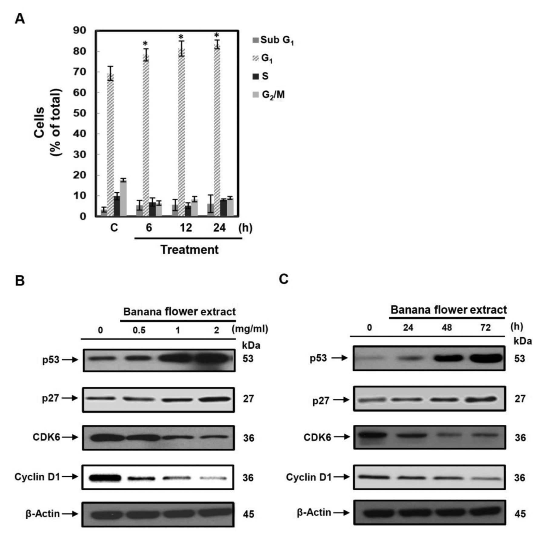
Effect of banana flower extract on cell-cycle distribution and expression of related proteins. A: BPH-1 cells were treated with banana flower extract (2 mg/ml) for the indicated time. The numbers of cells in different phases were determined by flow cytometry. The data are the mean±SEM of three independent experiments. BPH-1 cells treated with banana flower extract at 0.5, 1.0 and 2.0 mg/ml for 48 h. C. BPH-1 cells were treated with banana flower extract (2 mg/ml) for the indicated times. The levels of cyclin D1, cyclin-dependent kinase 6 (CDK6), p53, and p27 were determined by western blot analysis of whole-cell lysates. Western blot data are representative of at least three independent experiments. *Significantly different at p<0.05, compared with vehicle control.
Flow cytometric analysis. BPH-1 cells (1×10⁴) were collected by centrifugation, and the cell number was adjusted to a density of ~1×10⁶ cells/ml. Cells were then incubated with PI using flow cytometric kits (Abcam) according to the manufacturer's instructions. Finally, cell-cycle analysis was carried out by BD FACScan™ system (BD Biosciences, San Jose, CA, USA).
Measurement of prostaglandin E2 (PGE2) release. BPH-1 cells were seeded into 48-well plates at 1×10⁴ cells/well. Overnight, the medium was refreshed and a series of concentrations of banana flower extract were added to the medium for 24 h. According to the manufacturer's instructions, culture supernatants were analyzed using PGE2 Enzyme Immunoassay Kit (Cayman Chem., Ann Arbor, MI, USA).
In vivo study. The animal study was approved by the Institutional Animal Care and Use Committee of China Medical University (#105365). Moreover, the animal procedures were performed according to the Guide for the Care and Use of Laboratory Animals (14). Seven-week-old male Sprague Dawley rats (200-220 g) were purchased from The BioLasco Taiwan Co., Ltd. (Taipei, Taiwan). Rats were randomized and divided into four groups (five mice/group). The negative control (NC) group was injected s.c. with 100 μl corn oil and given 0.5 ml PBS p.o.; a BPH control group was injected s.c. with 10 mg/kg of testosterone propionate (TP) (Sigma Chemical Co., St. Louis, MO, USA) dissolved in corn oil and given 0.5 ml PBS p.o.; banana flower extract-treated groups were injected s.c. with 10 mg/kg of TP (Sigma Chemical Co.) and given either 200 or 500 mg/kg of banana flower extract p.o. All treatments were given 5 days a week for 4 weeks.
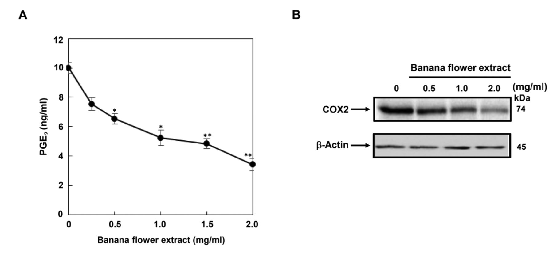
Effect of banana flower extract on inflammation in BPH-1 cells. A: BPH-1 cells were treated with vehicle, or banana flower extract for 24 h at the indicated concentration. Culture supernatants were analyzed for secreted prostaglandin E2 (PGE2). The data are the mean±SEM of three independent experiments. Significantly different at *p<0.05, and **p<0.01, compared to vehicle control. B: BPH-1 cells were treated with vehicle, or banana flower extract at 0.5, 1.0 and 2.0 mg/ml for 48 h. The level of cyclo-oxygenase-2 (COX2) was determined by western blot analysis of whole-cell lysates. Western blot data are representative of at least three independent experiments.
Measurement of DHT levels. After the animal study, the level of DHT in serum was determined using an ELISA kit according to the manufacturer's instructions (ALPCO Diagnostics, Salem, NH, USA) (15).
High-performance liquid chromatography (HPLC) analysis of the bioactive fraction. The HPLC separation was performed using WATERS HPLC system with 2487 dual λ U-V detector. The sample and mobile phase were filtered through 0.22 μm polyvinylidene difluoride filter before injecting to the column.
Statistical analysis. All values are presented as the mean±standard error of the mean (S.E.M) and were derived from at least three separate experiments for each group. Statistical analyses of data were performed with one-way analysis of variance with Dunnett's test. Differences from the control were considered significant at p<0.05.
Results
Banana flower extract reduced epithelial cell line BPH-1 cell viability in vitro. BPH is usually described as a pathological proliferation of epithelial cells. In order to evaluate the antiproliferative activities of banana flower extract on BPH, the epithelial cell line BPH-1 was treated with different concentrations of banana flower extract for 48 h. Our study found that banana flower extract significantly inhibited cell viability in a dose-dependent manner (Figure 1). The data showed that banana flower extract exhibited efficient cytotoxic effects on BPH-1 cells.
Banana flower extract induced cell-cycle arrest at G1 phase in BPH-1 cells. We next assumed that banana flower extract affects cell-cycle regulation in BPH-1 cells. To explore this hypothesis, the populations of BPH-1 cells in different cell-cycle stages were analyzed by flow cytometry. Treatment with 2 mg/ml of banana flower extract significantly induced cell-cycle arrest at G1 phase in BPH-1 cells (Figure 2A). To elucidate the arrest point of banana flower extract-treated BPH-1 cells in the G1 phase, the expression of cyclin D1, CDK6, p53 and CDK inhibitor p27 was determined by western blot analysis. Banana flower extract reduced the expression of cyclin D1 and CDK6, while it increased the expression of p53 and p27 in a dose- (Figure 2B) and time-dependent manner (Figure 2C).
Banana flower extract suppressed BPH-related inflammatory responses in vitro. Inflammation plays a crucial role in the BPH growth and proliferation. To test whether banana flower extract inhibited inflammatory responses, we examined the production of PGE2, which is a potent inflammatory mediator. Banana flower extract significantly suppressed PGE2 production a dose-dependent manner (Figure 3A). Moreover, treatment with banana flower extract inhibited the expression of COX2, an inducible enzyme that produces PGE2 during inflammation (Figure 3B). Our results suggest that banana flower extract suppresses BPH-related inflammatory responses.

Effects of banana flower extract on prostate weight and serum dihydrotestosterone (DHT) concentrations in a rat model of testosterone propionate (TP)-induced BPH. A: Change in relative ratio of prostate to body weight. NC: Negative control group; BPH TP-induced control group. B: Serum concentrations of DHT were measured using enzyme-linked immunosorbent assay. The data are the mean±SEM of three independent experiments (n=5). Significantly different at ##p<0.01 compared to the NC group; *p<0.05 and **p<0.01 compared with the BPH control group.
Effects of banana flower extract on prostate weight and serum DHT concentrations in the TP-induced rat model of BPH. Prostate weight is commonly used to examine the development of BPH. TP-induced rat model of BPH was used to evaluate the anti-BPH activity of banana flower extract in vivo. Our results showed that the prostate weight of rats in the TP-treated group (BPH group) significantly increased compared to the negative control group, which indicated that TP successfully induced BPH. Compared with the BPH control group, the groups treated with banana flower extract (200 mg/kg and 500 mg/kg) showed a significant trend for a reduction in prostate weight (Figure 4A). We next examined whether banana flower extract attenuated the production of DHT, a product that is catalyzed through 5αR activity. As shown in Figure 4B, a significant increase in serum DHT concentration was found in the groups treated with BPH compared to the NC group. In the banana flower extract-treated groups (200 and 500 mg/kg), the serum DHT concentrations were significantly lower than for the BPH control group. In summary, our results suggest a marked recovery of prostate hyperplasia after treatment with banana flower extract.
Active compounds of banana flower extract. Active compounds of banana flower extract were analyzed quantitatively by HPLC. HPLC revealed that banana flower extract contains citric acid, taurine, pantothenic acid and nicotinic acid components.
Discussion
A range of treatment options are currently available for BPH, including surgical interventions and medical treatment. Natural products have gained public interest in recent times due to their role in the prevention of BPH. Native communities have used banana flower as an important wild food and medicinal plant. Although recent studies have demonstrated the biological effects of banana flower, scientific study underlying banana flower action in BPH have not been carried out. To our knowledge, this is the first study to evaluate the efficacy and molecular mechanism of banana flower extract against BPH. Our study found that treatment with banana flower extract markedly inhibited BPH-1 cell proliferation through arrest at the G1 phase. Moreover, treatment with banana flower extract significantly inhibited PGE2 production through inhibition of COX2 expression.
BPH is defined by hyperproliferation of stromal and epithelial cells of the prostate gland (16). Here, we showed that banana flower extract inhibited BPH-1 cell proliferation. Cell proliferation is governed by the cell cycle, which is comprised of four distinct phases: G1 phase, Synthesis phase (S), G2 phase and mitosis (M). The transitions of G1 to S and G2 to M are regulated by various cell cycle-regulatory molecules such as CDKs, CDK inhibitors and cyclins. Banana flower extract reduced the expression of CDK6, while it increased the expression of p27. In summary, our results indicate that banana flower extract arrested cells at the G1 phase of the cell cycle.
Up-regulation of pro-inflammatory cytokines in prostate tissues of patients with BPH has been widely reported (17-19). Moreover, there are many studies which have suggested that chronic prostatic inflammation may be involved in the pathogenesis of BPH (20). COX2 is an enzyme that has been detected in all types of inflammatory cells, and it is increased in epithelial cells from BPH samples, generating pro-inflammatory prostaglandins (21). One recent study evaluated the antioxidant potential of banana flower and the results suggested the banana flower contains potent antioxidants extractable with aqueous and organic solvents (11). In our study, treatment with banana flower extract suppressed COX2 expression and PGE2 production. These data suggest that the anti-BPH effects of banana flower extract might be related to its anti-inflammatory activity.
In this study, the active compounds of banana flower extract were measured by HPLC. HPLC revealed that banana flower extract contains citric acid, taurine, pantothenic acid and nicotinic acid components. Recent studies reported that citric acid has various pharmacological properties, including anti-inflammatory, neuroprotective and hepatoprotective effects (22, 23). In addition, taurine has epithelial–mesenchymal transition-inhibitory effects on human prostate cancer cells (24). Pantothenic acid may contribute to the inflammatory process through increasing CoA level and promoting glutathione synthesis, thereby reducing oxidative stress (25, 26). Our findings of the anti-BPH effects of banana flower extract might be related to its antiproliferative, anti-inflammatory and antioxidant activities consistent with previous studies.
Acknowledgements
The efforts of Yun-Chi Wang, Huai-Mei Hsu and Hsin-Ting Li are appreciated. This study was supported partially by TCI Co., Ltd., (Taipei, Taiwan, ROC).
Footnotes
This article is freely accessible online.
Conflicts of Interest
The banana flower extract was obtained from TCI Co., Ltd., and supported partially by TCI Co., Ltd., (Taipei, Taiwan, ROC).
- Received July 31, 2018.
- Revision received September 19, 2018.
- Accepted September 25, 2018.
- Copyright© 2018, International Institute of Anticancer Research (Dr. George J. Delinasios), All rights reserved
In this issue


Jump to section
Related Articles
Cited By...
- No citing articles found.