


Abstract
Aim: Azoxymethane (AOM) is a potent carcinogen that induces colorectal cancer in mice. Intraluminal gel ultrasound is a technique based on the injection of gel into the rectum. This technique allows the colon to be straightened and to visualize and identify tumours. Materials and Methods: Twenty female C57Bl/6J mice were injected intraperitoneally with 10 mg/kg of AOM one time per week for six weeks. The mice were monitored by ultrasound with a Vevo 2100 system. We evaluated the tumour area and tumour vasculature with Ecocolor-Doppler (ECD). Histological examination of sacrificed mice was employed as the standard protocol. Results: After 40 weeks from the injection, ultrasound analysis revealed the presence of tumours in 50% of all mice. Ex vivo analysis revealed the presence of 57% true-positives and only one false-positive. In two mice, ultrasound did not reveale the presence of tumour due to its small dimension. This indicates that ultrasound is able to detect only tumours with sizes ≥3 mm2. Conclusion: Ultrasound is a rapid examination compared to other diagnostic techniques. It has a good sensitivity when the tumours reach the dimensions of 3 mm2 or more. Intraluminal gel allows for the tumour area to be evaluated when mice are still alive, while ECD allows for vasculature of intestinal walls and colorectal tumour to be evaluated.
Colorectal cancer (CRC) is a major health problem in industrialized countries (1). It represents one of the leading causes of cancer-related death in the United States and around the world. Each year, approximately 150,000 Americans are affected with CRC, and approximately 50,000 of them die. Many factors, including genetic, epigenetic, and lifestyle, act independently or together and are the cause of CRC (2). Whereas the 5-year survival rate for patients with early-stage disease is 90%, more than 57% of patients with newly-diagnosed CRC already have regional or distant metastasis at the time of diagnosis (4). Prognosis becomes increasingly worse with more advanced disease with a 5-year survival rate of only 10% for those with distant metastases. Thus, early detection of colonic cancer is vital to increase survival in CRC. The high disease burden has led to the development of experimental carcinogenesis models that allow the study of the biology of cancer and evaluation of pharmacological agents. Animal models have increasingly been valuable in elucidating the pathogenic mechanisms of CRC (5-31). Mouse models are especially important because of their relatively low maintenance cost, short gestation period, and ease of genetic manipulation, and because of the extensive information available on their genetic background (2). There are numerous CRC animal models that approximate some of the characteristics of human CRC, each with its own peculiar advantages and limitations (32). Chemical carcinogens widely used to induce colonic lesions similar to human malignancies are: heterocyclic amines, aromatic amines, alkylnitrosamide compounds, dimethylhydrazine and azoxymethane (32). The carcinogenic properties of these substances and their effects on histogenesis, cell proliferation kinetics, interaction between genetic susceptibility traits, and environmental factors have been already reported (5-10). Azoxymethane (AOM) is a potent carcinogen that induces colorectal cancer in rats and mice. The spectrum of AOM-induced epithelial lesions resembles those of the various types of neoplastic lesions in human CRC. In addition, AOM-neoplastic CRC appears to follow the concept in which tumour initiation is followed by tumour promotion and progression in a sequential manner (21). Specifically, AOM induces the onset of aberrant crypt foci (33), as the precursor lesion, followed finally by metastasis to mesenteric lymph nodes and liver. The molecular pathogenesis is characterized by K-RAS and/or β-catenin mutations (23). Unique to the AOM rat model is the co-occurrence of both adenomas and adenocarcinomas, and it has been estimated that 70% of colonic tumours are adenocarcinomas, while the remainder are adenomas. Histologically, adenomas of the colon are non invasive, with low to high-grade dysplasia (3). The present study was performed to establish if ultrasound (US) represents an accurate method to measure CRCs in vivo before sacrificing the mice at 40 weeks (4, 11). When the small or large bowel is distended with fluid, certain characteristic features can be seen on US which cannot be visualized when the bowel is collapsed (34) (Figure A and B). In this study, we evaluated the applications of intraluminal gel US and Ecocolor Doppler (ECD) in the assessment of CRC in mice models with AOM- induced cancer.

Representative image of collapsed bowel (A) and distended bowel wall (B).
Materials and Methods
Murine carcinogenesis protocol. Twenty-eight week-old female mice C57BL/6J (n=20) purchased from Harlan (San Pietro al Natisone, Italy) were injected with 10 mg/kg of AOM (Sigma-Aldrich) intraperitoneally, as described by Papanicolae and colleagues (35). All in vivo procedures were carried out in accordance with protocols approved by the European Animal Care and Use Committee. Standard safety precaution was taken during handling of AOM and the animal treated with AOM. The mice were intraperitoneally injected weekly for six consecutive weeks with 10 mg AOM/kg body weight from the age of eight weeks onwards. Terminal autopsy of the mice at 42 weeks after the first AOM treatment. One week before the autopsy, all mice underwent a intraluminal gel US, and ECD for preliminary morphological evaluation of the tumour. Tumour counts and measurements were performed in a blinded fashion under a stereo-dissecting microscope. Microscopic analysis was performed for severity of dysplasia on H&E-stained Swiss rolled colons by a gastrointestinal pathologist.
Intraluminal gel US and ECD. Imaging was performed by Vevo 2100 High Resolution Ultrasound Imaging System in B-mode using the MS-550d (center operating frequency of 40 MHz, axial resolution 40 μm) probe, which gives typical frame rates of 557, positioning the mice on the platform ventral side up. Ultrasound System Vevo 2100 (Visual Sonics Toronto Canada) is also equipped with MS200 of 9-18 MHz, MS250 of 13-24 MHz probes. Intraluminal gel ultrasound is a technique based on the injection of gel into the rectum and colon to straighten the colon and visualize the tumour. We evaluated the tumour area and tumour vasculature. Before introducing the intraluminal gel, the mice were anasthetized with zoletil i.p.; a 22–gauge gavage needle was inserted into the rectum to straighten the colon of the mouse until resistance was felt.
Histology and immunohistochemistry (IHC). Large bowel specimens of all mice and those with macroscopic signs of tumour were processed for microscopy by conventional methods, stained with H&E, and examined by stereomicroscopy. Macroscopic lesions were identified and measured. Five-micrometer sections were cut, dewaxed, hydrated and endogenous peroxidase activity quenched with 0.03% hydrogen peroxide in MeOH.

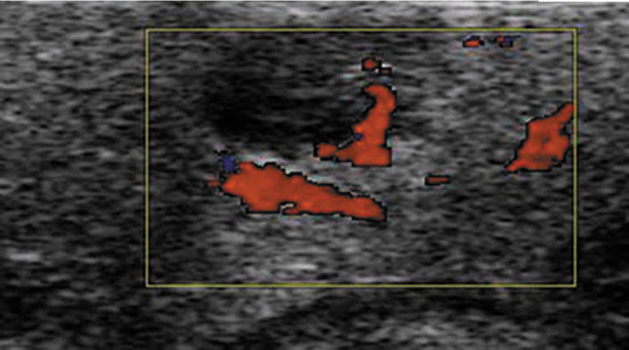
Representative image of colon axial section (A), measurement of thickness of tumour mass (B), Power-Doppler mode of tumour mass (C).

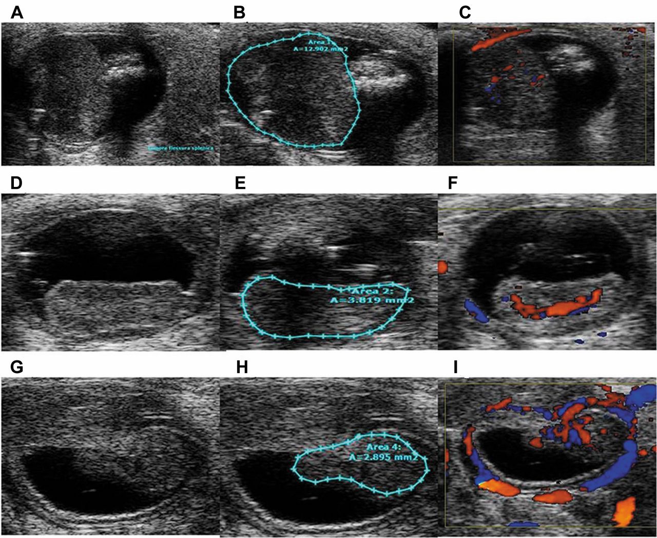
A, D, G: Representative images of tumour mass positive at ultrasound (US) evaluation and confirmed by histological analysis; B, E, H: Tumour area measurement; C, F, I: power Doppler mode of US for colorectal cancer.

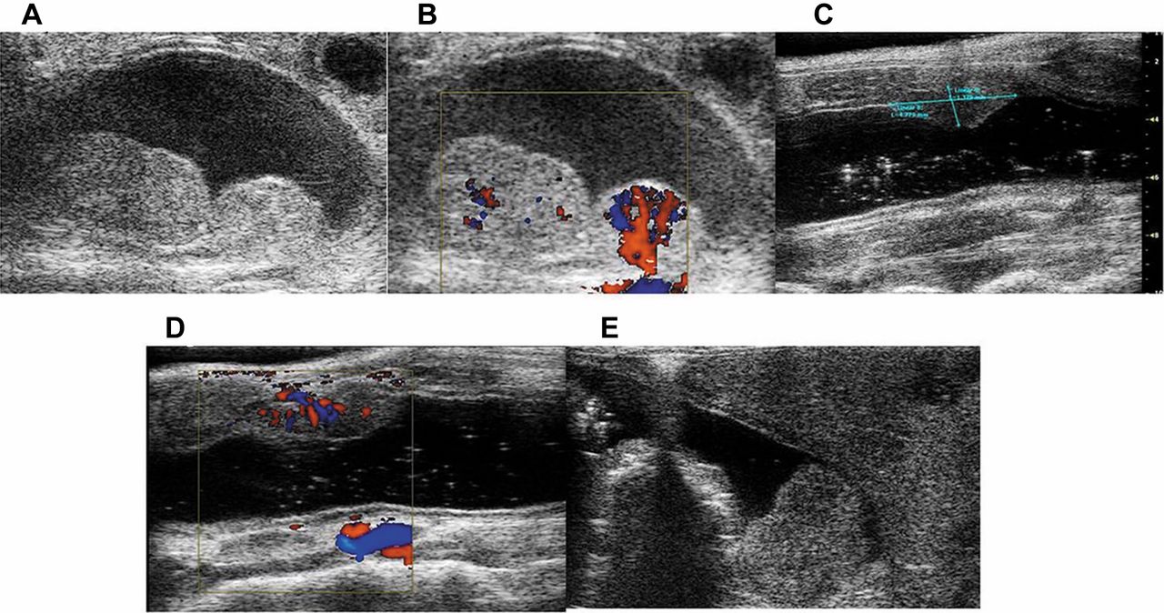
Representative images of longitudinal tumour mass that showing the presence of two tumour lesions not shown in coronal section in B mode (A), an image in Power Doppler mode (B), false-positive lesion in longitudinal mode of increased thickness of rectal wall in B-mode (C), an image in Power Doppler mode (D), fecal material present in a section of colon (E).
Results
Tumour burden analysis. Macroscopic examination of large bowels revealed tumours in six out of 14 mice, evaluated 40 weeks after the first AOM injection. Macroscopic inspection of the intestines demonstrated all six tumour in the large bowel, (n=4 distal part, n=2 mid large bowel).
Ultrasound assessment of tumour mass. Imaging the non-distended colon is difficult owing to the collapse of the colonic walls. In fact, without bowel distension, US imaging was insufficient to discriminate the wall from the luminal contents (34). The lumen of a mouse 39 weeks after exposure to AOM was clearly distended by rectal introduction of intraluminal gel, and the tumours were distinguishable along the explored section. During the period of observation, six mice died. After 40 weeks, US revealed seven tumours in 14 mice while ex vivo examination revealed eight tumours (Figures 2, 3, 4 and 5). In one out 14 mice, US revealed focal wall thickening that represented a false positive case (Figure 6A). In two out 14 mice, US did not reveal the tumour because the tumor dimensions were very small. These data show that US is able to detect only tumours of 3 mm2 or more.
Tumour grading. The standard endpoint for measuring tumours in this model is 40 weeks after exposure to AOM (33). At this time, colonic neoplasm adenocarcinomas can be seen histologically. We have sacrificed the animals 40 weeks after the last injection of AOM. Four out of the 6 analyzed animals had a tumor in 3 out of 4 cases were colon adenocarcinoma.
Discussion
Currently, there are several tests commonly used to screen for CRC, including the focal occult blood test, digital rectal examination, sigmoidoscopy, barium enema, colonoscopy, and, most recently, computed tomographic (CT) colonography. The focal occult blood test is limited in its sensitivity for early disease. Digital rectal examination and sigmoidoscopy examine only a limited anatomic section of the colon. Barium enema is a projection technique that is insensitive for early polyps. Colonoscopy allows for visualization of the entire colon and detection of pre-cancerous lesions such as aberrant crypt foci, polyps, and CRC. A particular advantage of colonoscopy is that biopsy can be performed during the examination. The disadvantages of colonoscopy include an unpleasant bowel preparation, the need for sedation, and potential perforation of the bowel. Moreover, the accuracy of a colonoscopy depends on the skill of the operator to visualize all surfaces of the colon. More recently, magnetic resonance (MR) and CT colonographies have been explored as complementary or alternatives for optical colonoscopy. Both MR and CT colonographies often include computer-generated fly-through of the colon referred to as virtual colonoscopy. Both offer a less demanding alternative for the patient and allow complete visualization of the colonic surface after the bowel is distended with an enema or with a gas such as air or carbon dioxide (34, 35). In recent years, research is focused on translational studies, so the possibility of studing tumourigenesis step by step in a carcinogenesis model of AOM represents an important strategy for new schedules of treatments. Thanks to animal models, it is possible detect the tumour with respect to the age of mice and to provide information on the number of stages and nature of intermediate steps in the tumourigenic process, and this helps to determine the nature of the effect of interventions (21, 31). These insights will be important for understanding the mechanism of therapeutic interventions and designing new approaches. The availability of animal models which reproduce human diseases has increased our knowledge of human physiopathology and led to new diagnostic and therapeutic approaches. This study, based on an animal model of colonic adenocarcinoma, was designed to validate intraluminal gel US study to gain live information before the endpoint of the experiments and to compare anatomopathological features with histology. Non-invasive, small animal in vivo imaging has taken on an increasingly important role in pre-clinical research. In the present study, we investigated whether intraluminal gel US visualized colorectal cancer lesions induced by AOM in an experimental mouse model. In this study, we showed for the first time the possibility of assessing the formation of tumour mass with intraluminal gel US, demonstrating that US is an examination of rapid execution compared to other diagnostic techniques. The use of the intraluminal gel study method and other diagnostic imaging techniques for the colon are emerging as key techniques of in vivo morphological imaging, given their ability to identify events with sufficient sensitivity, specificity and temporal and spatial resolution. Our preliminary findings suggest that US has a good sensitivity when the tumours are 3 mm2 or more in size, and US is also able to visualize other metastatic localizations (Figure 6). However, the limits of the method are the following: it is not always possible to achieve relaxation of the transverse colon (Figure 7A); there may be failure to distend of the right colon (Figure 7B); the presence of fecal material may affect the visualization of the tumour (Figure 7C). Subsequently, US implemented with intraluminal gel injection and ECD is effective for the in vivo visualization and monitoring of colonic tumour progression, carcinogenesis and treatment response. From the present study, the following conclusions can be drawn: AOM treatment leads to development of colonic adenocarcinomas; the AOM model is a valid model of CRC that allows for evaluation of intervention strategies with intraluminal gel US study as a valid outcome measure; intraluminal gel US is an examination of rapid execution compared to other diagnostic techniques; US has a good sensitivity when tumours are 3 mm2 or more in size and is also able to visualize other metastatic localizations

A: Macroscopic colorectal cancer lesion and histology. B: Gross image of ex vivo colorectal cancer. C: Haematoxylin and eosin–stained section of colorectal cancer lesion.
Representative image of liver metastasis by colorectal cancer.

A: Representative image of collapsed wall of the transverse colon. B: Failure of the right colon to distend. C: Presence of fecal material preventing visualization of the tumour mass.
Acknowledgements
A special thanks goes to Dr. Luca Vannucci for his valuable suggestions on animal models of carcinogenesis. We thank Lega Italiana Lotta Tumori - Naples and Banco di Napoli-Fondazione.
Footnotes
-
↵* These Authors contributed equally to this study.
- Received April 7, 2013.
- Revision received May 8, 2013.
- Accepted May 10, 2013.
- Copyright © 2013 International Institute of Anticancer Research (Dr. John G. Delinassios), All rights reserved


